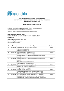
Review Article
Nitric Oxide and the Cardiovascular System: Cell Activation, Vascular
Reactivity and Genetic Variant
Rodrigo Gonçalves Dias1,2, Carlos Eduardo Negrão1, Marta Helena Krieger2
Instituto do Coração - InCor (HCFMUSP)1, São Paulo, SP; Labcardio - Universidade Estadual de Campinas (Unicamp)2, Campinas, SP - Brazil
Summary
Nitric oxide (NO), primarily identified as an endotheliumderived relaxing factor, is a free radical that signals different
biological processes. The identification of NO synthase
(NOS) isoforms and the subsequent characterization of the
mechanisms of cell activation of the enzymes permitted the
partial understanding of both the physiological interactions and
of the mechanisms of the diseases in which NO is involved.
Mainly expressed in the vascular endothelium, the endothelial
NOS isoform (eNOS) plays an important role in the regulation
of vascular reactivity and in the development and progression
of atherosclerosis. The purpose of this review is to contextualize
the reader about the eNOS structure and its mechanisms of cell
activation. In view of the advances in molecular biology, we
will also address the known mechanisms of gene expression
regulation and the role of variants on the genetic code of
eNOS associated with cardiovascular phenotypes. Although
the importance of NO as an atheroprotective molecule is
recognized, our focus will be the review of the literature on
NO and its participation in the modulation of the muscle
vasodilatation phenotype.
Introduction
The primary evidence that the endothelium is an
indispensable component in the regulation of the vascular
tone emerged from experimental analyses demonstrating that,
in the absence of this single layer of squamous epithelium,
acetylcholine-induced vasodilatation did not occur. In that
moment, Furchgott and Zawadzki1 documented the fact that,
when stimulated, the endothelium had the capacity to release
a vasoactive substance, which was named endotheliumderived relaxing factor (EDRF). Some years later, EDRF was
Keywords
Nitric oxide; nitric oxide synthase type III; polymorphism,
genetic.
Mailing address: Rodrigo Gonçalves Dias •
Unidade de Hipertensão - Laboratório de Genética e Cardiologia Molecular
Av. Dr. Enéas de Carvalho Aguiar, 44 (2º andar; Bloco II) - Cerqueira César 05403-000 - São Paulo, SP - Brazil
E-mail: [email protected]
Manuscript received February 12, 2009; revised manuscript received June
26, 2009; accepted August 14, 2009.
68
identified by Ignarro et al2 as nitric oxide (NO), a compound
characterized in 1977 by Ferid Murad which caused smooth
muscle cell relaxation, when released by nitrates. For this
finding, Robert F. Furchgott, Ferid Murad and Louis J. Ignarro
were awarded the Nobel Prize of Physiology or Medicine
in 1998 (Figure 1). A series of studies was responsible for
the demonstration that the endothelium releases other
EDRF such as prostacyclin (PGI2), and the endotheliumderived hyperpolarizing factor (EDHF), in addition to other
endothelium-derived contracting factors (EDCF) such as
endothelin (ET-1), cyclooxygenase pathway products such
as thromboxane A2 (TXA2) and reactive oxygen species such
as the superoxide anion (O2.-)3. In addition to characterizing
the endothelium as a biological sensor capable of detecting
any mechanical, physical or chemical stimulus - and respond
to it - these findings raised the endothelium to the position
of a multifunctional tissue that plays an important role in the
homeostasis of all physiological systems.
Nitric oxide and the cardiovascular system
NO, a gaseous molecule that acts in the signaling of
different biological processes, is a free radical presenting an
unpaired electron in the last layer and a half-life of 4 to 8
seconds in oxygenated aqueous medium4,5. It is described as
a labile gas, capable of free diffusion across cell membranes,
a characteristic that enhances its high biological activity6.
The multiple NO actions were further broadened by the
confirmation that the vascular endothelium is an active
organ and that its integrity promotes beneficial effects,
such as antioxidant, anti-inflammatory, anticoagulant, and
pro-fibrinolytic actions; inhibition of leukocyte adhesion
and migration; inhibition of smooth muscle cell migration
and proliferation; and inhibition of platelet aggregation
and adhesion 7 . Thus, this atheroprotective setting is
characterized by a harmony between substances released by
the endothelium, in which NO is cited as one of the most
relevant vasoactive compounds. Characterized as a systemic
disorder that precedes atherosclerosis and its complications,
endothelial dysfunction in atherosclerotic coronary arteries
was first described by Ludmer et al8 and later related to the
change in NO bioavailability9.
In the current literature, it is agreed that the reduced
NO biological activity, caused both by synthase reduction
and by increased degradation due to oxidation stress, has
been identified as the most relevant mechanism in the
multifactorial process in endothelial dysfunction and in the
participation in the main cardiovascular dysfunctions10. Thus,
reductions in NO bioavailability and subsequent endothelial
Dias et al
Genetics, nitric oxide and vascular reactivity
Review Article
Robert F. Furchgott, Louis J. Ignarro and Ferid Murad were awarded the
1998 Nobel Prize in Physiology or Medicine. The three pharmacologists were
acknowledged for their valuable contribution related to the discovery that Nitric
Oxide is a signaling molecule in the cardiovascular system.
Figure 1 - 1998 Nobel Prize winners in Physiology or Medicine. Source: Available at: <http://nobelprize.org>.
dysfunction trigger, in the vascular environment, events such
as tone alterations, thrombotic dysfunctions, smooth muscle
cell (SMC) proliferation and migration, as well as leukocyte
adhesion11. When endothelial dysfunction occurs, there
is also an increased production of reactive oxygen species
(ROS)12, and these can reduce endothelial NO availability via
different pathways: direct NO inactivation by superoxide with
peroxynitrite (ONOO-)13 formation; reduction in NO synthase
expression and activity due to changes in their substrate or
cofactors, and in the increase in asymmetric dimethylarginine
(ADMA) levels14; and also endothelial NOS uncoupling caused
by increased tetrahydrobiopterin (BH4) oxidation15.
The understanding of the complexity of endothelial
function has improved, and the difficulty in studying each
one of its components separately has been overcome. In
this context, animal models able to reproduce endothelial
dysfunction have been developed, thus enabling the system
to work under conditions of low or high NO bioavailability,
for instance. Additionally, studies in vivo in humans, using
intra-arterial infusion of compounds with the potential of
modulating endothelial-dependent or independent function,
permitted the investigation of modulatory mechanisms on the
vascular function under different physiological conditions and
more prevalent diseases.
Nitric oxide synthases
NO enzymatic production from the amino acid L-arginine
is mediated by a family of three nitric oxide synthases (NOS)
codified by distinct genes16. The isoforms share 50%-60%
homology in the amino acid sequence in the oxidase
and reductase domains 17. These isoforms have distinct
characteristics that reflect their specific functions in vivo18.
Endothelial NO synthase (eNOS or NOS III; 7q35-36) and
neuronal nitric oxide synthase (nNOS or NOS I; 12q24.2) have
a mechanism of constitutive activation (cNOS). The inducible
isoform (iNOS or NOS II; 17cen-q12) is expressed in abnormal
cell processes such as heart failure19, after induction by
cytokines and other inflammatory agents, which results in a high
NO flow20,21. Endothelial NOS is mainly located in endothelial
cell compartments named caveolae22, and is essential for
keeping the baseline vascular tone. This tone is partly mediated
by NO synthesis, a vasoactive compound that participates
in blood flow regulation in the different vascular beds and,
particularly, in the coronary blood flow23. The subcellular
location of NO synthesis has a great influence on its biological
activity. In the 1990’s, the initial identification of eNOS location
in the caveolae in the cell plasma membrane provided the
structural basis for the recognition of compartmentalization
in the mechanisms of cell signaling promoted by NO. The
subsequent observation that eNOS interacts directly with
structural proteins of the caveola - the caveolins, provided
biochemical evidence of the interaction between eNOS and
caveola and its implication with numerous signaling molecules
concentrated in this environment of the cell membrane24. A
great number of evidences revealed that the caveolae are
capable of recruiting numerous signaling molecules and to
regulate their activities instead of acting as a simple support for
cell exchange and transportation25. Thus, it has been described
that eNOS is located inside the caveolae and is kept in a less
active state via its interaction with caveolin-126.
eNOS structure
eNOS functions as a dimer consisting of two identical
monomers, which, in turn, can be functionally and structurally
Arq Bras Cardiol 2011; 96(1): 68-75
69
Dias et al
Genetics, nitric oxide and vascular reactivity
Review Article
divided into two major domains: a C-terminal reductase
domain, homologous to cytochrome P450 which contains
binding sites for NADPH, flavine mononucleotide (FMN)
and flavine adenine dinucleotide (FAD); and an N-terminal
oxidase, which subtracts one electron from the L-arginine
substrate and has binding sites for heme iron, for the
tetrahydrobiopterin cofactor (BH4) and for L-arginine20,21,27
(Figure 2). The catalysis reaction of constitutive NOS involves
two oxidation stages: L-arginine hydroxylation into NGhydroxy-L-arginine, followed by oxidation of this intermediate
compound with utilization of one NADPH electron, thus
forming L-citrulline and NO28. This reaction consumes 1.5
mol of NADPH and 2 moles of oxygen per mol of L-citrulline
formed16,29,30. Cofactors such as heme iron, BH4 and L-arginine
have been studied in depth, and their low bioavailability
induces the phenomenon of dysfunctional eNOS31-33. Heme
iron is essential for dimerization of the three isoforms34: low
concentrations or absence of L-arginine catalyze oxygen
reduction in superoxide (O2.-)35, and decreased levels of BH4
lead to the simultaneous production of NO e O2.-, products
that react with each other forming peroxynitrite (ONOO-)36.
Regulation of eNOS activity and gene
expression
Ever since it was verified that the endothelial cells contain
a baseline concentration of eNOS protein, the eNOS gene
was considered constitutively expressed. Interestingly, further
studies demonstrated that stable mRNA concentrations are
subject to a modest level of regulation37. The eNOS gene
promoter was cloned, and it was demonstrated that it has a
complex regulatory mechanisms of gene expression. Like the
promoter of constitutively expressed genes, the eNOS gene
promoter does not contain the TATA box sequence. However,
it has multiple cis-regulatory DNA sequences, including CCAT
box, Sp1 sites, GATA motifs, CACCC box, AP-1 and AP-2
sites, p53 binding sites, and NF-1 elements, in addition to
sequences responsive to sterol elements and shear stress38.
Bovine and human promoter sequences have a homology
of 75%, thus suggesting a high evolutionary preservation of
the gene transcriptional regulation. Located in the proximal
promoter, the positive regulatory domains I and II (PRD I
and PRD II) are involved in the baseline regulation of gene
Monomer
Arginine
N-terminal domain
Oxidase
Citrulline
C-terminal domain
Reductase
Arginine
Citrulline
N-terminal domain
Oxidase
C-terminal domain
Reductase
Figure 2 - A) Model proposed for the dimeric eNOS structure. B) Electron transfer between cofactors and substrates of the enzyme structure. The electron flows in
the direction NADPH → FAD → FMN in the reductase domain of one monomer to Fe in the oxidase domain of the contralateral monomer. In figure A, observe that
electron flow and arginine catalysis are shown in only one side of the enzyme. Electron transfer from one domain to another is mediated by calmodulin, which justifies
the need for its linking to the recognition site for enzyme activation and subsequent NO synthesis. FAD - flavine adenine dinucleotide; FMN - flavine mononucleotide;
BH4 - tetrahydrobiopterine; Fe - heme iron; CaM - calmodulin.
70
Arq Bras Cardiol 2011; 96(1): 68-75
Dias et al
Genetics, nitric oxide and vascular reactivity
Review Article
transcription, showing an affinity with the transcription factors
Sp-1, Sp-3, Ets-1, Elf-1, YY1 and MYC-associated zinc finger
protein39. Studies in vitro demonstrated that eNOS promoter
responsiveness to shear stress is dependent on sequences
located between -1,000 and -975, a region relative to
transcription start40,41. Besides, the binding of NF-κβ subunits
p50 and p65 to the responsive element GAGACC (-990; -984)
- located upstream of the transcription start site, is involved
in shear stress activation of the promoter42. Also, transcription
in bovine endothelial cells subject to laminar flow showed
a nine-fold increase in mRNA. This effect was mediated
by two distinct mechanisms: 1) transient increase in gene
transcription, and 2) subsequent prolonged mRNA half-life43.
The mechanism of eNOS activation has been described
as the most elaborated of the free isoforms, reflecting
the complexity of the physiologic control of the different
vascular beds17,44. The classical mechanism of constitutive
isoform activation is calcium (Ca++)-dependent, whereas
iNOS is independent of the elevation of intracellular Ca++
concentrations, because of the high affinity of the enzyme
binding with calmodulin31,39,44.
The complexity of the eNOS post-transcriptional regulatory
mechanisms has been considered in the dimerization of the
protein subunits and in the role of the protein caveolin in
the formation of the caveolar structure45. Endothelial NOS is
located inside the caveola and is kept in a less active state via
its interaction with caveolin-126. The mechanisms migration
from the cell membrane to the Golgi apparatus and Akt, PKA,
and AMPK-kinase-dependent phosphorylations are described
as responsible for the activation of this NOS isoform. Thus,
the interaction keeps eNOS inactive, and calmodulin acts
directly on the competition with caveolin in the promotion
of the calcium-dependent enzyme activation15. Endothelial
NOS activity is well known in the vascular environment,
and is regulated by six mechanisms after its translation:
lipid inclusion; calcium/modulin-dependent mechanism;
direct protein-protein interactions; several phosphorilations;
glycosylation; and substrate and cofactor availability. Thus,
eNOS can interact with several proteins in their “less active”
or “more active” states. The plasmalemma-bound eNOS
N-myristoylation and palmitoylation processes are required,
which, in this state, is associated with caveolin-1 and HSP
90. The protein that interacts with HSP70, named “CHIP”,
interacts with both HSP70 and HSP90, and downregulates
eNOS traffic to the Golgi apparatus, in contrast to “NOSIP”
and “NOSTRIN” which can downregulate eNOS location in
the plasma membrane. The main eNOS activation mechanism
occurs via phosphorylation of the amino acid serine at position
1.17746 by the Akt-kinase enzyme (or kinase protein B),
which increases eNOS sensitivity to baseline Ca++/calmodulin
concentrations47. Tonic or phasic eNOS activation in response
to blood flow is independent of Ca++ concentration changes
and constitutes the shear stress. Dimmeler et al48 demonstrated
that the exchange of the serine 1177/1179 residue for the
amino acid alanine makes eNOS incapable of responding
to phosphorylation and activation by the Akt enzyme, a
phosphatidylinositol-3-kinase (PI-3K)-dependent pathway.
Although phosphorylation of the serine1177 residue plays
a pivotal role in eNOS enzyme activation, its regulation is
known to be also dependent on the phosphorylation pattern
of other currently well-characterized sites49. Phosphorylation
of the serine633 residue, located in the binding domain of
flavine mononucleotide (FMN) also increases eNOS activity
and seems to be particularly important to keep NO synthesis
after serine1177 residue phosphorylation and activation by
Ca++/calmodulin. Phosphorylated by protein kinase C (PKC),
the threonin495 residue interferes with the calmodulin binding
domain, thus downregulating NO synthesis.
Recent studies have evaluated the capacity of certain drugs,
such as antioxidants and renin-angiotensin system blockers,
to reduce endothelial dysfunction by means mechanisms of
eNOS activation via phosphorylation of specific sites such as
the serine1177 residue. This phosphorylation may be affected
by the subcellular enzyme location, such as the caveolae,
intercellular junctions, Golgi apparatus, and cytosolic
compartments, as well as by protein kinases and phosphatases
associated with these structures. Recently, our group50
demonstrated that Telmisartan, an angiotensin II receptor
blocker, promotes reduction of endothelial dysfunction by
means of eNOS activation via phosphorylation of specific
sites, such as serine1177 and serine635 residues.
Nitric oxide, vascular tone and muscle
vasodilatation
After confirmation that NO is synthesized by endothelial
cells and that it participates in the cardiovascular hemodynamic
regulation, interest was focused on the quantification of its
participation in this homeostasis of this system. Studies in
vivo in healthy individuals demonstrated that the intra-arterial
administration of NG-monomethyl-L-arginine (L-NMMA) - an
unspecific NOS activity blocker, reduces local blood flow
by 25% to 50%51. Although the baseline vascular tone is the
product of vasoconstrictor versus vasodilator forces, these
results demonstrate that NO is, at least in part, the modulator
of the phenotype at issue.
During exercise and mental stress conditions, in addition
to the tachycardic response and blood pressure increase,
vasodilatation in the skeletal muscle bed is also observed as
part of the physiological responses of body adjustment. It was
hypothesized that part of this muscle vasodilator response
would be modulated by a neural component, and this was
later evidenced by the existence of cholinergic sympathetic
fibers in the skeletal muscles of some mammal species,
except for primates and humans. Electrical stimulation of
the sympathetic nerve was verified to lead to vasodilatation
in the human muscle bed when presynaptic norepinephrine
release was inhibited by intra-arterial drug infusion. However,
this vasodilator response was attenuated when a muscarinic
antagonist was administered52,53. Later, it was evident that NO
is, at least in part, the modulator of the vasodilator response
verified when cholinergic sympathetic fibers are stimulated54. In
fact, Blair et al55 had already shown that forearm vasodilatation
in humans during physiologic maneuvers is mediated by
a neural component. During application of mental stress,
blood flow in the sympathectomized limb did not change in
comparison to the blood flow in the control limb. In addition,
intra-arterial atropine infusion in the control limb reduced
Arq Bras Cardiol 2011; 96(1): 68-75
71
Dias et al
Genetics, nitric oxide and vascular reactivity
Review Article
the increase in blood flow by approximately 50%. At that
moment, using indirect evidence, the authors suggested the
existence of cholinergic sympathetic enervation in the skeletal
musculature of humans. Mimicking animal experiments,
Dietz et al’s studies56,57 later showed that part of the muscle
vasodilator response, as measured in the forearm during
exercise or mental stress, is attenuated by the intra-arterial
administration of L-NMMA. The mechanisms through which
acetylcholine and NO are synthesized and released during the
body defense reactions in humans are still not fully understood.
Evidences obtained with pharmacological blockade permit
only the suggestion of the existence of cholinergic sympathetic
fibers in the skeletal musculature. Due to these limitations,
the authors do not rule out the possibility of vasodilatation
being caused by a combination between circulating and local
factors. A small part of endothelial cells could synthesize
and release acetylcholine58. In addition, activation of β2adrenergic receptors located in the vascular smooth muscle
would result in relaxation of this tissue and, consequently, in
vasodilatation. However, Majmudar et al59 verified that part
of the vasodilatation resulting from β2-adrenoceptor activation
is mediated by NO. Although the authors do not explain the
mechanism responsible for this phenomenon, approximately
25% of the vasodilatation observed in the forearm with
infusion of Ritodrine (a selective β2-adrenergic agonist) was
attenuated with the concomitant administration of L-NMMA.
These results suggest the existence of β2-adrenoceptors in the
vascular endothelium, thus contributing to the increase in
eNOS activity. In addition, increased mechanical stimulation
of the vascular endothelium would result in increased NO
synthesis via PI-3K-Akt kinase, with subsequent serine1177
residue phosphorylation.
eNOS polymorphisms and functional
studies on the G894T variant
Genotyped and sequenced in 1993 by Marsden et al38
(GenBank D26607), eNOS is located in chromosome 7q3536, and variations in its sequence have been described in the
promoter, exons and introns27. The gene (21-22 kbp) comprises
26 exons and 25 introns with 133 kDa. The polypeptide
sequence generated contains 1,203 amino acids38. The
existence of three single nucleotide polymorphisms (SNP)
in the promoter, in non-binding locations of transcription
factors has already been described in the literature39. SNP
were found in introns 2, 11, 12, 18, 22 and 2360, and repeat
sequence polymorphisms in introns 2, 4, 8, and 1338,61. Of
the polymorphisms found in exons 6 and 7, replacement of
the nitrogenous base guanine for thymine (G→T) at position
894 located in exon 7 results in a change of the amino acid
glutamate (GAG) to aspartate (GAT) at position 298 of the
polypeptide sequence62. It is suggested that polymorphisms
located in the gene promoter have an influence on mRNA
transcription, whereas polymorphisms located in coding
regions may result in changes in enzymatic activity63.
Residue 298 is located outside in the oxidase domain of the
enzyme, binding sites for L-arginine or BH4. Enzymatic studies
using recombinant eNOS showed no difference in Michaelis
constant (km) or in Vmax between the two enzyme forms63.
72
Arq Bras Cardiol 2011; 96(1): 68-75
Although the enzymatic activity is seemingly not affected by
the Asp298 form of the enzyme, Tesauro et al64 showed that this
variant is more susceptible to proteolytic cleavage into 100 and
35 KDa fragments, more precisely in position Asp298-Pro299,
in comparison to the Glu298 variant. However, Fairchild et
al65 demonstrated that this proteolytic susceptibility occurred
because of an artifact of the sample preparation method. The
inconsistency of these results does not rule out the possibility
that an unknown proteolytic mechanism or even a change
in post-transcriptional regulation may be modulated by the
Asp298 variant of the enzyme in vivo.
Association between the G894T variant
of the eNOS gene and cardiovascular
phenotypes
Several diseases have been associated with abnormalities
in NO biosynthesis, and many of these conditions are related
to autonomic dysfunction. Population genetics studies
have demonstrated a significant association of eNOS gene
G894T polymorphism with coronary artery disease (CAD)6668
and also with acetylcholine (Ach)-induced coronary
vasospasm69. Among the cardiovascular dysfunctions, the
G894T variant was also demonstrated to be associated with
hypertension70,71, although this association had not been
verified in other populations72,73.
The correlation between genotype and clinical phenotype
varies both quantitatively and qualitatively, and the
inconsistent association between eNOS polymorphism and
several clinical phenotypes is a phenomenon commonly
observed in other phenotype-associated genes 27. This
inconsistency has been attributed to environmental factors,
independent alleles, interaction between genes and variability
in clinical phenotypes. The importance of environmental
factors in the genesis of diseases is reflected in morbidity
and mortality differences between genetically homogeneous
groups, however with different life styles.
A genetic variation may not be relevant to a determined
population, thus reflecting differences in the frequency
of allele distribution. One example is the frequency of
the allele 894T, which is significantly higher in white
populations in comparison to the Japanese population23.
The inconsistent association between eNOS polymorphism
and vascular changes is still being attributed to the variation
in eNOS distribution in different organs. Arteries of specific
organs are subject to different hemodynamic pressures,
determining the vessel wall response and subsequent degree
of endothelial dysfunction27.
Philip et al 62 studied patients undergoing surgical
revascularization and observed that vascular reactivity to
phenylephrine (PE) infusion is influenced by the G894T variant
of the eNOS gene. The dose-dependent vasoconstrictor
response to PE was significantly greater for the TT and GT
alleles in comparison to the homozygote GG group, thus
indicating that vascular reactivity to vasoconstrictor drugs
may be influenced by eNOS polymorphism in humans.
The greater dose-dependent response of patients with the
894T variant suggests reduced NO biosynthesis. Systemic
Dias et al
Genetics, nitric oxide and vascular reactivity
Review Article
administration of NG-nitro-L-arginine methyl esther (L-NAME)
leads to hypertension in humans74,75. According to Frandsenn
et al76, the administration of 4mg/kg of L-NAME in humans
reduces eNOS activity by 67%. The possibly reduced NO
bioavailability subsequent to eNOS gene variations is an
important candidate to the susceptibility to the development
of endothelial dysfunction62,63 and changes in modulation of
the sympathetic nervous activity on the vessel77.
Studies demonstrating the association between the G894T
variant of the eNOS gene and cardiovascular phenotypes point
to a possible reduction in NO bioavailability, thus suggesting
that eNOS transcribed from the mutant allele has an abnormal
enzymatic activity. The lack of evidence supporting this
rationale encouraged us to test the functionality of the genetic
variant on the muscle vasodilatation phenotype78. If the G894T
variant of the eNOS gene could reduce the enzymatic activity,
individuals with the mutant allele (T) would show a smaller
increase in muscle blood flow in response to the handgrip
isometric exercise. In order to test this hypothesis, 287
individuals were genotyped; from these, 33 healthy individuals
were selected to represent three genotypes: GG (wild type),
GT and TT. The results showed that attenuated reflex muscle
vasodilatation in response to exercise occurred only in the
TT genotype, since vasodilatation among heterozygotes (GT
genotype) was similar to that observed among homozygotes
for the G allele (GG genotype). These results suggest that the
presence of the G allele is sufficient to overcome a possible
T allele deficiency. Subsequent analyses in vivo proved that
the attenuated muscle vasodilatation observed in the TT
genotype is a consequence of the reduced eNOS-mediated
vasodilatation, since intra-arterial L-NMMA infusion did not
change the vasodilator response to exercise in this genotype.
In contrast, L-NMMA significantly reduced the vasodilator
response to exercise in the GG genotype to values similar
to those of the TT genotype. Also, our study demonstrated
that the attenuated muscle vasodilatation observed in the
TT genotype may not be accounted to a possibly increased
vasoconstrictor sympathetic tone. In fact, the muscle
sympathetic nervous activity, as measured directly in the fibular
nerve using the microneurography technique, increased in a
similar fashion among the genotypes during exercise. Although
these results do not confirm the functionality of the T allele
in the alteration of the enzymatic activity (this variant may be
in linkage disequilibrium with other functional variant on the
same gene or on a nearby gene on the same chromosome),
they may be used as a marker of the dysfunction observed
in the muscle vasodilatation phenotype. This is the first
demonstration that the G894T variant of the eNOS gene
is functionally associated with reduced eNOS-mediated
vasodilatation. Also, these results suggest that the reduced
endothelium-dependent vasodilatation may anticipate
vascular dysfunction in individuals with the TT-genotype.
Conclusion
NO synthases were proven to be constitutively expressed
(eNOS and nNOS) and NO plays an important role in the
regulation of cardiovascular activities. These facts aroused the
interest for the understanding of the molecular and cellular
mechanisms that regulate their functionality. Although these
mechanisms are complexo and difficult to access, some of
them have already been elucidated, thus permitting partial
knowledge of the NO biology. In this context, the advances
in molecular biology techniques enabled the identification
of variants in the human genetic code that could, at least in
part, explain the phenotypical response variation between
individuals. Genetic association studies are not easy to
understand. A single gene may have a small to moderate
participation in the regulation of a multigenic phenotype and,
in this situation, a determinate functional genetic variant in
this gene would explain only a small part of the phenotype
response variation. The screening of candidate genes, that
is, the identification of multiple genes and their respective
polymorphisms which trigger variations in the cardiovascular
function leads to the moment when part of the diagnosis and
management adopted for the treatment will be based on
genomic medicine.
Potential Conflict of Interest
No potential conflict of interest relevant to this article was
reported.
Sources of Funding
This study was funded by FAPESP.
Study Association
This article is part of the thesis of doctoral submitted by
Rodrigo Gonçalves Dias, from Universidade Estadual de
Campinas e Instituto do Coração - InCor (HCFMUSP) de
São Paulo.
References
1. Furchgott RF, Zawadzki JV. The obligatory role of endothelial cells in the
relaxation of arterial smooth muscle by acetylcholine. Nature. 1980; 288:
373-6.
2. Ignarro LJ, Buga GM, Wood KS, Byrns RE, Chaudhuri G. Endothelium-derived
relaxing factor produced and released from artery and vein is nitric oxide.
Proc Natl Acad Sci USA. 1987; 84: 9265-9.
3. Vila E, Salaices M. Cytokines and vascular reactivity in resistance arteries. Am
J Physiol Heart Circ Physiol. 2005; 288: H1016-21.
4. Moncada S, Palmer RM, Higgs EA. The biological significance of nitric oxide
formation from L-arginine. Biochem Soc Trans. 1989; 17: 642-4.
5. Kojda G, Harrison D. Interactions between NO and reactive oxygen species:
pathophysiological importance in atherosclerosis, hypertension, diabetes
and heart failure. Cardiovasc Res. 1999; 43: 562-71.
6. Palmer RM, Ferrige AG, Moncada S. Nitric oxide release accounts for the
biological activity of endothelium-derived relaxing factor. Nature. 1987;
327: 524-6.
Arq Bras Cardiol 2011; 96(1): 68-75
73
Dias et al
Genetics, nitric oxide and vascular reactivity
Review Article
7. Förstermann U. Janus-faced role of endothelial NO synthase in vascular
disease: uncoupling of oxygen reduction from NO synthesis and its
pharmacological reversal. Biol Chem. 2006; 387 (12): 1521-33.
8. Ludmer PL, Selwyn AP, Shook TL, Wayne RR, Mudge GH, Alexander RW, et
al. Paradoxical vasoconstriction induced by acetylcholine in atherosclerotic
coronary arteries. N Engl J Med. 1986; 315: 1046-51.
9. Verma S, Buchanan MR, Anderson TJ. Endothelial function testing as a
biomarker of vascular disease. Circulation. 2003; 108: 2054-9.
10.Pepine CJ The impact of nitric oxide in cardiovascular medicine: untapped
potential utility.Am J Med. 2009; 122 (5 Suppl): S10-5.
11.Madamanchi NR, Vendrov A, Runge MS. Oxidative stress and vascular
disease.Arterioscler Thromb Vasc Biol. 2005; 25: 29-38.
12.Cai H. NAD(P)H oxidase-dependent self-propagation of hydrogen peroxide
and vascular disease. Circ Res. 2005; 96: 818-22.
13.Gao L, Mann GE.Vascular NAD(P)H oxidase activation in diabetes: a doubleedged sword in redox signalling.Cardiovasc Res. 2009; 82 (1): 9-20.
14.De Gennaro Colonna V, Bianchi M, Pascale V, Ferrario P, Morelli F, Pascale
W, et al. Asymmetric dimethylarginine (ADMA): an endogenous inhibitor of
nitric oxide synthase and a novel cardiovascular risk molecule. Med Sci Monit.
2009; 15 (4): RA91-101.
15.Sessa WC. eNOS at a glance. J Cell Sci. 2004; 117: 2427-9.
16.Marletta MA. Nitric oxide synthase: aspects concerning structure and
catalysis. Cell. 1994; 78: 927-30.
17.Govers R, Rabelink TJ. Cellular regulation of endothelial nitric oxide synthase.
Am J Physiol Renal Physiol. 2001; 280: F193-206.
18.Stuehr DJ. Structure-function aspects in the nitric oxide synthases. Annu Rev
Pharmacol Toxicol. 1997; 37: 339-59.
19.Ferreiro CR, Chagas AC, Carvalho MH, Dantas AP, Scavone C, Souza LC, et
al. Expression of inducible nitric oxide synthase is increased in patients with
heart failure due to ischemic disease. Braz J Med Biol Res. 2004; 37: 1313-20.
20.Andrew PJ, Mayer B. Enzymatic function of nitric oxide synthases. Cardiovasc
Res. 1999; 43: 521-31.
21.Alderton WK, Cooper CE, Knowles RG. Nitric oxide synthases: structure,
function and inhibition. Biochem J. 2001; 357: 593-615.
33.Cai H, Harrison DG. Endothelial dysfunction in cardiovascular diseases: the
role of oxidant stress. Circ Res. 2000; 87: 840-4.
34.Klatt P, Pfeiffer S, List BM, Lehner D, Glatter O, Bachinger HP, et al.
Characterization of heme-deficient neuronal nitric-oxide synthase reveals a
role for heme in subunit dimerization and binding of the amino acid substrate
and tetrahydrobiopterin. J Biol Chem. 1996; 271: 7336-42.
35.Mayer B, John M, Heinzel B, Werner ER, Wachter H, Schultz G, et al. Brain
nitric oxide synthase is a biopterin- and flavin-containing multi-functional
oxido-reductase. FEBS Lett. 1991; 288: 187-91.
36.Beckman JS, Koppenol WH. Nitric oxide, superoxide, and peroxynitrite: the
good, the bad, and ugly. Am J Physiol. 1996; 271: C1424-37.
37.Searles CD. Transcriptional and posttranscriptional regulation of endothelial
nitric oxide synthase expression. Am J Physiol Cell Physiol. 2006; 291:
C803-16.
38.Marsden PA, Heng HH, Scherer SW, Stewart RJ, Hall AV, Shi XM, et
al. Structure and chromosomal localization of the human constitutive
endothelial nitric oxide synthase gene. J Biol Chem. 1993; 268: 17478-88.
39.Karantzoulis-Fegaras F, Antoniou H, Lai SL, Kulkarni G, D’Abreo C, Wong
GK, et al. Characterization of the human endothelial nitric-oxide synthase
promoter. J Biol Chem. 1999; 274: 3076-93.
40.Malek AM, Jiang L, Lee I, Sessa WC, Izumo S, Alper SL. Induction of nitric
oxide synthase mRNA by shear stress requires intracellular calcium and
G-protein signals and is modulated by PI 3 kinase. Biochem Biophys Res
Commun. 1999; 254: 231-42.
41.Silacci P, Formentin K, Bouzourene K, Daniel F, Brunner HR, Hayoz D.
Unidirectional and oscillatory shear stress differentially modulate NOS III
gene expression. Nitric Oxide. 2000;4:47-56.
42.Davis ME, Grumbach IM, Fukai T, Cutchins A, Harrison DG. Shear stress
regulates endothelial nitric-oxide synthase promoter activity through nuclear
factor kappaB binding. J Biol Chem. 2004; 279: 163-8.
43.Davis ME, Cai H, Drummond GR, Harrison DG. Shear stress regulates
endothelial nitric oxide synthase expression through c-Src by divergent
signaling pathways. Circ Res. 2001; 89: 1073-80.
22.Shaul PW, Anderson RG. Role of plasmalemmal caveolae in signal
transduction. Am J Physiol. 1998; 275: L843-51.
44.Michel T, Feron O. Nitric oxide synthases: which, where, how, and why? J
Clin Invest. 1997; 100: 2146-52.
23.Wang XL, Sim AS, Wang MX, Murrell GA, Trudinger B, Wang J. Genotype
dependent and cigarette specific effects on endothelial nitric oxide synthase
gene expression and enzyme activity. FEBS Lett. 2000; 471: 45-50.
45.Zhang Q, Church JE, Jagnandan D, Catravas JD, Sessa WC, Fulton D.
Functional relevance of Golgi- and plasma membrane-localized endothelial
NO synthase in reconstituted endothelial cells. Arterioscler Thromb Vasc Biol.
2006; 26: 1015-21.
24.Garcia-Cardena G, Martasek P, Masters BS, Skidd PM, Couet J, Li S, et al.
Dissecting the interaction between nitric oxide synthase (NOS) and caveolin:
functional significance of the nos caveolin binding domain in vivo. J Biol
Chem. 1997; 272: 25437-40.
25.Razani B, Lisanti MP. Caveolin-deficient mice: insights into caveolar function
human disease. J Clin Invest. 2001; 108: 1553-61.
26.Bucci M, Gratton JP, Rudic RD, Acevedo L, Roviezzo F, Cirino G, et al. In vivo
delivery of the caveolin-1 scaffolding domain inhibits nitric oxide synthesis
and reduces inflammation. Nat Med. 2000; 6: 1362-7.
27.Wang XL, Wang J. Endothelial nitric oxide synthase gene sequence variations
and vascular disease. Mol Genet Metab. 2000; 70: 241-51.
28.Albrecht EW, Stegeman CA, Heeringa P, Henning RH, van Goor H. Protective
role of endothelial nitric oxide synthase. J Pathol. 2003; 199: 8-17.
29.Griffith OW, Stuehr DJ. Nitric oxide synthases: properties and catalytic
mechanism. Annu Rev Physiol. 1995; 57: 707-36.
30.Korth HG, Sustmann R, Thater C, Butler AR, Ingold KU. On the mechanism
of the nitric oxide synthase-catalyzed conversion of N omega-hydroxyl-Larginine to citrulline and nitric oxide. J Biol Chem. 1994; 269: 17776-9.
31.Harrison DG. Cellular and molecular mechanisms of endothelial cell
dysfunction. J Clin Invest. 1997; 100: 2153-7.
32.Vasquez-Vivar J, Kalyanaraman B, Martasek P, Hogg N, Masters BS, Karoui
74
H, et al. Superoxide generation by endothelial nitric oxide synthase: the
influence of cofactors. Proc Natl Acad Sci USA. 1998; 95: 9220-5.
Arq Bras Cardiol 2011; 96(1): 68-75
46.Boo YC, Kim HJ, Song H, Fulton D, Sessa W. Coordinated regulation of
endothelial nitric oxide synthase activity by phosphorylation and subcellular
localization. Free Radic Biol Med. 2006; 41 (1): 144-53.
47.Fulton D, Gratton JP, McCabe TJ, Fontana J, Fujio Y, Walsh K, et al. Regulation
of endothelium-derived nitric oxide production by the protein kinase Akt.
Nature. 1999; 399: 597-601.
48.Dimmeler S, Fleming I, Fisslthaler B, Hermann C, Busse R, Zeiher AM.
Activation of nitric oxide synthase in endothelial cells by Akt-dependent
phosphorylation. Nature. 1999; 399: 601-5.
49.Mount PF, Kemp BE, Power DA. Regulation of endothelial and myocardial
NO synthesis by multi-site eNOS phosphorylation. J Mol Cell Cardiol. 2007;
42: 271-9.
50.Krieger MH, Di Lorenzo A, Sessa W. Telmisartan reverts endothelial
dysfunction. Am J Physiol Cell Physiol. 2009. In press.
51.Joyner MJ, Dietz NM. Nitric oxide and vasodilation in human limbs. J Appl
Physiol. 1997; 83: 1785-96.
52.Abrahams VC, Hilton SM. The role of active muscle vasodilatation in the
alerting stage of the defence reaction. J Physiol. 1964; 171: 189-202.
53.Bolme P, Fuxe K. Adrenergic and cholinergic nerve terminals in skeletal muscle
vessels. Acta Physiol Scand. 1970; 78: 52-9.
Dias et al
Genetics, nitric oxide and vascular reactivity
Review Article
54.Matsukawa K, Shindo T, Shirai M, Ninomiya I. Nitric oxide mediates cat
hindlimb cholinergic vasodilation induced by stimulation of posterior
hypothalamus. Jpn J Physiol. 1993; 43: 473-83.
55.Blair DA, Glover WE, Greenfield AD, Roddie IC. Excitation of cholinergic
vasodilator nerves to human skeletal muscles during emotional stress. J
Physiol. 1959; 148: 633-47.
56.Dietz NM, Rivera JM, Warner DO, Joyner MJ. Is nitric oxide involved in
cutaneous vasodilation during body heating in humans? J Appl Physiol. 1994;
76: 2047-53.
57.Dietz NM, Engelke KA, Samuel TT, Fix RT, Joyner MJ. Evidence for nitric
oxide-mediated sympathetic forearm vasodiolatation in humans. J Physiol.
1997; 498 (Pt 2): 531-40.
58.Milner P, Kirkpatrick KA, Ralevic V, Toothill V, Pearson J, Burnstock G.
Endothelial cells cultured from human umbilical vein release ATP, substance
P and acetylcholine in response to increased flow. Proc Biol Sci. 1990; 241:
245-8.
59.Majmudar NG, Anumba D, Robson SC, Ford GA. Contribution of nitric oxide
to beta2-adrenoceptor mediated vasodilatation in human forearm arterial
vasculature. Br J Clin Pharmacol. 1999; 47: 173-7.
60.Poirier O, Mao C, Mallet C, Nicaud V, Herrmann SM, Evans A, et al.
Polymorphisms of the endothelial nitric oxide synthase gene - no consistent
association with myocardial infarction in the ECTIM study. Eur J Clin Invest.
1999; 29: 284-90.
61.Miyahara K, Kawamoto T, Sase K, Yui Y, Toda K, Yang LX, et al. Cloning and
structural characterization of the human endothelial nitric-oxide-synthase
gene. Eur J Biochem. 1994; 223: 719-26.
62.Philip I, Plantefeve G, Vuillaumier-Barrot S, Vicaut E, LeMarie C, Henrion
D, et al. G894T polymorphism in the endothelial nitric oxide synthase gene
is associated with an enhanced vascular responsiveness to phenylephrine.
Circulation. 1999; 99: 3096-8.
63.Hingorani AD. Polymorphisms in endothelial nitric oxide synthase and
atherogenesis: John French Lecture 2000. Atherosclerosis. 2001; 154: 521-7.
64.Tesauro M, Thompson WC, Rogliani P, Qi L, Chaudhary PP, Moss J.
Intracellular processing of endothelial nitric oxide synthase isoforms
associated with differences in severity of cardiopulmonary diseases: cleavage
of proteins with aspartate vs. glutamate at position 298. Proc Natl Acad Sci
USA. 2000; 97: 2832-5.
65.Fairchild TA, Fulton D, Fontana JT, Gratton JP, McCabe TJ, Sessa WC. Acidic
hydrolysis as a mechanism for the cleavage of the Glu(298)-->Asp variant of
human endothelial nitric-oxide synthase. J Biol Chem. 2001; 276: 26674-9.
66.Hingorani AD, Liang CF, Fatibene J, Lyon A, Monteith S, Parsons A, et al.. A
common variant of the endothelial nitric oxide synthase (Glu298-->Asp) is
a major risk factor for coronary artery disease in the UK. Circulation. 1999;
100: 1515-20.
67.Hibi K, Ishigami T, Tamura K, Mizushima S, Nyui N, Fujita T, et al. Endothelial
nitric oxide synthase gene polymorphism and acute myocardial infarction.
Hypertension. 1998; 32: 521-6.
68.Shimasaki Y, Yasue H, Yoshimura M, Nakayama M, Kugiyama K, Ogawa H, et
al. Association of the missense Glu298Asp variant of the endothelial nitric oxide
synthase gene with myocardial infarction. J Am Coll Cardiol. 1998; 31: 1506-10.
69.Yoshimura M, Yasue H, Nakayama M, Shimasaki Y, Sumida H, Sugiyama S,
et al. A missense Glu298Asp variant in the endothelial nitric oxide synthase
gene is associated with coronary spasm in the Japanese. Hum Genet. 1998;
103: 65-9.
70.Miyamoto Y, Saito Y, Kajiyama N, Yoshimura M, Shimasaki Y, Nakayama M, et
al. Endothelial nitric oxide synthase gene is positively associated with essential
hypertension. Hypertension. 1998; 32: 3-8.
71.Lacolley P, Gautier S, Poirier O, Pannier B, Cambien F, Benetos A. Nitric
oxide synthase gene polymorphisms, blood pressure and aortic stiffness in
normotensive and hypertensive subjects. J Hypertens. 1998; 16: 31-5.
72.Kato N, Sugiyama T, Morita H, Nabika T, Kurihara H, Yamori Y, et al. Lack of
evidence for association between the endothelial nitric oxide synthase gene
and hypertension. Hypertension. 1999; 33: 933-6.
73.Benjafield AV, Morris BJ. Association analyses of endothelial nitric oxide
synthase gene polymorphisms in essential hypertension. Am J Hypertens.
2000; 13: 994-8.
74.Vallance P, Collier J, Moncada S. Effects of endothelium-derived nitric oxide
on peripheral arteriolar tone in man. Lancet. 1989; 2: 997-1000.
75.Haynes WG, Noon JP, Walker BR, Webb DJ. Inhibition of nitric oxide synthesis
increases blood pressure in healthy humans. J Hypertens. 1993; 11: 1375-80.
76.Frandsenn U, Bangsbo J, Sander M, Hoffner L, Betak A, Saltin B, et al. Exerciseinduced hyperaemia and leg oxygen uptake are not altered during effective
inhibition of nitric oxide synthase with N(G)-nitro-L-arginine methyl ester in
humans. J Physiol. 2001; 531: 257-64.
77.Fabi F, Argiolas L, Chiavarelli M, Del Basso P. Nitric oxide-dependent and
-independent modulation of sympathetic vasoconstriction in the human
saphenous vein. Eur J Pharmacol. 1996; 309: 41-50.
78.Dias RG, Alves MJ, Pereira AC, Rondon MU, Dos Santos MR, Krieger JE, et al.
Glu298Asp eNOS gene polymorphism causes attenuation in non-exercising
muscle vasodilatation. Physiol Genomics. 2009; 37 (2): 99-107.
Arq Bras Cardiol 2011; 96(1): 68-75
75
Baixar