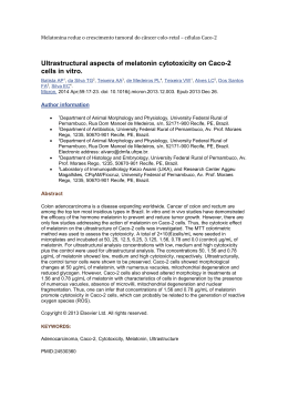
Original Article
Braz J Oral Sci.
October/December 2009 - Volume 8, Number 4
Evaluation of the in vitro biocompatibility
of orthodontic elastics
Matheus Melo Pithon1; Rogério Lacerda dos Santos1; Fernanda Otaviano Martins2;
Maria Teresa Villela Romanos3; Mônica Tirre de Souza Araújo4
1
DDS, MS, PhD Student in Orthodontics, Federal University of Rio de Janeiro, Brazil
2
BS, Federal University of Rio de Janeiro, Brazil
3
BS, Assistant Professor of Microbiology, Federal University of Rio de Janeiro, Brazil
4
DDS, MS, PhD, Assistant Professor of Orthodontics, Federal University of Rio de Janeiro, Brazil
Abstract
Aim: Latex has been extensively used in orthodontics since the advent of the specialty. Natural latex does not
fall into the category of materials known to be entirely inoffensive. The objective of the present in vitro study
is to test the hypothesis that there is no difference in the cytotoxicity between natural latex elastics of different
colors. Methods: The present article compared different latex intra-oral elastics (5/16 = 7.9 mm, mean load).
The samples were divided into six groups of 15 elastics according to their manufacturer: Groups N, Y, V, R, G and
P (Uniden, natural latex elastics and colored elastics, namely, yellow, violet, red, green and pink, respectively).
Cytotoxicity assays were performed by using cell culture medium containing cells from mouse fibroblast cell
line L929. The cytotoxicity was evaluated by using the “dye-uptake” test, which was employed at two different
moments (1 and 24 h). Data were compared by ANOVA and Tukey’s test (P < 0.05). Results: There was statistically
significant difference (P < 0.05) between the groups N, Y, V, R, G, P and the cell control at 1 h. After 24 h, a decrease
in cell viability was observed in all groups. There was statistically significant difference (P < 0.05) between all test
elastics groups and the cell control at 24 h. No statistically significant difference (P >0.05) was found among the
test elastics groups at 24 h. Conclusion: Latex elastics from natural, yellow, violet and red colors induced a
lowest amount of cell lysis compared to the elastics green and pink colors at 1 h, all latex elastics were found
to be highly cytotoxic, regardless of their color at 24 h.
Keywords: cytotoxicity, elastics, biocompatibility, orthodontics.
Introduction
Received for publication: May 27, 2009
Accepted: November 11, 2009
Correspondence to:
Mônica Tirre de Souza Araújo
Universidade Federal do Rio de Janeiro - UFRJ
Faculdade de Odontologia Departamento de Ortodontia
Av. Prof. Rodolpho Paulo Rocco325,
Ilha do Fundão
Rio de Janeiro - RJ - Brasil - CEP: 21941-617
E-mail: [email protected]
Latex elastic has been extensively used in orthodontics since the advent of the specialty. However,
natural latex does not fall into the category of materials known to be entirely inoffensive1-2.
Allergy caused by latex proteins has been well documented3, including immediate
hypersensitivity reactions4. Among the allergic reactions caused by orthodontic elastics, swelling
and stomatitis, erythematous oral lesions, respiratory reactions, and even anaphylactic shock,
the most severe form of allergy5-6, can be cited. Latex allergy occurs in 3-17% of the cases7.
Prevulcanized latex is produced by mixing pure natural latex, which has the highest
molecular weight8, with stabilizers such as zinc oxide and chemically vulcanized materials. The
resulting mixture is then heated until 70o C9. Although zinc is known to be neurotoxic10, the
amount released by orthodontic elastics can be ingested as research studies show no evidence
of harm11. Anti-ozone and anti-oxidant agents are also added to latex during the manufacture
of orthodontic elastics8. This process has the advantage of producing latex with higher mechanical
properties, thus increasing its strength and elasticity9,11.
The use of cell culture medium for testing the toxicity of dental products is a valid way of
understanding the biological behavior of such materials1. The objective of the present in vitro
study was to test the hypothesis that there is no difference in the cytotoxicity between natural
latex elastics of different colors.
Braz J Oral Sci. 8(4):171-174
172
Evaluation of the in vitro biocompatibility of orthodontic elastics
Table 1. Elastic and control groups used for the assays.
Groups
Trademark
Main Composition
Color
N
Uniden
Natural Látex
Natural
000-1204
Y
Uniden
Natural Látex
Yellow
000-1206
V
Uniden
Natural Látex
Violet
000-1206
R
Uniden
Natural Látex
Red
000-1206
G
Uniden
Natural Látex
Green
000-1206
P
Uniden
Natural Latex
Pink
000-1206
C+
Tween 80 (Polyoxyethylene-20-sorbitan, Sigma, St. Louis, MO, USA)
C-
PBS solution (phosphate-buffered saline, Cultilab, Campinas, SP, Brazil)
Material and methods
Latex intra-oral elastics of different colors (5/16" = 7.9 mm, mean
load), were selected for studying their cytotoxicity in cell culture (Table
1). The samples were divided into six groups of 15 elastics according
to their manufacturer: Groups N, Y, V, R, G and P (natural latex elastics
and colored elastics, namely, yellow, violet, red, green and pink,
respectively, Uniden, Sorocaba, SP, Brazil) (Figure 1).
Reference number
(Sigma), and 10% bovine fetal serum (Cultilab) for growth medium or
no bovine fetal serum for maintenance medium only. Next, the cell
culture medium was incubated at 37oC for 48 h.
The method for evaluating the cytotoxicity was the “dye-uptake”13
test. This method is based on neutral red dye incorporated into live
cells. It was used in this experiment only at two periods of evaluation:
1 and 24 h. The 1-h period represents the maintenance of the elastic
in the cell culture medium for 1 h after removal, whereas the 24-h
period represents the maintenance of the elastic in the cell culture
medium for 24 h after removal.
Dye-uptake
Fig. 1: Latex intraoral elastics evaluated in this study: N, Y, V, R, G and P
(Uniden, natural latex elastics and colored elastics, namely, yellow, violet, red,
green and pink, respectively).
All samples had recent manufacturing dates, from the same
production lot and came in sealed plastic packages. The elastics of
experimental and control groups had their both sides previously
sterilized with ultraviolet light (Labconco, Kansas City, MO, USA)
during 30 minutes12.
To verify the cell response to extreme situations, other three
groups were included in the study: Group CC (cell control), consisting
of cells not exposed to any material; Group C+ (positive control),
consisting of Tween 80 and Group C- (negative control), consisting of
PBS solution in contact with the cells (Table 1).
Cells from mouse fibroblast cell line L929 (American Type Culture
Collection - ATCC, Rockville, MD) were cultured in Eagles’ minimum
essential medium (MEM; Cultilab, Campinas, SP, Brazil) by adding
0.03 mg/mL of glutamine (Sigma, St. Louis, MO, USA), 50 µg/mL of
garamicine (Schering Plough, Kenilworth, New Jersey, USA), 2.5 mg/
mL of fungizone (Bristol-Myers-Squibb, New York, USA), 0.25% sodium
bicarbonate solution (Merck, Darmstadt, Germany), 10 mM of HEPES
Braz J Oral Sci. 8(4):171-174
Volumes of 100 µL of L-929 cells were distributed into 96-well
microplates. After 48 h, the growth medium was replaced with 100 µL
of MEM obtained following incubation in the different types of elastics
and positive and negative control at 1 and 24 h. Positive and negative
control groups consisted of culture medium put in contact with 100
µL of Tween 80 and 100 µL PBS solution, respectively.
After 24-h incubation, 100 µL of 0.01% neutral red dye (Sigma)
were added to the culture medium in the 96-well microplates, which
were incubated again for 3 h at 37oC so that the red dye could penetrate
the live cells. Following this period of time, 100 µL of 4% formaldehyde
solution (Vetec, Rio de Janeiro, RJ, Brazil) in PBS (130 mM of NaCl; 2
mM of KCl; 6 mM of Na2HPO4 2 H2O; 1 mM of K2HPO4 1 mM; pH 7.2)
were added in order to promote cell attachment to the plate. After 5
minutes, 100 µL of 1% acetic acid (Vetec) and 50% methanol (Vetec)
were added in order to remove the dye. After 20 minutes, a
spectrophotometer (BioTek Instruments, Winooski, VT, USA) at 492
nm wavelength (λ492 nm) was used for data reading. This test was
repeated 3 different times.
Data were compared by ANOVA, and Tukey’s multiple
comparison test was used for identifying differences between the groups.
Significance level was set at p<0.05.
Results
The results showed significant difference (p< 0.05) between the groups
N, Y, V, R, G, P and the cell control at 1 h (Table 2). Group G produced
the lowest value (34.8% ± 2.9%) and group N produced the more
viability (49.8% ± 10.5%), whereas the viability of the Tween 80 was
8.8% ± 10.5% (Table 2). The results showed statistically significant
differences (p<0.05) between the groups N, Y, V, R and the groups G
and P at 1 h. No significant difference was found between the groups N
and Y, N and V, N and R, Y and V, Y and V, Y and R, V and R, and G and
P tested (p>0.05) at 1 h (Table 2).
Evaluation of the in vitro biocompatibility of orthodontic elastics
173
Table 2. Descriptive statistics for optical density of latex elastics.
Time
Groups
CC
CC+
N
Y
V
R
G
P
1h
24 h
Mean
Median
S. D.
0.874ª
0.866
0.076
0.435 b
0.403cb
0.434dbc
0.379ebcd
0.304f
0.329gf
0.910
0.898
0.084
0.458
0.442
0.452
0.402
0.338
0.346
0.062
0.007
0.008
0.046
0.042
0.013
0.011
0.009
0.008
Viable cells
100.0
99.1
8.8
49.8
46.2
49.7
43.4
34.8
35.7
(%)Mean
Median
0.610ª
0.603
0.054
0.115b
0.102 b
0.106 b
0.101b
0.096 b
0.100 b
0.642
0.634
0.058
0.134
0.127
0.129
0.119
0.108
0.114
S. D.
0.049
0.012
0.007
0.008
0.014
0.007
0.006
0.005
0.007
Viable cells (%)
100.0
98.9
9.0
18.9
16.8
17.5
16.7
15.9
16.4
N=15.Values followed by same letters are not significantly different (p>0.05) for the same time. SD: standard deviation.
After 24 h, a decrease in cell viability was observed in all groups.
Viability ranged from 15.9% to 18.9%, relative to the cell control (Table
2). The lowest viability (15.9% ± 5.2%) corresponded to group G,
whereas the viability of the Tween 80 (positive cytotoxicity control)
was 9.0% ± 12.9%. The results showed statistically significant
differences (p<0.05) between all test elastics groups and the cell control
at 24 h. On the other hand, no statistically significant difference (p>0.05)
was found among the test elastics groups at 24 h (Table 2).
Discussion
Although case reports on latex allergy is not so frequently seen in the
literature, allergic reactions have been relatively prevalent as latexbased products become commercially available. Most of the allergic
reactions14 have been related to the use of orthodontic elastics15, which
is characterized by presence of small vesicles or acute edema and
complaints of itching and burning.
The most serious consequence of natural rubber latex allergy
commonly takes place during the mucosal absorption of natural
rubber latex proteins during intraoperative medical or dental
procedures when health care workers or others already sensitized
become patients16.
Because natural latex rubber has been increasingly used as dental
material, many cytotoxicity issues have been reported as well17.
Preservatives such as sulfur and zinc oxide as well as antioxidants
such as di-thio-carbohydrates, N-nitrosodibutylamine, and Nnitrosopiperidine are all known to be cytotoxic substances18.
Allergy to natural latex occurs because of the presence of many
types of proteins, and the powder covering the orthodontic elastics
works as a transporter for these proteins. Therefore, the development
of non-latex elastics has become increasingly important for clinical
usage17.
As sterilization is a prerequisite for cytotoxicity assay,
ultraviolet light was used in the present study for sterilizing both
sides of the elastics1,12 during 30 minutes. All the elastics were found
to have the same color and malleability following UV light sterilization.
It was demonstrated by Franz et al.19 and Schedle et al.20 that
L929 mouse fibroblasts show comparable results to primary human
gingival fibroblasts and therefore might represent a model for gingival
toxicity in vitro21-22.
The percentage of viable cells was obtained by comparing
the mean optical density (OD) of control cells (no contact with the
materials) with that of cell cultures put in contact with different elastics,
resulting in 50% toxicity for the cell cultures (CC50) (Table 2).
Evidence of this cytotoxic feature was shown following exposition
of the elastics to cell culture medium. It was used in this experiment
only two times of evaluation 1 and 24 h, a time that, usually these
elastics are changed to each 24 h for the patient.
Natural latex elastics from all colors induced a greater amount
of cell lysis at 24 h compared to the time of 1 h. latex elastics from
natural, yellow, violet and red colors induced a lowest amount of cell
lysis compared to the elastics green and pink colors at 1 h.
After 24 h, a lowest viability was observed in all groups regardless
of their color, which is accordance with the findings of Santos, et al.12,23.
This suggests a greater release of toxic ingredients at 24 h, due to a
possible latex degradation and release of allergenic proteins, which
was not shown in 1 h.
Holmes, et al.2 have verified whether the colorants used in the
fabrication of colored latex could have some toxic effect. Their results
showed that these colorants had low toxicity. However, such an effect
is clinically inoffensive. Variations occur in the composition of the
latex elastics and this could explain the different results between the
elastics.
According to Schmalz1, the great danger is that potentially
cytotoxic intra-oral elastics could release substances that might be
ingested by the patient over time, thus causing diseases resulting from
a cumulative effect. It is known that latex is not entirely biocompatible
as it may interact with foods7,24 and medications25.
As these materials are widely used in clinical orthodontics, care
regarding the cytotoxicity of orthodontic elastics should be taken,
mainly with regard to intraoral elastics as they have a very close
contact with gingival and mucosa. Thus, clinically proven biocompatible
materials should be used whenever possible.
It is important for the practitioner to know how to manage
patients presenting latex allergy and how to deal with this problem26.
The safety biocompatibility of silicone has been well proved through
the use of mouth guards in dentistry27. An alternative for patients with
allergy to latex is to use non-latex elastics, which can be used in
orthodontics without jeopardizing the orthodontic treatment28.
It may be concluded that natural, yellow, violet and red latex
orthodontic elastics induced less cell lysis than green and pink elastics
at 1 h. At 24 h, latex elastics were found to be highly cytotoxic, regardless
of their color.
Braz J Oral Sci. 8(4):171-174
174
Evaluation of the in vitro biocompatibility of orthodontic elastics
References
1.
2.
3.
4.
5.
6.
7.
8.
9.
10.
11.
12.
13.
14.
15.
Schmalz G. Use of cell cultures for toxicity testing of dental materials—
advantages and limitations. J Dent. 1994; 22 Suppl 2: S6-11.
Holmes J, Barker MK, Walley EK, Tuncay OC. Cytotoxicity of orthodontic elastics.
Am J Orthod Dentofacial Orthop. 1993; 104: 188-91.
Palosuo T, Alenius H, Turjanmaa K. Quantitation of latex allergens. Methods.
2002; 27: 52-8.
Wakelin SH, White IR. Natural rubber latex allergy. Clin Exp Dermatol. 1999;
24: 245-8.
Everett FG, Hice TL. Contact stomatitis resulting from the use of orthodontic
rubber elastics: report of case. J Am Dent Assoc. 1974; 88: 1030-1.
Tomazic VJ, Withrow TJ, Fisher BR, Dillard SF. Latex-associated allergies and
anaphylactic reactions. Clin Immunol Immunopathol. 1992; 64: 89-97.
Turjanmaa K, Alenius H, Makinen-Kiljunen S, Reunala T, Palosuo T. Natural
rubber latex allergy. Allergy. 1996; 51: 593-602.
Weiss ME, Hirshman CA. Latex allergy. Can J Anaesth. 1992; 39: 528-32.
Perrella FW, Gaspari AA. Natural rubber latex protein reduction with an
emphasis on enzyme treatment. Methods. 2002; 27: 77-86.
Lobner D, Asrari M. Neurotoxicity of dental amalgam is mediated by zinc. J
Dent Res. 2003; 82: 243-6.
Hanson M, Lobner D. In vitro neuronal cytotoxicity of latex and nonlatex
orthodontic elastics. Am J Orthod Dentofacial Orthop. 2004; 126: 65-70.
Santos RL, Pithon MM, Oliveira MV, Mendes GS, Romanos MTV, Ruellas ACO.
Cytotoxicity of intraoral orthodontic elastics. Braz J Oral Sci. 2008; 24: 1520-5.
Neyndorff HC, Bartel DL, Tufaro F, Levy JG. Development of a model to
demonstrate photosensitizer-mediated viral inactivation in blood.
Transfusion. 1990; 30: 485-90.
Snyder HA, Settle S. The rise in latex allergy: implications for the dentist. J Am
Dent Assoc. 1994; 125: 1089-97.
Neiburger EJ. A case of possible latex allergy. J Clin Orthod. 1991; 25: 559-60.
Braz J Oral Sci. 8(4):171-174
16.
17.
18.
19.
20.
21.
22.
23.
24.
25.
26.
27.
28.
Sussman GL, Beezhold DH, Liss G. Latex allergy: historical perspective.
Methods. 2002; 27: 3-9.
Hwang CJ, Cha JY. Mechanical and biological comparison of latex and silicone
rubber bands. Am J Orthod Dentofacial Orthop. 2003; 124: 379-86.
Fiddler W, Pensabene J, Sphon J, Andrzejewski D. Nitrosamines in rubber
bands used for orthodontic purposes. Food Chem Toxicol. 1992; 30: 325-6.
Franz A, Konig F, Skolka A, Sperr W, Bauer P, Lucas T et al. Cytotoxicity of resin
composites as a function of interface area. Dent Mater. 2007; 23: 1438-46.
Schedle A, Samorapoompichit P, Rausch-Fan XH, Franz A, Fureder W, Sperr
WR et al. Response of L-929 fibroblasts, human gingival fibroblasts, and human
tissue mast cells to various metal cations. J Dent Res. 1995; 74: 1513-20.
Franz A, Konig F, Lucas T, Watts DC, Schedle A. Cytotoxic effects of dental
bonding substances as a function of degree of conversion. Dent Mater. 2009;
25: 232-9.
Schmid-Schwap M, Franz A, Konig F, Bristela M, Lucas T, Piehslinger E et al.
Cytotoxicity of four categories of dental cements. Dent Mater. 2009; 25: 360-8.
Santos RL, Pithon MM, Mendes GS, Romanos MTV, Ruellas ACO. Cytotoxicity
of intermaxillary orthodontic elastics of different colors: An in vitro study. J
Appl Oral Sci. 2009; 4: 326-9.
Carey AB, Cornish K, Schrank P, Ward B, Simon R. Cross-reactivity of alternate
plant sources of latex in subjects with systemic IgE-mediated sensitivity to
Hevea brasiliensis latex. Ann Allergy Asthma Immunol. 1995; 74: 317-20.
Towse A, O’Brien M, Twarog FJ, Braimon J, Moses AC. Local reaction secondary
to insulin injection. A potential role for latex antigens in insulin vials and
syringes. Diabetes Care. 1995; 18: 1195-7.
Hain MA, Longman LP, Field EA, Harrison JE. Natural rubber latex allergy:
implications for the orthodontist. J Orthod. 2007; 34: 6-11.
Chauvel-Lebret DJ, Pellen-Mussi P. Evaluation of the in vitro biocompatibility
of various elastomers. Biomaterials. 1991; 20: 291-9.
Gandini P, Gennai R, Bertoncini C, Massironi S. Experimental evaluation of latexfree orthodontic elastics’ behaviour in dynamics. Prog Orthod. 2007; 8: 88-99.
Baixar