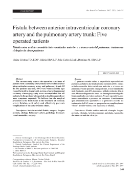
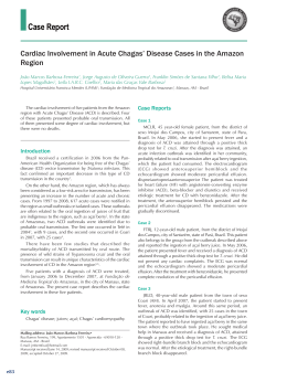
Original Article
Experimental Myocardium Infarction in Rats: Analysis of the Model
Leonardo A. M. Zornoff, Sergio A. R. Paiva, Marcos F. Minicucci, Joel Spadaro
Faculdade de Medicina de Botucatu, Botucatu, SP - Brazil
Summary
Method development
One of the most often used strategies to study the
physiopathological alterations caused by coronary occlusion
is the use of the experimental infarction model in rats.
Among other factors, this is due to the similarities in
the physiopathological alterations that occur after the
infarction in humans. One must consider, however, that
this model has characteristics that can hinder the use as
well as the interpretation of eventual outcomes. Thus, this
review aims at analyzing the main characteristics of the
experimental infarction model in rats, discussing the coronary
occlusion technique, the consequences and the methods of
morphological and functional assessment of the infarction
and its clinical implications.
The experimental AMI model in rats due to left coronary
occlusion was initially described by Heimburger in 19463. With
the passing time, the technique was progressively modified
by Johns and Olson4, Kaufman et al5 and Seyle et al6. In the
beginning of the 1980s, the method was introduced in Brazil
at the School of Medicine of Botucatu7, and from there it
spread to other Services. In short, after the anesthesia, a
left thoracotomy is performed, between the 4th and the 5th
intercostal spaces. The heart is exteriorized through lateral
compression of the chest and the left coronary artery is ligated
with 5-0-size suture thread at approximately 2 mm from its
origin, between the left atrium border and the pulmonary
artery sulcus (Figure 1). Then, the heart is rapidly returned
to the thoracic cavity, the lungs are expanded with positive
ventilation with 100% oxygen and the surgical wound is closed
3-6
. The method described above has several advantages. First,
the animals can be raised specifically for the protocols, at a
lower cost than with larger animals. Second, the surgery is
extremely fast, with a duration ranging from 2 to 5 minutes.
Another aspect is that due to the rat metabolism characteristics,
the phases involved in the infarction evolution such as necrosis,
healing and remodeling occur rapidly, which decreases the
time of study observation. Finally, the morphological and
functional alterations caused by the infarction are similar to
those found in humans8,9.
Introduction
The acute myocardial infarction (AMI) is defined as a
focus of necrosis that results from low tissue perfusion,
with signs and symptoms that are the consequence of the
cardiac cell death.
It is estimated that this syndrome can occur in epidemic
proportions worldwide. Few pathologies had their evolution
altered so radically as the AMI, with an accentuated decrease
in mortality due to the changes regarding its treatment in the
last 30 years1,2. The change in the treatment was the result of
advances obtained in the study of the pathogenesis of the AMI
and its complications. This fact emphasizes the importance of
a better understanding of the physiopathological mechanisms
of the acute coronary syndromes.
One of the most often used models to study the
physiopathological alterations caused by coronary occlusion
is the experimental infarction model in rats. However, the
model presents several particular characteristics that can
make it difficult to handle. Thus, the objective of this review
is to discuss the main methodological aspects related to the
experimental infarction model in the rats.
Animals and surgery
The most often used rat strains are Wistar and SpragueDawley, depending on the Service and animals weighing 200
to 250g are usually used in the studies. In this weight range,
a rat is considered a young adult, when its evolution phase
is characterized by slow growth and presents lower surgical
mortality than older animals. It is also recommended to use
animals of the same sex, as this variable can be an important
regulation factor of the cardiac adaptations in response to
several stimuli10,11.
Myocardial infarction; animals, laboratory; rats; coronary
occlusion.
Regarding the anesthesia, several agents have been
used, and among them, inhalation agents such as ether
and isoflurane, as these drugs are relatively inexpensive
plus the advantage of having a short-term action, in
addition to allowing the variation of anesthesia intensity
during surgery10.
Mailling address: Leonardo Antônio Mamede Zornoff •
Faculdade de Medicina de Botucatu, Departamento de Clínica Médica,
Rubião Jr, 18.618-000, Botucatu, SP - Brasil
E-mail: [email protected], [email protected]
Manuscript received April 18, 2008; revised manuscript received May 29,
2008; accepted May 29, 2008.
Barbiturates or the association of ketamine chloride
(50 mg/kg) and xylidine chloride (1mg/kg) are also frequently
used via intraperitoneal or intramuscular routes. These agents
are fast-acting and can produce anesthesia within 5 minutes.
It is important to remember that the agents with short half-life
are preferable to the long-action ones10.
Key Words
403
Zornoff et al
Experimental infarction in rats
Original Article
Aspects related to the size of the infarction
Figure 1 - Experimental infarction. Exteriorization of the heart for coronary
suture at 2 mm from the origin, between the border of the left atrium and the
pulmonary artery sulcus.
During the procedure, it is recommended that the animals
be artificially ventilated, with oxygen supplementation.
Therefore, the animals can be ventilated by nasal compression,
with catheter, or be submitted to mechanical ventilation after
orotracheal intubation with a number 16 catheter10.
After the surgery, the most commonly used analgesic drugs
are morphine, dipyrone or paracetamol. Another aspect is
that the environmental conditions must be kept constant, as
temperature variations, for instance, can increase the mortality
after the infarction.
Mortality
The observed mortality within the first 24 hrs after surgery
is usually 40 to 60%. Among the causes of death in this period
are mainly the factors related to the surgical procedure,
such as pneumothorax and respiratory depression as well as
factors associated to the cardiac pump failure, such as acute
pulmonary edema10,12. The main cause of death in this model
is, however, the high prevalence of malignant arrhythmias,
such as sustained ventricular tachycardia and ventricular
fibrillation. Opitz and cols. found a mortality of 65% in the first
48 hours after the infarction. Through continuous monitoring,
the authors demonstrated that 96% of the animals submitted
to the AMI presented hundreds of episodes of ventricular
tachycardia and at least 20 episodes of ventricular fibrillation.
Additionally, two distinct periods of arrhythmia were
demonstrated: the first period included the first 30 minutes
after the coronary occlusion and the second, responsible for
65% of the deaths, included the period between 1.5 and 9
hours after the infarction13.
Regarding the chronic period after the AMI, the mortality
observed in this model is extremely variable, with the size
of the infarction being the main determinant of this event.
In this sense, Pfeffer et al14, following infarcted animals for
a period of one year, verified that for small (5 to 19.9%
of the left ventricle), moderate (20 to 39.9%) and large
infarctions (≥ 40%), the mortality rates were around 50%,
75% and 85%, respectively14.
404
Arq Bras Cardiol 2009; 93(3) : 403-408
One of the most relevant aspects of this model is related
to the size of the infarction. It is accepted that this parameter
resulting from the coronary occlusion, in its proximal region,
is not uniform, varying from 4% to 65%12. This fact is a
consequence of the incapacity of occluding the coronary
exactly at the same point in all animals, as well as of eventual
anatomic variations among them. Thus, this model is not
adequate for the verification of the reduction effect of infarct
size through different interventions, as eventual differences in
the infarction size might be inherent to the variability of the
method. Consequently, it is recommended first the analysis
of the risk area affected by the coronary artery ligation in a
certain point, through the injection of dyes. Subsequently,
starting from the risk area, the size of the final infarcted area
is verified15,16.
Another factor to be considered is the site of the occlusion
of the coronary artery. When the occlusion occurs too close
to the origin, there is the septal artery involvement and the
size of the infarction can be higher than 65%. In this case,
however, the mortality of the animals is 100%12,14.
Another relevant characteristic is that, in this experimental
model of AMI, the percentage of infarction at the apex is higher
than that at the basis of the left ventricle. For this reason, the
most often used method to determine the size of the infarction
is the one that uses several transversal cuts. The size of the
infarction is determined by the mean of all cuts. Different
authors, however, have observed that the medial transversal
cut of the left ventricle, between 5 and 6 mm from the apex,
reflects the size of the infarction of the entire left ventricle17,18.
Therefore, the use of this region alone would simplify the
measurement of the infarction size.
Regarding the methods used to determine the infarction
size, this variable has been assessed, preferentially, by 4
different methods: 1) measurement of the infarcted area in
relation to the left ventricle area, determined by histology
or planimetry; 2) histology with the measurement of the
internal perimeter of the infarcted area in relation to the total
perimeter of the cavity; 3) histology with the measurement
of epicardial and endocardial circumferences of the infarcted
and non-infarcted segments and 4) echocardiogram with the
measurement of the internal perimeter of the infarcted region
in relation to the total perimeter of the cavity.
The most often recommended method for determining
the infarction size is the measurement of the epicardial
and endocardial circumferences of the infarcted and noninfarcted segments (Figure 2)12,14,19. A potential limitation to
the use of different techniques is, as mentioned before, the
fact that the size of the infarction can vary, depending on the
method used.
Regarding the measurement of the infarction size by the
area, one must consider that, after the infarction, dynamic
alterations occur in the infarcted segment as well as in the
non-infarcted area. In the infarcted region, the necrotic
tissue is substituted by fibrous scar tissue. At the later phases
of the healing process, the contraction of the fibrotic area
occurs8. On the other hand, in the non-infarcted area of the
left ventricle, different degrees of cardiac hypertrophy occur,
Zornoff et al
Experimental infarction in rats
Original Article
such as adaptation to the loss of the myocytes. Due to these
alterations, the measurement of the infarction size by volume
or area can result in error, as the resorption and retraction
of the infarcted area, added to the hypertrophy of the noninfarcted region, can result in the underestimation of the
infarction size in relation to the original infarcted area12,14,20.
Another method used to determine the size of the
infarction is the measurement of the internal perimeter of
the infarcted segment, in relation to the total perimeter of
the ventricular cavity. This analysis can be attained by two
methods: echocardiogram and histology. However, similarly
to the estimate made by area, these methods can present
important limitations. Simultaneously to the necrosis of the
myofibrils, there is interfibrillar collagen disintegration by
the activation of proteolytic enzymes. This fact causes loss of
the support tissue, which makes the region more distensible
and, consequently, more susceptible to deformations. Thus,
the slippage of muscular necrotic areas can occur, with the
realignment of the myocytes on the infarcted wall. As a
result, there is a narrowing of the region and dilation of the
infarcted segment. This acute dilation, characterized by the
thinning and distension of the infarcted region is called the
infarction expansion21-23. Therefore, as a consequence of the
expansion, the measurement of the infarction size by the
internal perimeters can overestimate the size of the AMI20.
Morphological characteristics
Regarding the anatomic characteristics of the rat’s heart,
the left coronary artery originates between the border of the
left atrium and the pulmonary artery sulcus. Additionally, it
was observed that the left coronary artery, in its proximal
region, is usually intramyocardial, returning to the surface
epicardially at approximately 3 to 4 mm from its origin4. The
rat does not have a true circumflex artery6. The proximal
region of the left coronary artery, practically at the ostium, gives
origin to the septal branch and, farther down, to the branch
that corresponds to the circumflex artery17. This anatomic
characteristic ensures that the septal branch originates above
the site where the coronary occlusion is performed. Thus,
this model is characterized by presenting infarction of the left
ventricular free wall, without involving the interventricular
septum. Consequently, this region is used as control for
morphological and biochemical studies17.
Another anatomic characteristic of this model is that the
rat has scarce collateral circulation, similarly to humans. The
coronary occlusion invariably causes, therefore, transmural
infarctions, making the subendocardial infarction a rare event,
of around 3%24.
Another pertinent aspect of this model is related to the
involvement of the papillary muscle. Contrary to the dog
model, in which the coronary occlusion leads to necrosis
of the papillary muscle in 85% of the cases, the infarction
model in rats is characterized by the preservation of posterior
papillary muscle. The histological analysis showed that, in rats,
the coronary occlusion does not affect or minimally affects
this muscle. The explanation for this phenomenon is that the
irrigation of the posterior papillary muscle is carried out by the
septal branch of the left coronary artery, which, as discussed
above, is not affected by the coronary occlusion17.
Figure 2 - Evaluation of the infarction size through the measurement of the
epicardial and endocardial circumferences of the infarcted segments (right)
in relation to the total epicardial and endocardial circumferences of the left
ventricle (left).
Regarding the healing characteristics after the coronary
occlusion, the rat model also shows some peculiarities25-31. The
activation of metalloproteases (MMP), proteolytic enzymes
responsible for collagen degradation, was identified 1 hour
after the coronary occlusion28. Initially, the MMP degrades
the fibrillar collagen and, subsequently, the MMP-2, MMP3 and MMP-9 degrade these fragments26. A previous study
suggests that, 3 hours after the coronary occlusion, around
50% of the collagen has been degraded28. This proteolytic
activity ceases one week after the infarction27. Concomitant
to the proteolytic activity, other alterations have been
identified. After 24 hours, the muscular necrosis becomes
evident, initially followed by a neutrophilic infiltration and
later, by a lymphocytic and monocytic infiltrate30. Three to
four days after the coronary occlusion, the accumulation of
fibroblast-like cells start, but with actin in its composition,
the myofibroblasts.
It is accepted that the myofibroblasts are fibroblasts
modified by the TGF-β secreted by the monocytes and
responsible for the synthesis of collagen I and III, which
starts to accumulate in the peripheral region of the infarction
on the third day and becomes well organized 14 days after
the AMI. The healing process is complete 21 days after the
coronary occlusion25-31.
Another aspect that must be considered is that, in animals
with large infarctions, the non-infarcted areas of the left
ventricle and the of the right ventricle show an increase in
the expression of pre-collagen types I and III mRNA, starting
between 4 to 7 days after the AMI, which can result in
the accumulation of progressive collagen. Therefore, large
infarctions are often accompanied by different degrees of
fibrosis in the non-infarcted areas26.
One of the main characteristic of myocardial infarction
is associated to the fact that the loss of contractile tissue
triggers an adaptive cell growth process in the non-infarcted
tissue32-37. In the rat model, the left ventricular hypertrophy
is characterized for having an eccentric pattern, being an
early event and being detectable on the third day after the
coronary occlusion.
After three to four weeks, the degree of hypertrophy
increases between 30% and 60%. Hypertrophy can
also occur in the right ventricle, characteristically with
a concentric pattern and correlated to increases in the
left-ventricular end-diastolic pressure and right ventricular
Arq Bras Cardiol 2009; 93(3) : 403-408
405
Zornoff et al
Experimental infarction in rats
Original Article
systolic pressure12. However, similarly to what occurs in
the infarcted ventricle, the right ventricular hypertrophy
is present on the third day and affects around 30% at the
end of one month38,39.
Finally, similarly to what occurs in the infarcted area, the
non-infarcted area, mainly the one that separates the infarcted
tissue from the non-affected tissue, can also be the target
of MMP activation. As a consequence, bundles of viable
myocytes can undergo muscular slippage and realignment
processes (side-to-side slippage)40.
Thus, as a consequence of the processes of expansion,
of ventricular hypertrophy (of eccentric characteristic) and
of cell slippage in the region that is borderline with the
infarction area, the infarcted cavity can increase in diameter
and lose its normal elliptical geometry, taking on a spherical
configuration. These changes in size, mass and ventricular
geometry clinically characterize the process of cardiac
remodeling after the infarction41-47.
Functional characteristics
The hemodynamic consequences caused by the coronary
occlusion in the rat have been well documented. Different
studies have shown that the infarction causes the decrease of
several functional variables, such as systolic volume, cardiac
output, left ventricular systolic pressure, first positive pressure
derivative and negative pressure derivative. In parallel, there
is an increase in the left ventricular-end diastolic pressure and
of the decline time constant of the isovolumetric pressure.
The infarction is accompanied, therefore, by systolic as well
as diastolic dysfunctions, which are identified as early as 3
hours after the coronary occlusion12,48-51.
Regarding the mechanisms involved in the ventricular
dysfunction, there is evidence that up to the three first weeks
after large infarctions, the non-infarcted muscle function
is normal, although the chamber function is depressed37.
On the other hand, after 6 weeks, the muscular function
is depressed52. Thus, this evidence suggests that, initially,
the ventricular dysfunction is a consequence of the loss of
contractile tissue, secondary to the infarction. Chronically,
however, the remaining muscle becomes dysfunctional,
probably due to the post-infarction remodeling process.
One of the main characteristics of this model is that the
functional alterations are closely related to the size of the
infarction. Therefore, rats with infarctions < 30% did not
present hemodynamic abnormalities. Animals with moderate
infarctions (31-46%) had normal basal hemodynamic values,
but reduced pressure-generating capacity. On the other hand,
rats with large infarctions (> 46%) presented heart failure,
with elevated filling pressures and cardiac output decrease12.
Therefore, this model allows the study of different degrees of
ventricular dysfunction.
Finally, the experimental infarction model in rats allows
the evaluation of the right ventricular function. Thus, in a
previous study, in which an isolated heart preparation was
used, perfused with a nutrient solution, a decrease in the
systolic pressure was identified, which correlated, in linear
form, with the increase in the right ventricular mass53.
406
Arq Bras Cardiol 2009; 93(3) : 403-408
Morphological, functional and clinical
evaluation of heart failure
To evaluate hypertrophy in different models of cardiac
injury, the ratio between the left ventricular weight,
adjusted by the body weight of the animal of by the tibia,
is commonly used.
In the infarction model, however, the complex interaction
of events such as the resorption of the necrotic tissue and the
amount of collagen of the scar can interfere with the weight
of the infarcted ventricle so that it won’t reflect the actual
cell growth. Therefore, the use of the myocyte transversal
diameter is preferable for the evaluation of the degree of left
ventricular hypertrophy in this model54. On the other hand, to
determine the right ventricular hypertrophy, ratios with body
weight can be used55.
To evaluate the amount of collagen of the non-infarcted
tissue, the most commonly used methods are: hydroxyproline
measurement56, interstitial collagen fraction57 and detection of
collagens I and III26 through the RNA analysis or anti-collagen
I and III antibodies. An important fact is that, although it is an
indirect method, a close correlation between the biochemical
method and the amount of collagen analyzed by morphometry
was verified58,59.
For the functional assessment, several methods are
available: analysis by the papillary muscle 60, isolated
heart61,62 and invasive hemodynamic evaluation12,63. In the
recent years, however, the echocardiogram has become
increasingly important in the morphological and functional
assessment of infarcted rats. There are several functional
variables that can be used, but the most frequent ones are:
variation in area fraction, shortening fraction, cardiac output,
segmental wall motion score, transmitral flow and cardiac
performance index51,64-67.
There are several clinical variables that can be used for
the diagnosis of heart failure in this model, among which
are: general appearance, slow movements, alterations in fur
texture, delayed growth, body weight and dyspnea. These
variables, however, can be little sensitive for the diagnosis
of congestion or low cardiac output12. The first studies for
the clinical detection of heart failure signs in rats were
carried out by Bing and cols. that studied spontaneously
hypertensive rats with heart dysfunction 68-70. The data
on the incidence of post-infarction heart failure and the
possibility of its clinical recognition in rats, however, are
scarce. Recently, using the rat model with moderate and
large infarctions, the observed prevalence of clinical and
anatomopathological signs of heart failure was tachypnea,
(46%); liver congestion, (21%); thrombus in the left atrium,
(21%); ascites, (25%); pleural-pericardial effusion, (71%)
and right ventricular hypertrophy, (100%)71.
Implications
The first implication of this model is related to the fact that,
as it frequently results in large transmural infarctions located
in the anterior wall, the infarction model in rats is ideal for the
study of the physiopathology of post-infarction remodeling,
Zornoff et al
Experimental infarction in rats
Original Article
as these variables are the great determinants of the presence
and intensity of the remodeling process41-47.
Another implication is the similarity with the
physiopathological alterations that occur after infarctions in
humans52. For this reason, this model is ideal for the study
of therapeutic interventions to minimize the morphological
and functional alterations that can occur after the infarction.
Thus, many of the interventions used in infarction patients
were initially analyzed in the rat model, such as: angiotensinconverting enzyme inhibitors14,41, angiotensin-II receptor
antagonists72, aldosterone antagonists73 and betablockers74.
As demonstrated, the experimental infarction model in rats
has been widely used for the study of the outcomes that occur
after the coronary occlusion. Due to the animal’s inherent
characteristics, we believe that the discussion of the main
aspects of this model is extremely useful for those dedicated
to the study of the acute myocardial infarction.
Potential Conflict of Interest
No potential conflict of interest relevant to this article was
reported.
Sources of Funding
There were no external funding sources for this study.
Study Association
This study is not associated with any post-graduation
program.
References
1. Braunwald E. Evolution of the management of acute myocardial infarction:
a 20th century saga. Lancet. 1998; 352: 1771-4.
2. Antman EM, Hand M, Armstrong PW, Bates ER, Creen LA, Halasyamani
LK, et al. 2007 focused update of the ACC/AHA 2004 guidelines for the
management of patients with ST-elevation myocardial infarction. Circulation.
2008; 117: 296-329.
al. High-dose folic acid pretreatment blunts cardiac dysfunction during
ischemia coupled to maintenance of high-energy phosphates and reduces
postreperfusion injury. Circulation. 2008; 117: 1810-9.
17.Spadaro J, Fishbein MC, Hare C, Pfeffer MA, Maroko PR. Characterization of
myocardial infarcts in the rat. Arch Pathol Lab Med. 1980; 104: 179-83.
3. Heimburger RF. Injection into pericardial sac and ligation of coronary artery
of the rat. Arch Surg. 1946; 52: 677-89.
18.Oh B-H, Ono S, Rockman HR, Ross J Jr. Myocardial hypertrophy in the
ischemic zone induced by exercise in rats after coronary reperfusion.
Circulation. 1993; 87: 598-607.
4. Johns TNP, Olson BJ. Experimental myocardial infarction: I. Method of
coronary occlusion in small animals. Ann Surg. 1954; 140: 675-82.
19.Maclean D, Fishbein MC, Maroko PR, Braunwals E. Hyaluronidase-induced
reduction in myocardial infarction size. Science.
�����������������������������
1976; 194: 199-200.
5. Kaufman N, Gavan TL, Hill RW. Experimental myocardial infarction in the
rat. Arch Pathol Lab Med. 1959; 67: 482-8.
20.Minicucci MF, Azevedo OS, Duarte DR, Matsubara BB, Matsubara AO,
Campana AO, et al. Comparison
��������������������������������������������������������
of different methods to measure experimental
chronic infarction size in the rat model. Arq Bras Cardiol. 2007; 89: 83-7.
6. Selye H, Bajusz E, Grassos S, Mendell P. Simple techniques for the surgery
occlusion of coronary vessels in the rat. Angiology. 1960; 11: 398-407.
7. Spadaro J, Hashimoto LM, Franco RSS, Bregagnollo EA, Tucci PJF. �����������
Efeitos da
administração prévia de amiodarona na incidência precoce de fibrilação
ventricular durante isquemia miocárdica experimental. ������������������
Arq Bras Cardiol.
1984; 42: 25-9.
8. Fishbein MC, Maclean D, Maroko PR. Experimental myocardial infarction in
the rat. Am J Pathol. 1978; 90: 57-70.
9. Klocke R, Tian W, Kuhlmann MT, Nikol S. Surgical animal models of heart
failure related to coronary heart disease. Cardiovasc Res. 2007; 74: 29-38.
10.Litwin SE. The rat model of postinfarction heart failure. Heart Fail. 1995; 11:
182-95.
11.Jain M, Liao R, Podesser BK, Ngoy S, Apstein CS, Eberli FR. Influence of gender
on the response to hemodynamic overload after myocardial infarction. Am
J Physiol Heart Circ Physiol. 2002; 283: H2544-50.
12.Pfeffer MA, Pfeffer JM, Fishbein MC, Fletcher PJ, Spadaro J, Kloner RA, et
al. Myocardial infarct size and ventricular function in rats. Circ Res. 1979;
44:503-12.
13.Opitz CF, Mitchell GF, Pfeffer MA, Pfeffer JM. Arrhythmias and death after
coronary artery occlusion in the rat: continuous telemetric ECG monitoring
in conscious, untethered rats. Circulation. 1995; 92: 253-61.
14.Pfeffer MA, Pfeffer JM, Steimberg BS, Finn P. Survival after an experimental
myocardial infarction: beneficial effects of long-term therapy with captopril.
Circulation. 1985; 72: 406-12.
15.Li Y, Kloner RA. Is there a gender difference in infarct size and arrhythmias
following experimental coronary occlusion and reperfusion? J Thromb
Thrombolysis. 1995; 2: 221-5.
16.Moens AL, Champion HC, Claeys MJ, Tavazzi B, Kaminski P, Wolin M, et
21.Hockman JS, Bulkley BH. Expansion of acute myocardial infarction: an
experimental study. �������������������������������
Circulation. 1982; 65: 1446-50.
22.Matsubara BB, Zornoff LAM. Matriz colágena intersticial e sua relação com a
expansão miocárdica no infarto agudo. Arq
�����������������������������������
Bras Cardiol. 1995; 64: 559-63.
23.Hutchins GM, Bulkley BH. Infarction expansion versus extension: two
different complications of acute myocardial infarction. Am J Cardiol. 1978:
73: 843-9.
24.Cooper CJ, Pfeffer JM, Finn PV, Pfeffer MA. Characteristics of a model
of myocardial infarction produced by coronary artery ligation in the rat.
Cardiovasc Pathol. 1995; 4: 189-94.
25.Ertl G, Frantz S. Healing after myocardial infarction. Cardiovasc Res. 2005;
66: 22-32.
26.Sun Y, Weber KT. Infarct scar: a dynamic process. Cardiovasc Res. 2000; 46:
250-6.
27.Sun Y, Kiani MF, Postlethwaite AE, Weber KT. Infarct scar as living tissue. Bas
Res Cardiol. 2002; 97: 343-7.
28.Holmes JW, Borg TK, Cowel JW. Structure and mechanics of healing
myocardial infarction. Ann Rev Biomed Eng. 2005; 7: 223-53.
29.Whittaker P. Unravelling the mysteries of collegen and cicatriz after myocardial
infarction. Cardiovasc Res. 1995; 29: 758-62.
30.Fishbein MC, Maclean D, Maroko PR. The histopathologic evolution of
myocardial infarction. Chest. 1978; 73: 843-9.
31.Jugdutt BI. Ventricular remodeling after infarction and the extracellular collagen
matrix: when is enough enough? Circulation. 2003; 108: 1395-403.
32.Olivetti G, Capasso JM, Meggs LG, Sonnenblic EH, Anversa P. Cellular basis
of chronic ventricular remodeling after myocardial infarction in rats. Circ Res.
Arq Bras Cardiol 2009; 93(3) : 403-408
407
Zornoff et al
Experimental infarction in rats
Original Article
1991; 68: 856-69.
33.Rubin SA, Fishbein MC, Swan HJC. Compensatory hypertrophy in the heart
after myocardial infarction in the rat. J Am Coll Cardiol. 1983; 6: 1435-41.
34.Capasso JM, Li Peng, Zhang X, Anversa P. Heterogeneity of ventricular
remodeling after acute myocardial infarction in rats. Am J Physiol. 1992; 262:
H486-95.
35.Anversa P, Loud AV, Levicky V, Guideri G. Left ventricular failure induced
by myocardial infarction. I. Myocyte hypertrophy. Am J Physiol. 1985; 248:
H876-82.
56.Mill JG, Milanez MC, Busatto VC, Moraes AC, Gomes MG. Ativação
������������
da
enzima conversora da angiotensina no coração após infarto do miocárdio e
suas repercussões no remodelamento ventricular. Arq Bras Cardiol. 1997;
69: 101-10.
57.Zornoff LAM, Duarte DR, Minicucci MF, Azevedo PS, Matsubara BB,
Matsubara LS, et al. ���������������������������������������������������������
Effects of beta-carotene and smoking on heart remodeling
after myocardial infarction. Arq Bras Cardiol. 2007; 89: 135-41.
36.Anversa P, Olivetti G, Capasso JM. Cellular basis of ventricular remodeling
after myocardial infarction. Am J Cardiol. 1991; 68: D7-16.
58.Nicoletti A, Heudes D, Hinglais N, Appay MD, Philippe M, Sassy-Prigent C, et
al. Left ventricular fibrosis in renovascular hypertensive rats: effect of losartan
and spironolactone. Hypertension. 1995; 26: 101-11.
37.Bing OH, Brooks WW, Conrad CH, Weinstein KB, Spadaro J, Radvany P.
Myocardial mechanics of infarcted and hypertrophied non-infarcted myocardium
following experimental coronary artery occlusion. International Erwin Riesch
Symposium. Tuebingen, Germany. Stein-Kopf Darmstadt, 1983. p. 235-44.
59.Weber KT, Janicki JS, Shroff SG, Pick R, Chen RM, Bashey RI. Collagen
remodeling of the pressure-overload, hypertrophied nonhuman primate
myocardium. Circ Res. 1988; 62: 757-65.
38.Spadaro J, Cicogna AC, Tucci PJF, Cury PR, Montenegro MR. Morphometric
evaluation of the time course of right ventricular hypertrophy after left
coronary artery ligation in rats. Braz J Med Biol Res. 1989; 22: 517-22.
39.Anversa P, Sonnenblic EH. Isquemic cardiomyopathy: pathophysiologic
mechanisms. Prog Cardiovasc Dis. 1990; 33: 49-70.
40.Olivetti G, Capasso JM, Sonnenblic EH, Anversa P. Side-to-side slippage of
myocites participates in ventricular wall remodeling acutely after myocardial
infarction in rats. Circ Res. 1990; 67: 23-34.
41.Pfeffer JM, Pfeffer MA, Braunwald E. Influence of chronic captopril therapy
on the infarcted left ventricle of the rat. Circ Res. 1985; 57: 84-95.
42.Pfeffer MA, Braunwald E. Ventricular remodeling after myocardial infarction:
experimental observations and clinical implications. Circulation. 1990;
81:1161-72.
43.Cohn JN, Ferrari R, Sharpe N. Cardiac remodeling-concepts and clinical
implications: a consensus paper from an international forum on cardiac
remodeling. ������������������������������������
J Am Coll Cardiol. 2000; 35: 569-82.
44.Zornoff LAM, Spadaro J. Remodelação ventricular após infarto agudo do
miocárdio: conceitos, fisiopatologia e abordagem terapêutica. ���������
Arq Bras
Cardiol. 1997; 68: 453-60.
45.Swynghedauw B. Molecular mechanisms of myocardial remodeling. Physiol
Rev. 1999; 79: 215-62.
46.Udelson JE, Patten RD, Konstam MA. New concepts in post-infarction
ventricular remodeling. Rev Cardiovasc Med. 2003; 4 (Suppl 3): S3-12.
60.Mill JG, Zornoff LAM, Okoshi MP, Okoshi K, Padovani CR, Sugizaki MM, et
al. The early administration of growth hormone results in deleterious effects
on ventricular remodeling after acute myocardial infarction. Arq Bras Cardiol.
2005; 84: 115-21.
61.Zornoff LA, Paiva SA, Matsubara BB, Matsubara LS, Spadaro J. Combination
therapy with angiotensin converting enzyme inhibition and AT1 receptor
inhibitor on ventricular remodeling after myocardial infarction in rats. ��J
Cardiovasc Pharmacol Ther. 2000; 5: 203-9.
62.Paiva SAR, Matsubara LS, Matsubara BB, Minicucci MF, Azevedo PS, Campana
AO. ����������������������������������������������������������������������
Retinoic acid supplementation attenuates ventricular remodeling after
myocardial infarction in rats. J Nutr. 2005; 135: 2326-8.
63.deFelice A, Frering R, Horan P. Time course of hemodynamic changes in rats
with healed severe myocardial infarction. Am J Physiol. 1989; 257: H289-96.
64.Solomon SD, Greaves SC, Ryan M, Finn P, Pfeffer MA, Pfeffer JM. Temporal
dissociation of left ventricular function and remodeling following experimental
myocardial infarction in rats. J Card Fail. 1999; 5: 213-23.
65.Morgan EE, Faulx MD, McElfresh TA, Kung TA, Zawaneh MS, Stanley WC,
et al. Validation of echocardiographic methods for assessing left ventricular
dysfunction in rats with myocardial infarction. Am J Physiol Heart Circ Physiol.
2004; 287: H2049-53.
66.Staastad I, Sejersted OM, Ilebekk A, Bjornerheim R. Echocardiographic
criteria for detecting of postinfarction congestive heart failure in rats. J Appl
Physiol. 2000; 89: 1445-54.
47.Tiyyagura SR, Pinney S. Left ventricular remodeling after myocardial
infarction: past, present, and future. Mt Sinai J Med. 2006; 73: 840-51.
67.Saraiva RM, Kanashiro-Takeuchi RM, Antonio EL, Campos O, Tucci PJ, Moisés
VA. Rats with high left ventricular end-diastolic pressure can be identified by
Doppler echocardiography one week after myocardial infarction. Braz J Med
Biol Res. 2007; 40: 1557-65
48.Fletcher PJ, Pfeffer JA, Pfeffer MA, Braunwald E. Left ventricular diastolic
pressure-volume relations in rats with healed myocardial infarction: effects
on systolic function. Circ Res. 1981; 49: 618-26.
68.Bing OHL, Brooks WW, Robinson KG, Slawsky MT, Hayes JA, Litwin SE, et al. The
spontaneously hypertensive rat as a model of the transition from compensated
left ventricular hypertrophy to failure. J Mol Cell Cardiol. 1995; 27: 383-96.
49.Raya TE, Gay RG, Lancaster L, Aguirre M, Moffett C, Goldman S. Serial
changes in left ventricular relaxation and chamber stiffness after large
myocardial infarction in rats. Circulation. 1988; 77: 1424-31.
69.Conrad CH, Brooks WW, Hayes JA, Sen S, Robinson KG, Bing OHL.
Myocardial fibrosis and stiffness with hypertrophy and heart failure in the
spontaneously hypertensive rat. Circulation. 1995; 91: 161-70.
50.Litwin SE, Raya TE, Anderson PG, Litwin CM, Bressler R, Goldman S.
Induction of myocardial hypertrophy after coronary ligation in rats decreases
ventricular dilation and improves systolic function. Circulation. 1991; 84:
1819-27.
70.Brooks WW, Bing OHL, Robinson KG, Slawsky MT, Chaletsky DM, Conrad
CH. Effect of angiotensin-converting enzyme inhibition on myocardial
fibrosis and function in hypertrophied and failing myocardium from the
spontaneously hypertensive rat. Circulation.
�������������������������������
1997; 96: 4002-10.
51.Litwin SE, Katz SE, Morgan JP, Douglas PS. Serial echocardiographic
assessment of left ventricular geometry and function after large myocardial
infarction in the rat. Circulation. 1994; 89: 345-54.
71.Martinez PF, Zornoff LAM, Campos DHS, Oliveira Jr AS, Damato RL, Gosuen
G, et al. Caracterização clínica e anátomo-patológica da insuficiência
cardíaca induzida por infarto do miocárdio em ratos. Arq
������������������������
Bras Cardiol. 2007;
88 (supl): 14.
52.Goldman S, Raya TE. Rat infarct model of myocardial infarction and heart
failure. J Card Fail. 1995; 1: 169-77.
53.Stefanon I, Martins MA, Vassallo DV, Mill JG. Analysis of right and left
ventricular performance of the rat heart with chronic myocardial infarction.
Braz J Med Biol Res. 1994; 27: 2667-79.
54.Zornoff LAM, Matsubara BB, Matsubara LS, Minicucci MF, Azevedo PS,
Campana AO, et al. Cigarette smoke exposure intensifies ventricular remodeling
process following myocardial infarction. Arq Bras Cardiol. 2006; 86: 276-82.
55.Zornoff LAM, Matsubara BB, Matsubara LS, Paiva SAR, Spadaro J. Early rather
408
than delayed administration of lisinopril protects the heart after myocardial
infarction in rats. Basic Res Cardiol. 2000; 95: 208-14.
Arq Bras Cardiol 2009; 93(3) : 403-408
72.Zornoff LAM, Matsubara LS, Matsubara BB, Paiva SAR, Spadaro J. Effects of
losartan on ventricular remodeling in experimental infarction in rats. Arq
���������
Bras
Cardiol. 2000; 75: 459-70.
73.Mill JG, Milanez MC, Resende MM, Gomes MG, Leite CM. Spironolactone
���������������
prevents cardiac collagen proliferation after myocardial infarction in rats. Clin
Exp Pharmacol Physiol. 2003; 30: 739-44.
74.Fishbein MC, Li-Quan L, Rubin S. Long-term propranolol administration alters
myocyte and ventricular geometry in rat hearts with and without infarction.
Circulation. 1988; 78: 369-75.
Baixar