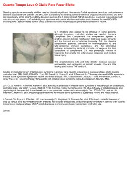
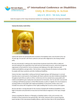
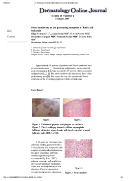
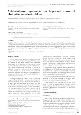
CLINICIAN’S CORNER
Sturge-Weber syndrome in an orthodontic
patient
Matheus Melo Pithon,a Ana Carolina Dias Viana de Andrade,b Ana Paula Dias Viana de Andrade,c
rio Lacerda dos Santosd
and Roge
Jequie, Bahia, and Patos, Paraiba, Brazil
The aim of this article was to describe the Sturge-Weber syndrome in a patient with orthodontic requirements.
Pathologies involved in this syndrome affect facial cranial growth. (Am J Orthod Dentofacial Orthop
2011;140:418-22)
S
turge-Weber syndrome, also known as encephalotrigeminal angiomatosis or encephalofacial
angiomatosis,1,2 is a rare, nonhereditary,
congenital development, characterized by hamartomatous proliferations of vascular cells that involve the
tissues of the brain and face.3-5
It is believed that Sturge-Weber syndrome results
from a developmental defect in the first months of gestation, represented by the persistence of a vascular
plexus around the cephalic portion of the neural tube.6
This plexus develops in the sixth week of intrauterine
life but normally undergoes regression during the ninth
week.7 The association between cerebral angiomatosis
and facial nevus, characteristic of the syndrome, is attributed to the development of the skin of the face
from the ectoderm that covers this vascular plexus.5,8,9
According to Roach,10 Sturge-Weber syndrome is
classified into 3 types. Type I presents facial and
leptomeningeal angiomas and glaucoma. Intercranial
angioma can be confirmed by histologic examination
or typical radiographic findings. Epileptic convulsions
or encephalographic alterations allow a presumptive
diagnosis in the child with typical nevus. Type II exhibits
facial angioma and glaucoma, without evidence of intercranial disease. Type III, which is rare, is characterized by
a
Professor orthodontics, Southwest Bahia University UESB, Jequie, Bahia, Brazil.
Professor dermatology, Southwest Bahia University UESB, Jequie, Bahia, Brazil.
c
Professor radiology, Southwest Bahia University UESB, Jequie, Bahia, Brazil.
d
Professor of Health and Technology, Rural Center at the Federal University of
Campina Grande, Patos, Paraiba, Brazil.
The authors report no commercial, proprietary, or financial interest in the products or companies described in this article.
Reprint requests to: Matheus Melo Pithon, Av Otavio Santos, 395, sala 705, Centro Odontomedico Dr Altamirando da Costa Lima, Vit
oria da Conquista, Bahia,
Brazil, CEP: 45020-750; e-mail, [email protected].
Submitted and accepted, October 2009.
0889-5406/$36.00
Copyright Ó 2011 by the American Association of Orthodontists.
doi:10.1016/j.ajodo.2009.10.049
b
418
isolated leptomengingeal angioma and absence of
glaucoma.11
Clinically, a patient with Sturge-Weber syndrome has
a port-wine stain, or nevus flammeus, which can be flat
or slightly elevated, distributed along the first branch of
the trigeminal nerve (ophthalmic), with or without
involvement of the other 2 branches (maxillary and mandibular).7,10 Abnormalities in the central nervous system,
such as cerebral atrophy, leptomeningeal angiomas, and
cortical calcifications also form part of the syndrome.
Such disturbances can lead to convulsions, mental
retardation, and hemiparesia1 or hemiplegia on the
side opposite the nevus flammeus.3-5
The treatment and prognosis of Sturge-Weber syndrome depend on the nature and intensity of the clinical
characteristics. Patients with nevus flammeus can be
treated with laser therapy; in some, surgical removal
of the meningeal angiomatous lesions might be
necessary.12-15
The aim of this article was to describe the clinical case
of an orthodontic patient with Sturge-Weber syndrome,
with neurologic, ophthalmic, facial, and oral involvement; orthodontic considerations are emphasized.
CLINICAL CASE
The patient, a boy aged 9 years 6 months, came to the
office in Vit
oria da Conquista, Bahia, Brazil, for orthodontic treatment, with the main complaint of crooked
teeth and difficulty in chewing food (Figs 1 and 2). His
mother related that he was born with a problem in the
left eye, which was diagnosed as congenital glaucoma
at the time. According to her, he was never able to see
with this eye. She was asked about the stain on the left
side of the face and said that she had been told that it
was a hemangioma. She related no other complications.
In the clinical examination, an irregular eruptive
pattern was noted between the left and right sides; the
Pithon et al
419
Fig 1. Pretreatment photographs.
Fig 2. Pretreatment dental models.
teeth on the left (affected side) were more developed,
with the eruption of permanent teeth including the second molars. The teeth on the right side were at a stage of
eruption compatible with the patient’s chronologic age.
In an intrabuccal front view, the occlusal plane had
an accentuated deviation as a result of the accelerated
maxillary alveolar bone growth on the left side. The
superior midline had an accentuated deviation.
Starting with the information collected during
anamnesis and the clinical examination, the patient
was referred to a dermatologist and a radiologist for
diagnosis of the pathologies involved. Periapical,
cephalometric, and panoramic radiographs and tomography of the cranium and face were requested (Fig 3).
The panoramic and periapical radiographs showed an
irregular pattern of development between the left and
right sides. The dental elements on the left side were
all erupted and had closed root apices. On the right
side, the dentition was in the mixed stage with some
deciduous and some permanent teeth, with open root
apices. The bones also differed between the right and
left sides. The alveolar process on the left side was
more developed, with more mineralized and mature
bone tissue when compared with the right side.
American Journal of Orthodontics and Dentofacial Orthopedics
September 2011 Vol 140 Issue 3
Pithon et al
420
Fig 3. Pretreatment radiographs.
Cephalometric analysis showed a skeletal Class II pattern (ANB, 6 ) with deficient mandibular growth (SNB,
79 ; SND, 76 ), a convex profile (LS-S, 110; LI-S, 19
mm), and a horizontal facial growth pattern (SNGOGN,
33 ). With regard to the dental pattern, the incisors
were projected and vestibularized (1.NA, 30 ; 1-NA, 10
mm; 1.NB, 40 ; 1-NB, 8 mm) (Fig 4).
The computerized tomography images of the cranium
in the axial plane showed irregular linear cortical calcifications along the spirals of the left occipital and parietal
lobes, and a discrete volumetric reduction in these cerebral
spirals was noted when compared with the contralateral
spirals. After endovenous injection of iodinated contrast,
a more pronounced highlight of the left choroid plexus
was noted when compared with the right (Fig 5).
In the clinical examination, the patient had a Class II
malocclusion with open bite on the right side, superior
midline with a 4-mm deviation to the right, and an
occlusal plane with a marked inclination. The alveolar
mucosa on the right side had a redder color (nevus flammeus). The maxillary arch had a parabolic shape and was
slightly contracted, and the mandibular arch also had
a parabolic shape.
September 2011 Vol 140 Issue 3
The patient’s port-wine stain was notable; it was
slightly elevated on the left side of the face, extending
from the cervical region to the scalp, with greater evidence in the middle and superior regions. The left eye
was affected by glaucoma. On the right side, a convex
profile was especially apparent, with anteroposterior
mandibular deficiency.
From the radiographic, tomographic, and clinical
evaluations made by the orthodontist, dermatologist,
and radiologist, the diagnosis of Sturge-Weber syndrome was reached.
DISCUSSION
The aim of this article was to report a clinical case of
a patient with Sturge-Weber syndrome who sought
orthodontic treatment. When seeking orthodontic treatment, the patient and mother did not know that a syndrome was involved, only that he had isolated problems
in the eye and hemangioma in the face. When the patient
was examined, it was thought that Sturge-Weber syndrome was involved because the classic form of the syndrome includes a port-wine stain and congenital
glaucoma. To prove that it really was a port-wine stain
American Journal of Orthodontics and Dentofacial Orthopedics
Pithon et al
421
Fig 5. Computerized tomography image of the cranium
after endovenous injection of iodinated contrast.
Fig 4. Pretreatment cephalometric tracing.
and not merely a facial hemangioma, the patient was
referred to a dermatologist for evaluation. The dermatologist confirmed that the facial lesion was a port-wine
stain and that there were strong indications that
Sturge-Weber syndrome was involved.
Although the port-wine stain is not an essential condition of the syndrome, it is the most frequent finding,
generally present from birth and affecting half the
face; it can extend up to the neck, with characteristic
darkening with age: red at first and becoming more
purple over time. Only those with nevus flammeus along
the ophthalmic ramus develop the syndrome in its classic
form.16 This information was compatible with the findings for this patient.
To finalize the diagnosis, computerized tomography
of the cranium was requested to evaluate cerebral involvement, since the mother reported that the patient
had never had a convulsive crisis or a learning deficiency.
The tomographic images showed irregular linear cortical calcifications along the spirals of the left occipital
and parietal lobes, and a discrete volumetric reduction
in the cerebral spirals was noted when compared with
the contralateral spirals. After endovenous injection of
iodinated contrast, a more pronounced highlight of
the left choroid plexus was noted when compared with
the right. These findings confirmed the diagnosis of
Sturge-Weber syndrome. The absence of both convulsions and learning deficit were compatible with the
tomographic images, which showed that involvement
was present but discrete. This condition is rare. In the
other clinical cases of Sturge-Weber syndrome in the
literature, the intracranial manifestations were more
evident, compromising the patients’ learning abilities.1,10,11,16,17
Based on the clinical and tomographic findings, the
diagnosis of Sturge-Weber syndrome was made.
Sturge-Weber syndrome has neurologic manifestations
(cerebral anomalies), and cutaneous, ocular, and oral
anomalies, which may or may not be associated; nevertheless, the most evident clinical manifestation is nevus
flammeus in the face or port-wine stain, which normally
follows the trajectory of the V1 and V2 branches of the
trigeminal nerve.3
There are oral manifestations in approximately 38%
of patients that can include hemangiomatous lesions
in the lip, oral mucosa, gingiva, tongue, and palatine region.18 They are generally unilateral and terminate
abruptly at the midline. Ipsilateral gingival hyperplasia
is observed, characterized by an increase in the vascular
component and gingival hemorrhage at the least traumatism. The accumulation of food and the presence of
bacterial plaque can intensify gingival inflammation
and hyperplasia.18,19
Macroglossia and hypertrophy of the maxillary bone,
found in some patients, might result in malocclusion
and facial asymmetry.
Dentists must be on the lookout for this syndrome,
because traumatic surgical interventions in the region
could result in hemorrhages that are difficult to resolve.
Moreover, surgical excision of the gingival hyperplasias
American Journal of Orthodontics and Dentofacial Orthopedics
September 2011 Vol 140 Issue 3
Pithon et al
422
is not always the procedure of choice, since anticonvulsant drugs might determine the maintenance and recurrence of the lesions.
When we examined the oral cavity of this patient, we
noted that the nevus flammeus was also present, which
would require a series of precautions before the start of
orthodontic treatment. Because it involved a vascular
lesion, the tissues had greater susceptibility to hemorrhages; therefore, any procedure involving blood in
this region could set off a hemorrhage that would be
difficult to resolve. Orthodontic procedures that should
be avoided include placement of mini-implants, orthognathic surgery, and extractions. This patient had an
accentuated inclination of the occlusal plane. If he had
sought treatment as an adult, the resolution would be
more difficult because mini-implants or orthognathic
surgery would be needed. But alternative and conservative mechanics were available for this adolescent patient,
such as the placement of a unilateral left bite-block, with
the goal of restricting growth of the alveolar process on
the affected side, allowing the other side to develop
normally.
Another occlusal problem noted was the skeletal Class
II relationship. An extraoral appliance with combined
pull to restrict maxillary growth while mandibular
growth occurs could be used to correct the Class II
relationship. Associated with the orthopedic effect, the
dental effect is achieved with distalization of the molars,
gaining space in the maxillary arch to improve the biprotrusion and convex facial profile, and correct the superior
midline, all at the expense of replacement of the incisors.
The posterior crossbite could be corrected with palatal disjunction. In this patient, the screw would be activated only once every 2 days. This care would help to
prevent rupture of small blood vessels on the affected
side that might have invaded the palatine suture region.
Although gingival hyperplasia is common in patients
with this syndrome, this was not seen in the patient, and
we believe that this was because he did not use anticonvulsant medications.20
The absence of greater cerebral compromise helps in
orthodontic treatment, since it is necessary to have
cooperation for auxiliary orthodontic appliances and
oral hygiene.
With regard to the facial nevus flammeus, the orthodontist could do little; this deformity must be treated by
a dermatologist, with the preferred treatment based on
laser therapy.
CONCLUSIONS
Patients with Sturge-Weber syndrome have neurologic,
ophthalmic, facial, and oral involvement. Therefore,
September 2011 Vol 140 Issue 3
orthodontic treatment must be planned carefully to avoid
compromising the patient’s health.
REFERENCES
1. Smith Pearl M, Abdalla WM, Lin DD, Comi AM, Boltshauser E,
Gailloud P, et al. Sturge-Weber syndrome with cerebellar involvement. J Neuroradiol 2009;36:57-60.
2. Mutalik SS, Bathi RJ, Naikmasur VG. Sturge-Weber syndrome: physician’s dream; surgeon’s enigma. N Y State Dent J 2009;75:44-5.
3. Parsa CF. Sturge-Weber syndrome: a unified pathophysiologic
mechanism. Curr Treat Options Neurol 2008;10:47-54.
4. Pascual-Castroviejo I, Pascual-Pascual SI, Velazquez-Fragua R,
Viano J. Sturge-Weber syndrome: study of 55 patients. Can J Neurol Sci 2008;35:301-7.
5. Rodriguez-Bujaldon AL, Vazquez-Bayo C, Jimenez-Puya RJ,
Moreno-Gimenez JC. Sturge-Weber syndrome and type 1 neurofibromatosis: a chance association? Actas Dermosifiliogr 2008;99:313-4.
6. Nathan N, Thaller SR. Sturge-Weber syndrome and associated congenital vascular disorders: a review. J Craniofac Surg 2006;17:724-8.
7. Comi AM. Update on Sturge-Weber syndrome: diagnosis, treatment, quantitative measures, and controversies. Lymphat Res
Biol 2007;5:257-64.
8. Hennedige AA, Quaba AA, Al-Nakib K. Sturge-Weber syndrome
and dermatomal facial port-wine stains: incidence, association
with glaucoma, and pulsed tunable dye laser treatment effectiveness. Plast Reconstr Surg 2008;121:1173-80.
9. Slasky SE, Shinnar S, Bello JA. Sturge-Weber syndrome: deep venous occlusion and the radiologic spectrum. Pediatr Neurol
2006;35:343-7.
10. Roach ES. Neurocutaneous syndromes. Pediatr Clin North Am
1992;39:591-620.
11. Dilber C, Tasdemir HA, Dagdemir A, Incesu L, Odaci E. SturgeWeber syndrome involved frontoparietal region without facial
nevus. Pediatr Neurol 2002;26:387-90.
12. Arulrajah S, Ertan G, Comi AM, Tekes A, Lin DL, Huisman TA. MRI
with diffusion-weighted imaging in children and young adults
with simultaneous supra- and infratentorial manifestations of
Sturge-Weber syndrome. J Neuroradiol 210;37:51-9.
13. Morrow SA, Campbell C. The cutaneous angioma of Sturge-Weber
syndrome. Can J Neurol Sci 2008;35:506-7.
14. Jordan LC, Wityk RJ, Dowling MM, DeJong MR, Comi AM.
Transcranial Doppler ultrasound in children with Sturge-Weber
syndrome. J Child Neurol 2008;23:137-43.
15. Jacobs J, Levan P, Olivier A, Andermann F, Dubeau F. Late-onset
epilepsy in a surgically-treated Sturge-Weber patient. Epileptic
Disord 2008;10:312-8.
16. Freilinger T, Peters N, Remi J, Linn J, Hacker M, Straube A, et al. A
case of Sturge-Weber syndrome with symptomatic hemiplegic
migraine: clinical and multimodality imaging data during a prolonged attack. J Neurol Sci 2009;287:271-4.
17. Welty LD. Sturge-Weber syndrome: a case study. Neonatal Netw
2006;25:89-98.
18. Mukhopadhyay S. Sturge-Weber syndrome: a case report. J Indian
Soc Pedod Prevent Dent 2009;2:S29-31.
19. Comi AM. Sturge-Weber syndrome and epilepsy: an argument for
aggressive seizure management in these patients. Expert Rev
Neurother 2007;7:951-6.
20. Pithon MM, Ruellas ACO. Clinical and radiographic evaluation of
phenobarbital (GardenalÒ) influence on orthodontic movement:
a study in rabbits. Rev Dent Press Ortodon Ortop Facial 2008;13:
34-42.
American Journal of Orthodontics and Dentofacial Orthopedics
Baixar