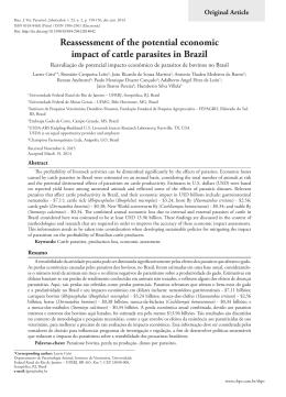
DOI: 10.5433/1679-0359.2014v35n6p3139
Molecular and serological detection of Babesia Bovis and Babesia
Bigemina in cattle in the Rio de Janeiro, Brazil
Detecção molecular e sorológica de Babesia Bovis e Babesia
Bigemina em bovinos no Rio de Janeiro, Brasil
Jenevaldo Barbosa da Silva1*; Priscilla Nunes dos Santos2;
Adivaldo Henrique Fonseca3
Abstract
Bovine babesiosis is an important disease of cattle where Rhipicephalus microplus acts as a vector for
the two causal organisms Babesia bovis and Babesia bigemina. A total of 22 calves were randomly
monitored during three years by semi-nested PCR assay and ELISA test to determine prevalence of B.
bovis and B. bigemina. The overall prevalence of B. bovis and B. bigemina was 30% and 35% by nested
PCR (nPCR), and 70% and 75% by ELISA, respectively. Statistical analysis of the characteristics of
animals showed that age and tick infestations (p<0.05) might play an important role in the spread of
babesiosis as animal less than 6 months old. A high correlation (Kappa index of 0.70 for B. bovis and 0.65
for B. bigemina, respectively) between serological and molecular tests suggests that the combination of
the utilized techniques in the present study is suitable for babesiosis diagnosis in an endemic unstable
area.
Key words: Babesiosis, Brazil, diagnosis, ELISA, PCR
Resumo
A babesiose bovina é uma importante doença do gado, onde Rhipicephalus microplus atua como um
vetor para os dois organismos causais Babesia bovis e Babesia bigemina. Um total de 22 animais foram
aleatoriamente e monitorados durante três anos, por nested PCR e teste de ELISA para determinar a
prevalência de B. bovis e B. bigemina. A prevalência global de B. bovis e B. babesia foi de 30% e 35%,
em nPCR, e 70% e 75% por ELISA, respectivamente. A análise estatística das características dos animais
mostrou que a idade e carrapatos (p <0,05) podem desempenhar um papel importante na propagação de
babesiose em animais com menos de 6 meses de idade. Uma correlação elevada (índice Kappa de 0,70
para B. bovis e B. babesia 0,65, respectivamente) entre os testes sorológicos e moleculares sugere que
a combinação das técnicas utilizadas no presente estudo é adequado para o diagnóstico da babesiose
numa área de instabilidade endémica.
Palavras-chave: Babesiose, Brasil, diagnóstico, ELISA, PCR
Discente da Faculdade de Ciências Agrárias e Veterinária, Universidade Estadual Paulista, FCAV/UNESP, Jaboticabal SP, Brasil.
E-mail: [email protected]
2
Discente da Universidade Federal Rural do Rio de Janeiro, UFRRJ, Seropédica, RJ, Brasil. E-mail: priscillanunes@rocketmail.
com
3
Prof., UFRRJ, Seropédica, RJ, Brasil. E-mail: [email protected]
*
Author for correspondence
1
Recebido para publicação 15/08/13 Aprovado em 08/08/14
Semina: Ciências Agrárias, Londrina, v. 35, n. 6, p. 3139-3146, nov./dez. 2014
3139
Silva, J. B. da; Santos, P. N. dos; Fonseca, A. H.
Introduction
Rhipicephalus microplus is considered the most
important vector of pathogens that cause disease
in cattle. Rhipicephalus microplus, are distributed
in tropical and subtropical regions of the world,
where they economically impact cattle industry by
reducing weight gain and milk production, and by
transmitting pathogens (PETER et al., 2005).
Bovine babesiosis is a tick-borne disease of
cattle that is caused by one or more intraerythrocytic
protozoa of the genus Babesia, order Piroplasmida,
phylum Apicomplexa and is generally characterized
by significant morbidity and mortality worldwide
(McCOSKER, 1981). Although outbreak reports
and localized epidemiological studies have been
published in the last decade, comprehensive and
detailed descriptions of the epidemiology of bovine
piroplasmoses are still lacking in Brazil.
The disease is clinically manifested by anemia,
fever, hemoglobinuria, and marked splenomegaly
and sometimes causes death. Of the species affecting
cattle, Babesia bovis and Babesia bigemina are the
most economically important species worldwide.
The economic losses from the infection are
incurred not only from mortality, loss of milk/meat
production, and cost of control measures but also
through its impact on the international cattle trade
(BOCK et al., 2004).
Definitive diagnosis of babesial infection is
generally made by microscopic identification of
Giemsa or Wright stained blood smears. This
technique is not reliable for detecting the infection
in carrier cattle and for differentiating closely related
infections (TERKAWI et al., 2011). Molecular
detection based on PCR technique has been proven
to provide reliable results with high sensitivity
and specificity in detection of babesial DNA in
blood, particularly when the parasitemia is very
low or no detectable in microscopic examination
(FIGUEROA et al., 1993). Serological tests, the
enzyme-linked immunosorbent assays (ELISA) are
capable of detecting the infection in carrier animals
and have been broadly used for surveillance and
export certification (GOFF et al., 2006; SILVA et
al., 2014).
Consequently, combination of molecular
and serological test for the infection provides
powerful tools for accurate diagnosis as well as for
epidemiological investigations (TERKAWI et al.,
2011). Thus, we investigated in a longitudinal study
the detection of B. bovis and B. bigemina by ELISA
test and PCR assay in cattle.
Materials and Methods
Background
Field activities were conducted from September
2008 to August 2011, at the Dairy Cattle Division
of Seropédica Experimental Station, Agricultural
Research Corporation of the State of Rio de Janeiro
(Pesagro- Rio). The experimental area was located
in the microregion of the Metropolitan Region of
Rio de Janeiro (latitude 22° 45’ S and longitude 43°
41’ W and altitude 33 m).
Field blood samples
By means of proportional stratified sampling, 22
calves were randomly selected. Blood samples were
collected every thirty days from birth until the three
year of age. The herds are usually moved to pasture
during the study. Cattle between 1 and 36 months
old were divided into three groups based on their
age; young (1 year), old (1-2 years) and older (2-3
years).
Blood was collected from the caudal or jugular
vein of individual cattle with EDTA or without,
incubated at room temperature for 2 h, and then
centrifuged at 3000rpm for 10 min. The sera were
collected and then stored at –20 ºC until use. The
genomic DNA samples were extracted from the
whole blood using a commercial kit (QIAamp DNA
Blood Mini-Kit, Madison, WI, USA) according to
the manufacturer’s instructions.
3140
Semina: Ciências Agrárias, Londrina, v. 35, n. 6, p. 3139-3146, nov./dez. 2014
Molecular and serological detection of Babesi Bovis and Babesia Bigemina in cattle in the Rio de Janeiro, Brazil
ELISA
The assay was then performed as earlier
described (Machado et al., 1997). Briefly, 100 µl
of antigen diluted in 0.05 M carbonate/bicarbonate
buffer, pH 9.6, was added to each well of a microELISA plate (Immulon®; Dynatech Laboratories
Inc.) and protein concentration was adjusted to 10
µg/m1-1. The plates were sealed and incubated
overnight at 4 °C. Plates were blocked for l h at
37 °C in a humid chamber with 3% ovalbumin in
carbonate/bicarbonate buffer. After five washes
with buffer (phosphatebuffered saline, pH 7.2, and
0.05% Tween 20, PBS-Tween), 100 µl of diluted
bovine sera (1:400) in PBS-Tween plus 5% normal
rabbit serum were added in duplicate to the ELISA
plate. Plates were incubated at 37 °C in a humid
chamber for 90 min and then washed five times with
PBS-Tween. A 100 µl aliquot of a 1:10000 dilution
of alkaline phosphatase conjugated anti-bovine IgG
(Sigma Chemical Co.) was added to each well and
the plates were incubated at 37 °C under the same
conditions for 90 min. Plates were washed five
times with PBS-Tween. The appropriate substrate
(p-nitrophenyi phosphate) was added and the plates
were sealed and incubated for 40 min at room
temperature. The plates were then read at 405 nm
wavelength on a micro-ELISA reader (B.T.-100;
Embrabio, São Paulo, Brazil). The cut-off values
were calculated based on 10 non-B. bovis and B.
bigemina-infected calves sera.
Nested-PCR
The reaction was conducted in a 25µl reaction
mixture containing 5µl of extracted genomic DNA,
12.5µl of PCR master mix (Roche), and a 1.6 µM
concentration of each primer set. Briefly, one universal
forward primer and two unique reverse primers
were designed to amplify the 18S rRNA gene. The
primer sequences are as follows: B. bovis primers
forward: 5’-AGTTGTTGGAGGAGGCTAAT-3’
and reverse: 5’-TCCTTCTCGGCGTCCTTTTC-3’
and B. bigemina primers forward 5’-
GAGTCTGCCAAATCCTTAC-3’ and reverse
5’-TCCTCTACAGCTGCTTCG-3’ (TERKAWI et
al., 2011). PCR amplifications were performed at 94
ºC for 3 min followed by 34 repetitive cycles of 94
ºC for 1 min, 55 ºC for 1 min, and 72 ºC for 2 min,
followed by a final extension at 72 ºC for 7 min. The
nPCR conditions were the same as described above,
1 µl of the primary PCR product was used as template
and amplified with 10 pmol of each of the primer (B.
bovis forward 5’-GAAATCCCTGTTCCAGAG-3’
and reverse 5’-TCGTTGATAACACTGCAA-3’
and
B.
bigemina
forward
5’-AGCTTGCTTTCACAACTCGCC-3’
and
reverse 5’- TTGGTGCTTTGACCGACGACAT-3’).
PCR products (primary as well as nested) were
checked for amplification by electrophoresis
on a 2.0% agarose gel and visualized using gel
documentation system (Syngene, UK).
Tick
To count the ticks, the animals were restrained
individually, and all the fully or partially engorged
females of R. microplus measuring between 4.5 and
8.0 mm that were found on the right side of each
animal were counted as described by Wharton,
Utech and Turner (1970). The result from each
count was multiplied by 2 to obtain the monthly
average for each animal. The chi-square test was
used to evaluate significant differences (P < 0.05)
of infection rate in animals by B. bovis and B.
bigemina. The kappa coefficient was calculated to
evaluate the agreement among the nPCR assay and
ELISA test. The operational procedures were done
using the R statistical software (R Foundation for
Statistical Computing, version 2.12.2, 2011).
Results and Discussion
The fluctuation of the prevalence of B. bovis and
B. bigemina was compared on the basis in animals
of different months old (Figure 1). Analysis of
serological data for B. bovis revealed a prevalence
of 51, 60% and 70% among animals of age ≥ 1 to
3141
Semina: Ciências Agrárias, Londrina, v. 35, n. 6, p. 3139-3146, nov./dez. 2014
Silva, J. B. da; Santos, P. N. dos; Fonseca, A. H.
≤ 12 months, > 12 to ≤ 24 and over 24 months,
respectively. For B. bigemina the seroprevalence
was 50%, 68% and 76% for the three age groups
examined. In a study on dairy cattle, Tembue et al.
(2011) observed that animal less than 1 year old
and those having ticks present on them were more
infected with B. bovis and B. bigemina.
Figure 1. Serological detection of Babesia bovis and Babesia bigemina in cattle from Rio de Janeiro, Brazil. Twenty
two calves were evaluated by ELISA every three months during the three year of life.
Source: Elaboration of the authors.
Statistically
significant
differences
Source: Elaboration
of the
authors. (P < 0.05) SUKANTO; PAYNE; PARTOUTOMO, 1993).
were essentially observed in aged cattle for both In general, young and adults are susceptible to
while calves
are naturally
resistant
parasiticStatistically
infections significant
for both agents
(Figure
differences
(P <2).
0.05)babesiosis,
were essentially
observed
in aged cattle
for due
both
This was consistent with related studies that to the strong innate immune response with high
parasitic infections for both agents (Figure 2). This was consistent with related studies that noted an increase
noted an increase in the prevalence in older cattle concentration of nitric oxide in the spleen (ZINTL
in(APPLEWHAITE;
the prevalence in older
cattleWAGNER,
(APPLEWHAITE;
CRAIG;
WAGNER, 1981; SUKANTO; PAYNE;
al., 2005).
CRAIG;
1981; et
PARTOUTOMO, 1993). In general, young and adults are susceptible to babesiosis, while calves are
naturally resistant due to the strong innate immune response with high concentration of nitric oxide in the
spleen (ZINTL et al., 2005).
3142
Semina: Ciências Agrárias, Londrina, v. 35, n. 6, p. 3139-3146, nov./dez. 2014
Figure 2. Comparisons of the prevalence serological (ELISA) and molecular (nPCR)
Molecular and serological detection of Babesi Bovis and Babesia Bigemina in cattle in the Rio de Janeiro, Brazil
Figure 2. Comparisons of the prevalence serological (ELISA) and molecular (nPCR) of Babesia bovis and Babesia
bigemina infections on the basis of age of calves in state of Rio de Janeiro, Brazil. Each asterisk indicates a significant
difference (*p < 0.05).
Source: Elaboration of the authors.
Source: Elaboration of the authors.
In this study the tick infestation showed to be risk when introduced to high endemicity or places
In this
study
thefor
tickbabesiosis
infestation
showed
an important
risk factor
for babesiosis
(Figure
an important
risk
factor
(Figure
3).to be
where
tick populations
fluctuate
during the
year3).
Regarding
are agree with
what was
observed by2008).
Andrade
Regardinganimal
animalage,
age,the
theresults
resultsfrom
fromthe
thepresent
presentstudy
(JONSSON;
BOCK;
JORGENSEN,
It etisal.
study
are
agree
with
what
was
observed
by
Andrade
possible
that
in
the
studied
herds
in
a
low
vector
(1998), who noticed a linear increase in the number of ticks as the animals became older. Conversely, highly
et al. (1998), who noticed a linear increase in population by insufficient for maintaining constant
specialized susceptible cattle from low endemicity areas are subject to great risk when introduced to high
the number of ticks as the animals became older. transmission of B. bovis and B. bigemina. In this
endemicity
places specialized
where tick populations
duringantheimbalance
year (JONSSON;
BOCK;
JORGENSEN,
Conversely,orhighly
susceptible fluctuate
cattle case,
caused by
the parasite-host
from low
arestudied
subjectherds
to great
relationship
due to infrequent
transmission
creates
2008).
It is endemicity
possible thatareas
in the
in a low
vector population
by insufficient
for maintaining
artificial
endemic
instability
constant transmission of B. bovis and B. bigemina. Inanthis
case, an
imbalance
causedcondition.
by the parasite-host
relationship due to infrequent transmission creates an artificial endemic instability condition.
Semina:
Ciências Agrárias,
Londrina,
v. 35, n.
6, p. 3139-3146,
2014bovis
Figure
3. Comparisons
of the
prevalence
molecular
(nPCR)nov./dez.
of Babesia
and Babesia bigemina with Rhipicephalus microplus count in cattle of Rio de
3143
Silva, J. B. da; Santos, P. N. dos; Fonseca, A. H.
Figure 3. Comparisons of the prevalence molecular (nPCR) of Babesia bovis and Babesia bigemina with Rhipicephalus
microplus count in cattle of Rio de Janeiro, Brazil.
Source: Elaboration of the authors.
Elaboration ofassay
the authors.
AlthoughSource:
this serological
detected higher positive rates of both parasites than the molecular
detection method, the results obtained from the nPCR assay were highly concordant with this of ELISA, as
Source: Elaboration of the authors.
Although
assay
detected
with
high
theB.diagnosis
determined
by this
the serological
kappa values,
which
were higher
calculatedinvestigations
to be 0.65 and
0.70
foraccuracy
B. bovis inand
bigemina,
positive rates of both parasites than the molecular of Babesia sp. in cattle.
this
serological
assay detected
highertopositive
ratesby
of Terkawi
both parasites
the molecular
respectively Although
(Figure 4).
While
these findings
are similar
that found
et al. than
(2011).
In a nutshell,
detection method, the results obtained from the
The
present
work
showedwith
thatthis
water
cattle from
detection
method,
the
results
obtained
from
the
nPCR
assay
were
highly
concordant
of
ELISA,
our
results
showed
a combination
of both
and serological techniques is useful tool as
in for
nPCR
assay
were that
highly
concordant with
thismolecular
of
state
of
Rio
de
Janeiro,
Brazil,
are
exposed
to
bovine
determined
byinvestigations
the kappa
which
were which
calculated
to be 0.65ofand
0.70 for
and B. bigemina,
ELISA,
as determined
by values,
thewith
kappa
values,
epidemiological
high
accuracy
in thebabesiosis
diagnosis
Babesia
sp.B.inbovis
cattle.
agents,
although
do not show clinical
respectively
(Figure
4).0.65
While
these
findings
similar to that found by Terkawi et al. (2011). In a nutshell,
were
calculated
to bework
and
0.70
B.are
bovis
The present
showed
thatforwater
cattle from
Rio deThe
Janeiro,
Brazil,
are diagnosis
exposed to
signsstate
of theofdisease.
PCR and
ELISA
andourB.results
bigemina,
(Figure of4).both
While
showedrespectively
that a combination
molecular
and
serological
techniques
is
useful
tool
in for
B. bovis
B. bigemina
blood
as a
bovine babesiosis agents, although do not show clinicalofsigns
of the and
disease.
The PCRutilizing
and ELISA
diagnosis
these
findings
are
similar
to
that
found
by
Terkawi
epidemiological investigations with high accuracy in thesource
diagnosis
Babesia
sp. in cattle.
to ofobtain
template
DNA proved efficient
ofetB.al.bovis
andInB.abigemina
blood
as a source
(2011).
nutshell, utilizing
our results
showed
that to obtain template DNA proved efficient in detecting
in detecting
hemoparasites
in asymptomatic
The present work showed that water cattle from
state of Rio
de Janeiro, Brazil,
are exposedcattle
to
a combinationin of
both molecular
hemoparasites
asymptomatic
cattleand
bredserological
in the regionsbred
studied.
bovine babesiosis agents, although do not show clinical signsinofthe
theregions
disease.studied.
The PCR and ELISA diagnosis
techniques is useful tool in for epidemiological
of B. bovis and B. bigemina utilizing blood as a source to obtain template DNA proved efficient in detecting
Figure
4. Molecular
andbred
serological
detection
of B. bovis and B. bigemina using
hemoparasites
in asymptomatic
cattle
inofthe
studied.
Figure
4. Molecular
and serological
detection
B. regions
bovis and
B. bigemina using nPCR assay and ELISA test. The
nPCR assay and ELISA test. The results of nPCR were cross-tabulated with these
results of nPCR were cross-tabulated with these of ELISA. Twenty two calves were evaluated by ELISA every three
of ELISA. Twenty calves were evaluated by ELISA every three months during the
months during the three year of life.
three
year 4.
of Molecular
life.
Figure
and serological detection of B. bovis and B. bigemina using
nPCR assay and ELISA test. The results of nPCR were cross-tabulated with these
of ELISA. Twenty calves were evaluated by ELISA every three months during the
three year of life.
Source: Elaboration of the authors.
Source: Elaboration of the authors.
3144
Source: Elaboration of the authors.
Semina: Ciências Agrárias, Londrina, v. 35, n. 6, p. 3139-3146, nov./dez. 2014
Molecular and serological detection of Babesi Bovis and Babesia Bigemina in cattle in the Rio de Janeiro, Brazil
Acknowledgements
We are grateful to Dra. Rosangela Zacarias
Machado by kindly supplied the antigen used. We
also thank the Coordination for the Improvement of
Higher Level of Education Personnel (CAPES) for
financial support.
References
ANDRADE, A. B. F.; SILVA, R. G.; COSTA, A.
J.; ROCHA, U. F.; LANDIM, V. J. C. Genetic and
environmental aspects of the resistance of Zebu cattle to
the tick Boophilus microplus. In: WORLD CONGRESS
ON GENETICS APPLIED TO LIVESTOCK
PRODUCTION, 6, 1998, Armidale. Proceedings…
Armidale: NSW, 1998. p. 339-342.
APPLEWHAITE, L. M.; CRAIG, T. M.; WAGNER,
G. G. Serological prevalence of bovine babesiosis
in Guyana. Tropical Animal Health and Production,
Edinburgh, v. 13, n. 1, p. 13-18, feb. 1981.
BOCK, R.; JACKSON, L.; de VOS, A.; JORGENSEN,
W. Babesiosis of cattle. Parasitology, Cambridge, v. 129,
n. 1, p. 247-269, 2004.
FIGUEROA, J. V.; CHIEVES, L. P.; JOHNSON, G. S.;
BUENING, G. M. Multiplex polymerase chain reaction
based assay for the detection of Babesia bigemina,
Babesia bovis and Anaplasma marginale DNA in bovine
blood. Veterinary Parasitology, Amsterdam, v. 50, n. 1,
p. 69-81, oct. 1993.
GOFF, W. L.; JOHNSON, W. C.; MOLLOY, J. B.;
JORGENSEN, W. K.; WALDRON, S. J.; FIGUEROA,
J. V.; MATTHEE, O.; ADAMS, D. S.; MCGUIRE, T. C.;
PINO, I.; MOSQUEDA, J.; PALMER, G. H.; SUAREZ,
C. E.; KNOWLES, D. P.; MCELWAIN, T. F. Validation
of a competitive enzyme-linked immunosorbent assay for
detection of antibodies against Babesia bovis. Clinical
and Vaccine Immunology, Washington, v. 13, n. 1, p.
1212-1216, 2006.
JONSSON, N. N.; BOCK, R. E.; JORGENSEN, W.
K. Productivity and health effects of anaplasmosis and
babesiosis on Bos indicus cattle and their crosses, and the
effects of differing intensity of tick control in Australia.
Veterinary Parasitology, Amsterdam, v. 155, n. 1, p. 1-9,
aug. 2008.
MACHADO, R. Z.; MONTASSIER, H. J.; PINTO, A.
A.; LEMOS, E. G.; MACHADO, M. R. F.; VALADÃO,
I. F. F.; BARCI, L. G.; MALHEIROS, E. B. An enzymelinked immunosorbent assay (ELISA) for the detection
on antibodies against Babesia bovis in cattle. Veterinary
Parasitology, Amsterdam, v. 71, n. 1, p. 17-26, jul. 1997.
McCOSKER, P. J. The global importance of babesiosis.
In: RISTIC, M.; KRIER, J. P. (Ed.). Babesiosis. New
York: Academic Press, 1981. n. 1, p. 1-24.
PETER, R. J.; VAN DEN BOSSCHE. P.; PENZHORN.
B. L.; SHARP, B. Tick, fly, and mosquito controllessons from the past, solutions for the future. Veterinary
Parasitology, Amsterdam, v. 132, n. 3, p. 205-15, sept.
2005.
SILVA, J. B.; CORDEIRO, M. D.; CASTRO, G. N. S.;
SANTOS, P. N.; FONSECA, A. H.; REIS, A. B.; SILVA,
N. S.; BARBOSA, J. D. Ocorrência sorológica de Babesia
bovis, Babesia bigemina e Anaplasma marginale em
bovinos e Bubalinos no estado do Pará, Brasil. Semina:
Ciências Agrárias, Londrina, v. 35, n. 5, 2014.
SUKANTO, I. P.; PAYNE, R. C.; PARTOUTOMO, S.
Bovine babesiosis in Indonesia. Preventive Veterinary
Medicine, Colorado, v. 16, n. 1, p. 151-156, jul. 1993.
TEMBUE, A. A. M.; SILVA. F. J. M.; SILVA, J. B.;
SANTOS, T. M.; SANTOS, H. A.; SOARES, C. O.;
FONSECA, A. F. S. Risk factors associated with the
frequency of antibodies against Babesia bovis and
Babesia bigemina in cattle in southern Mozambique.
Pesquisa Veterinária Brasileira, Seropédica, v. 31, n. 8,
p. 663-666, aug. 2011.
TERKAWI, M. A.; HUYEN, N. X.; SHINUO, C.;
INPANKAEW, T.; MAKLON, K.; ABOULAILA,
M.; UENO, A.; GOO, Y. K.; YOKOYAMA, N.;
JITTAPALAPONG, S.; XUAN, X.; IGARASHI, I.
Molecular and serological prevalence of Babesia bovis
and Babesia bigemina in water buffaloes in the northeast
region of Thailand. Veterinary Parasitology, Amsterdam,
v. 178, n. 3, p. 201-207, jun. 2011.
WHARTON, R. H.; UTECH, K. B. W.; TURNER, H. G.
Resistance to cattle tick, Boophilus microplus in a herd
of Australian Illawarra Shorthorn Cattle – its assessment
and heritability. Australian Journal of Agricultural
Research, Oxford, v. 21, n. 1, 163-180, 1970.
ZINTL, A.; GRAY, J. S.; SKERRETT, H. E.; MULCAHY,
G. Possible mechanisms underlying age-related
resistance to bovine babesiosis. Parasite Immunology,
Oxford, v. 27, n. 4, p. 115-120, apr. 2005.
3145
Semina: Ciências Agrárias, Londrina, v. 35, n. 6, p. 3139-3146, nov./dez. 2014
Baixar