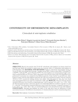
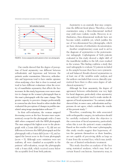
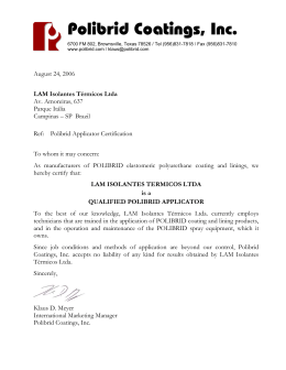
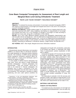
ONLINE ONLY
Biofilm retention by 3 methods of ligation on
orthodontic brackets: A microbiologic and optical
coherence tomography analysis
Aguinaldo S. Garcez,a Selly S. Suzuki,a Martha S. Ribeiro,b Edson Y. Mada,a Anderson Z. Freitas,b
and Hideo Suzukia
Campinas and S~ao Paulo, S~ao Paulo, Brazil
Introduction: The aim of this study was to evaluate biofilm retention around orthodontic brackets related to the
method of ligation by using optical coherence tomography (OCT) and microbiologic sampling. Methods:
Seventy-five plastic central incisors for dentures were divided into 3 groups and used with metal brackets
with a 0.022-in slot with elastomeric ligature (n 5 25), metal brackets with a 0.022-in slot with steel wire
ligature (n 5 25), and self-ligating brackets with a 0.022-in slot (n 5 25). The samples were submersed in
a suspension of Streptococcus mutans, genetically engineered to express green fluorescent protein, at 37 C
for 72 hours to allow biofilm formation. The samples were then submitted to microbiologic analysis and OCT
imaging. Results: The microbiologic analysis and the OCT showed significant differences in biofilm formation
depending on the ligating method. Brackets ligated with elastomeric rings held more S mutans biofilm, and steel
wire ligation had less biofilm retention compared with the other brackets. Conclusions: This study provided
validation that OCT can be used as a potential qualitative marker of total plaque bacteria that can be rapidly
and reliably visualized around orthodontic brackets. (Am J Orthod Dentofacial Orthop 2011;140:e193-e198)
O
rthodontic treatment with fixed appliances leads
to increased biofilm accumulation and elevated
levels of cariogenic and periodontal bacteria,1-3
mainly because orthodontic brackets make good oral
hygiene difficult, resulting in plaque accumulation and
significantly increased risks for enamel demineralization
or periodontal disease.4
Fixed orthodontic appliances create new retention
areas that are suitable for bacterial colonization and
lead to increases in the absolute numbers and the percentages of Streptococcus mutans and lactobacilli.
Many studies have evaluated the effect of fixed orthodontic appliances on microbial flora and periodontal
status5-8; however, only a few studies evaluated the
method of ligation as an additional factor.9,10
a
Assistant professor, Department of Orthodontics, St Leopoldo Mandic Dental
Research Center, Campinas, S~ao Paulo, Brazil.
b
Researcher, Center for Lasers and Applications, Nuclear and Energy Research
Institute, S~ao Paulo, S~ao Paulo, Brazil.
The authors report no commercial, proprietary, or financial interest in the products or companies described in this article.
Reprint requests to: Aguinaldo S. Garcez, Rua Nossa Senhora da Lapa, 671 sl 53,
05072-000 Lapa, S~ao Paulo, S~ao Paulo, Brazil; e-mail, garcez.segundo@gmail.
com.
Submitted, August 2010; revised and accepted, April 2011.
0889-5406/$36.00
Copyright Ó 2011 by the American Association of Orthodontists.
doi:10.1016/j.ajodo.2011.04.019
Conventionally, to connect the wire to the bracket, 3
methods are used: ligation with steel wires, elastomeric
ligatures, and self-ligating brackets. All of these methods
have advantages and disadvantages, but, concerning
biofilm retention, the literature suggests that elastomeric ligatures increase the retention of dental biofilm
compared with the 2 other methods.4,9
In this study, we used a noninvasive method to visualize the biofilm formed around the brackets related to
the ligating method: optical coherence tomography
(OCT). This technique provides a 3-dimensional (3D)
way to visualize the biofilm and allows a quantitative
analysis of the retention area. For in-vitro studies, it
can provide a reliable method for measuring biofilm
area and thickness without destruction or alteration of
the samples. Typically, the methods for plaque identification in vitro use microbiologic sampling11 or scanning
electron microscopy12 that are invasive and destructive.
In vivo, the procedures include biologic dyes and digital
images to visualize and quantify the biofilm; however,
this is normally uncomfortable for patients, since the
dyes can stain oral tissues or orthodontic appliances.
In addition, the dye has difficulty in staining thin
biofilms. OCT does not need dyes to identify the biofilm
and can provide image enhancement in a clinical
environment that is impossible to obtain without
a microscope.
e193
Garcez et al
e194
OCT has been developed for cross-sectional imaging
in biologic systems. It uses low-coherence interferometry and explores the optical properties of scattering
samples to produce images from internal tissue microstructures in a way that is analogous to ultrasound
pulse-echo imaging.13 This technique is a noninvasive,
harmless diagnostic method and can depict the microstructure of biologic samples.14,15 The use of OCT to
reveal biofilms on brackets is innovative. Image
software can process the data obtained by OCT to
quantify the biofilm retention area and identify in
a 3D analysis the site of retention.
The aims of this study were to evaluate biofilm retention around orthodontic brackets as function of the
method of ligation by using OCT and to compare the
results with conventional microbiologic sampling.
MATERIAL AND METHODS
Seventy-five plastic central incisors for dentures
(Biotone, Dentsply, Rio de Janeiro, Brazil) were used.
The samples were divided into 3 groups: conventional
metal brackets with a 0.022-in slot with elastomeric ligature (n 5 25), the same type of brackets with steel wires
ligature (n 5 25), and 25 self-ligating brackets with
a 0.022-in slot for the straight-arch technique (respectively, Ovation and in-Ovation brackets; Dentsply/GAC,
Bohemia, NY). All brackets used were maxillary central
incisor brackets, bonded with composite bonding material (TPH, Dentsply). The samples were disinfected before
the experiment by immersion in 1 mL of 70% ethanol for
30 minutes.
Genetically modified bacteria, S mutans (ATCC
25175), to express the green fluorescent protein were
aerobically grown in brain-heart infusion broth at
37 C with shaking (150 rpm) for 48 hours, until they
formed a stationary growth phase suspension. Genetically modified fluorescent bacteria were used to easily
confirm, by image analysis, the formation of biofilms
and the identification of the main site of biofilm retention on the brackets.
Bacterial cells were suspended in broth nutrient medium of brain-heart infusion to achieve a cell concentration of 109 colony-forming units per milliliter
(confirmed by photospectroscopy at l 5 540 nm).
One-milliliter aliquots were added to a petri dish with
approximately 20 mL of brain-heart infusion broth
with the samples inside. The petri dish was then sealed,
kept upright, and incubated aerobically for 72 hours at
37 C with shaking (100 rpm) to allow biofilm formation.
The brain-heart infusion broth was changed each 24
hours, and the presence of a microbial biofilm rather
than planktonic bacteria was confirmed by the failure
of irrigation with saline solution to significantly diminish
October 2011 Vol 140 Issue 4
Fig 1. A, Three-dimensional reconstruction of a confocal
sequence of S mutans biofilm fluorescent images at the
interface of the bracket and the artificial teeth (note the intense fluorescence of the biofilm); B, fluorescent image of
S. mutans biofilm over the bracket in a buccal view over
the bracket wing after irrigation with 10 mL of saline solution, indicating the adherence of the 3-day-old biofilm.
the fluorescence signal analyzed at a wavelength of 520
nm by fluorescence microscopy (Fig 1).
The configuration used in the OCT system was a fiber
Michelson interferometer (OCP930SR; Thorlabs, Newton, NJ) that uses a superluminescent diode at 930 nm
with 2 mW of optical power in the sample arm. The
system can produce up to 8 frames per second with
2000 3 512 pixels with axial and lateral resolutions in
air of 6.0 mm. The system has a hand probe implemented
with optical fiber, making data collection easier.
For tridimensional image reconstruction, a precise displacement of the sample in the y-axis (orthogonal to the
image acquisition by the OCT system) during the image
acquisition was done by a computer-controlled linear
translation stage (T25-XYZ, Thorlabs) with a minimum
step of 0.05 mm. The 3D images were reconstructed by
using VGStudioMax (version 1.2; Volume Graphics, Heidelberg, Germany) and analyzed with the software Image
J (National Institutes of Health, Bethesda, Md). For the
analysis, all areas of light scattering on each image,
showing the presence of biofilm, were selected, and the
software analyzed the values in square millimeters.
For the microbiologic analysis, after 72 hours, each
plastic tooth was washed by immersion in phosphatebuffered saline solution to remove the brain-heart
American Journal of Orthodontics and Dentofacial Orthopedics
Garcez et al
e195
Fig 2. Microbiologic analysis of biofilm retention on different brackets. Squares represent mean values; boxes represent standard deviations. Bars indicate ranges, and the
horizontal lines in the boxes correspond to the medians.
Steel wire (SW) ligating showed less biofilm retention
than self-ligating (SL) brackets and elastomeric rings
(ER) (P \0.05).
Fig 3. Quantification of biofilm area on different brackets
based on OCT images analyzed with the Image J software. Squares represent mean values; boxes represent
standard deviations. Bars indicate ranges, and the horizontal lines in the boxes correspond to the medians. Steel
wire (SW) ligation showed less biofilm area retention than
self-ligating (SL) brackets and elastomeric rings (ER)
(P \0.05).
infusion broth and placed in a microcentrifuge tube with
1 mL of phosphate-buffered saline solution, that was
subsequently sealed and vortexed for 1 minute and, after
that, challenged with a low-output ultrasonication to
unbind the biofilm.16 One hundred-microliter aliquots
were added to the wells of a 96-well plate for serial
dilution and streaking on square brain-heart infusion
agar plates. The plates were incubated by using standard
aerobic conditions at 37 C for 24 hours so that the
colony-forming units could be counted according to
the method of Jett et al.17 After we counted the
colony-forming units, we computed the mean values
from samples in each group.
For comparison and validation of the image area
analysis, the data from both experiments were compared.
RESULTS
Statistical analysis
Medians and means for bacterial counts and biofilm
areas with the corresponding standard deviations were
calculated. The mean bacterial counts (in colonyforming units per milliliter) and the biofilm area determination (in square millimeters) from teeth with steel
wire ligation, self-ligating brackets, and elastomeric
rings were tested for significant differences by using
analysis of variance (ANOVA) followed by Tukey tests.
P \0.05 was considered statistically significant. Statistical comparisons between means were performed with
software (Origin, version 8.5; OriginLab, Northampton,
Mass).
Biofilm was detected on all orthodontic brackets by
fluorescence and OCT images. The addition of 1 mL of
a suspension containing 109 cells of S mutans into the
petri dish with brain-heart infusion broth and the samples followed by 3 days of incubation at 37 C in aerobic
conditions reliably and reproducibly produced fluorescent biofilms (Fig 1, A). Irrigation with 10 mL of saline
solution did not affect significantly the fluorescence
signal when analyzed by fluorescence microscopy.
Figure 1, A, shows the fluorescent image that confirms
the biofilm presence on the brackets, and Figure 1, B,
shows a bracket with an elastomeric ring after the
biofilm challenge.
The microbiologic analysis showed a significant difference on biofilm formation depending on the ligating
method. Brackets ligated with elastomeric rings collected more S mutans biofilm than did self-ligating
brackets and steel wire ligation. Figure 2 gives the data
obtained from the biofilms removed from each type of
ligation. The initial bacterial burden did vary between
individual brackets. For elastomeric rings, the mean
value was 6.09 log colony-forming units per milliliter
(range, 7.19-5.62). Self-ligating brackets had less
biofilm compared with elastomeric rings, an average of
4.14 log colony-forming units per milliliter (range,
4.93-3.09). However, steel wire brackets showed still
less biofilm, with a mean value of colony-forming units
per milliliter of 3.4 log (range, 4.17-2).
American Journal of Orthodontics and Dentofacial Orthopedics
October 2011 Vol 140 Issue 4
Garcez et al
e196
Fig 4. OCT images from elastomeric ring A, before and B, after 3 days of incubation with S mutans.
The asterisks indicate the biofilm. Note the light scattering from the S mutans biofilm.
Figure 3 shows the area of biofilm on an OCT image
measured by Image J software. The area measured confirms that elastomeric rings retain more biofilm than
self-ligating brackets and steel wire ligation, showing
good correlation between the methods.
By using image amplification and 3D reconstruction,
we can observe the main sites of biofilm retention of
each ligating method. As shown in Figure 4, A and B,
for elastomeric ligating brackets, the site of biofilm
retention is around the elastomeric ring and in the
slot; for self-ligating brackets, it is mainly in the slot
and under the clip. The steel wire ligating bracket had
the least biofilm retention. Both Figures 4 and 5 show
the OCT images from brackets that were more
representative of each group, and the asterisks identify
the main sites of biofilm retention. As seen in the
image, the area of light scattering from the biofilm is
greater on the elastomeric ligating brackets compared
with the self-ligating and steel wire ligating brackets.
DISCUSSION
The aim of this study was to evaluate biofilm retention around orthodontic brackets as a function of the
method of ligation by using images from OCT and
microbiologic sampling. OCT is a noninvasive and
real-time biofilm study method, facilitating bacterial
analysis without altering the sample. Current methods
for in-vitro analysis use microbiologic sampling or
scanning electron microscopy, which are invasive and
destructive methods.
Biofilms were detected on all orthodontic brackets
independent of ligation method by microbiologic analysis and OCT images, and confirmed by fluorescence microscopy. These data could indicate good correlations
October 2011 Vol 140 Issue 4
among biofilm image areas and counts of colonyforming units.
S mutans was selected for this study because it is an
acidogenic and aciduric bacterium that is considered to
be the primary organism responsible for enamel caries.18
Furthermore, many studies have demonstrated increased
retention of S mutans and lactobacilli in saliva and dental
plaque during orthodontic treatment.5,7,8 The expression
of a fluorescent protein by S mutans facilitated the
biofilm visualization, avoiding the use of others
methods that could interfere with the biofilm structure.
Despite recent advances in orthodontic materials,
there has been no decrease in the prevalence of enamel
demineralization near the bracket-tooth junction;
orthodontic materials create a favorable substratum for
biofilm formation. The formation of dental plaque might
exacerbate preexisting periodontal diseases and cause
enamel decalcification, affecting some patients undergoing orthodontic treatment.19
Concerning the method of ligation, Forsberg et al9
studied the effect of biofilm retention around brackets
ligated with steel ligatures and elastomeric ties in
patients. They found that brackets attached to archwires
with elastomeric rings had more bacteria than those
ligated with steel wires. They recommended avoiding
the use of elastomeric ligatures in patients with poor
oral hygiene because elastomeric ligation rings can
significantly increase microbial accumulation on tooth
surfaces adjacent to brackets, leading to predisposition
for dental caries and gingivitis. Additionally, Pellegrini
et4 showed in an in-vivo study that, in most cases, teeth
bonded with self-ligating attachments had fewer bacteria in plaque than did teeth bonded with elastomeric
brackets after 1 to 5 weeks of bonding.
American Journal of Orthodontics and Dentofacial Orthopedics
Garcez et al
e197
Fig 5. OCT images from the brackets: A, steel wire ligation on Ovation bracket; B, elastomeric ring on
Ovation bracket; C, self-ligating In-Ovation bracket. Asterisks indicate the main sites of biofilm retention
on each bracket.
Based on the results of Forsberg et al9 who found
reduced bacterial retention around brackets ligated with
steel ligatures compared with elastomeric rings, and
Pellegrini et al,4 who showed greater biofilm on brackets
with elastomeric ties as opposed to self-ligating appliances, we compared the 3 methods of ligating with respect
to biofilm retention. In contrast, Tukkahraman et al10
found no significant differences in the numbers of microorganisms from teeth ligated with either elastomeric rings
or steel ligature wires. Also, Pandis et al20 established that
the levels of S mutans in whole saliva of orthodontically
treated patients did not seem to be significantly different
between conventional and self-ligating brackets.
Our results from the microbiologic analysis and the
OCT images clearly indicated that around steel wire ligatures there was less biofilm compared with self-ligating
brackets and elastomeric ligating brackets. The images
showed that elastomeric and self-ligating brackets
have more microbial shelters, resulting in enhanced accumulation of plaque compared with brackets with steel
ligature wires. Pellegrini et al4 hypothesized that the
complete absence of a ligature in a self-ligating mechanism would presumably be equally as hygienic as, if not
better than, a stainless steel ligature. However, the image in Figure 5, C, show that, because of the clip and
other retention areas on self-ligating brackets, they retain significantly more biofilm than steel wire ligatures.
For self-ligating brackets, on the other hand, the main
sites of biofilm retention will be filled with the archwire,
decreasing the space for retention.
This study has also provided validation that OCT images can be used as a potential qualitative and quantitative marker of total plaque bacteria that can be rapidly
and reliably visualized around orthodontic brackets.
We thank Ilka Tiemy Kato and Renato Araujo Prates
for their professional reading of the manuscript.
REFERENCES
1. Papaioannou W, Gizani S, Nassika M, Kontou E, Nakou M. Adhesion of Streptococcus mutans to different types of brackets. Angle
Orthod 2007;77:1090-5.
2. Balenseifen Jw, Madonia JV. Study of dental plaque in orthodontic
patients. J Dent Res 1970;49:320-4.
3. Pandis N, Vlachopoulos K, Polychronopoulou A, Madianos P,
Eliades T. Periodontal condition of the mandibular anterior dentition in patients with conventional and self-ligating brackets. Orthod Craniofac Res 2008;11:211-5.
4. Pellegrini P, Sauerwein R, Finlayson T, McLead J, Covell DA Jr,
Maier T, et al. Plaque retention be self-ligation vs elastomeric
orthodontic brackets: quantitative comparison or oral bacteria
detection with adenosine triphosphate-driven bioluminescence.
Am J Orethod Dentofacial Orthop 2009;135:426.e1-9:discussion,
426-7.
5. Scheie AA, Arneberg P, Krogstad O. Effect of orthodontic treatment on prevalence of Streptococcus mutans in plaque and saliva.
Scand J Dent Res 1984;92:211-7.
6. Rosenbloom RG, Tinanof N. Salivary Streptococcus mutans levels
in patients before, during, and after orthodontic treatment. Am J
Orthod Dentofacial Orthop 1991;100:35-7.
7. Chang HS, Walsh LJ, Freer TJ. The effect of orthodontic treatment
on salivary flow, pH, buffer capacity, and levels of mutans streptococci and lactobacilli. Aust Orthod J 1999;15:229-34.
8. Sukontapatipark W, el-Agroudi MA, Selliseth NJ, Thunold K,
Selvig KA. Bacterial colonization associated with fixed orthodontic
appliances. A scanning electron microscopy study. Eur J Orthod
2001;23:475-84.
9. Forsberg CM, Brattstrom VF, Malmberg E, Nord CE. Ligature wires
and elastomeric rings: two methods of ligation, and their association with microbial colonization of Streptococcus mutans and lactobacilli. Eur J Orthod 1991;13:416-20.
10. Turkkahraman H, Sayin MO, Bozkurt FY, Yetkin Z, Kaya S, Onal S.
Archwire ligation techniques, microbial colonization, and periodontal status in orthodontically treated patients. Angle Orthod
2005;75:231-6.
American Journal of Orthodontics and Dentofacial Orthopedics
October 2011 Vol 140 Issue 4
Garcez et al
e198
11. van Gastel J, Quirynen M, Teughels W, Pauwels M, Coucke W,
Carels C. Microbial adhesion on different bracket types in vitro.
Angle Orthod 2009;79:915-21.
12. Brusca MI, Chara O, Sterin-Borda L, Rosa AC. Influence of different
orthodontic brackets on adherence of microorganisms in vitro.
Angle Orthod 2007;77:331-6.
13. Freitas AZ, Amaral MM, Raele MP. Optical coherence tomography:
development and applications. In: Duarte FJ, editor. Laser pulse phenomena and applications. Available at: http://www.intechopen.
com/articles/show/title/optical-coherence-tomography-develop
ment-and-applications. Accessed February 22, 2011.
14. de Melo LS, de Araujo RE, Freitas AZ, Zezell D, Vieira ND, Girkin J,
et al. Evaluation of enamel dental restoration interface by optical
coherence tomography. J Biomed Opt 2005;10:064027.
15. Velasco VM, Baby AR, Sarruf FD, Kaneko TM, Samad RE, Vieira ND,
et al. Prospective ultramorphological characterization of human
hair by optical coherence tomography. Skin Res Technol 2009;5:
440-3.
October 2011 Vol 140 Issue 4
16. Larsen T, Fiehn NE. Development of a flow method for susceptibility testing of oral biofilms in vitro. APMIS 1995;103:
339-44.
17. Jett BD, Hatter KL, Huycke MM, Gilmore MS. Simplified agar plate
method for quantifying viable bacteria. Biotechniques 1997;23:
648-50.
18. Garcia-Godoy F, Hicks MJ. Maintaining the integrity of the enamel
surface: the role of dental biofilm, saliva and preventive agents in
enamel demineralization and remineralization. J Am Dent Assoc
2008;139(Suppl):25S-34.
19. Mei L, Busscher HJ, van der Mei HC, Chen Y, de Vries J, Ren Y. Oral
bacterial adhesion forces to biomaterial surfaces constituting the
bracket-adhesive-enamel junction in orthodontic treatment. Eur
J Oral Sci 2009;117:419-26.
20. Pandis N, Papaioannou W, Kontou E, Nakou M, Makou M,
Eliades T. Salivary Strptococcus mutans levels in patients with
conventional and self-ligating brackets. Eur J Orthod 2010;
32:94-9.
American Journal of Orthodontics and Dentofacial Orthopedics
Baixar