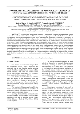
75
Combined treatment by antibiotic therapy and surgery of chronic
mandibular osteomyelitis: a case report
Daniel Humberto Pozza 1
Nelson Ribeiro Neto 2
João Batista Macedo Sobrinho 3
Jean Nunes Santos 4
João Batista Blessmann Weber 5
Marília Gerhardt de Oliveira 6
Abstract
Chronic osteomyelitis of the mandible is usually a difficult pathology to resolve. Treatment has included long-term antibiotics,
and surgical debridement, aggressive in some cases. This paper reports a 54-year-old female with an eight-month-mandibular
chronic osteomyelitis who was successfully treated with the combination of medication and surgical intervention.
Keywords: osteomyelitis; oral surgery; clindamycin; mandible
INTRODUCTION
Osteomyelitis is an inflammatory process acute, sub-acute or chronic - which reaches the
medullar spaces and/or the osseous corticals and
might extend from the original place, arising usually
from an odontogenic infection, among others. The
osseous spaces are usually filled with exudates, which
can lead to pus formation. Chronic osteomyelitis
can be the result of a non-treated acute mild
inflammation or emerge without a precursor. When
osteomyelitis happens in the mandible, it is usually
more diffuse and widespread.1-5
This infection usually affects bones such as
sternum, clavicles, ribs, spinal bones, pelvis, as well
as the peripheral long bones. Involvement of the
mandible alone occurs less frequently. In the
mandible, it can be found as a single lesion or
multiple lesions.6-8
Clinical examination alone is enough to
diagnose chronic mandibular osteomyelitis due to
the progression of this disease. Computed tomography
is the preferred imaging method, since it presents
advantages over conventional methods, such as the
possibility of visualizing soft tissues and multiplanar
reconstruction. Bone scintigraphy can also be used
to detect the initial inflammation, and magnetic
resonance imaging can be used to check for
1
Doctor in Dentistry. Laboratory of Laser. School of Dentistry - UFPB/UFBA. João Pessoa – PB / Salvador - BA
Doctoral Student - Laboratory of Laser. School of Dentistry - UFPb/UFBA. João Pessoa – Pb / Salvador – BA. Specialist in CTBMF
– UFCE. Fortaleza – CE. Specialist in Implantodontology - EAP/ABO/BA. Salvador - BA
3
Doctor in Dentistry. Laboratory of Laser. School of Dentistry - UFPB/UFBA. João Pessoa – PB / Salvador – BA. Professor and
Coordinator of the Specialization Course in CTBMF – CEBEO. Salvador – BA
4
Doctor in Oral Pathology. Adjunt Professor and Titular of Laboratory of Pathologic Anatomy. School of Dentistry – UFBA. Salvador
- BA
5
Doctor in CTBMF. Adjunt Professor of Pediatric Dentistry – PUCRS. Porto Alegtre - RS
6
Titular Professor of the School of Dentistry, PUCRS. Porto Alegre. Productivity Scholarship Holder- CNPq
2
Correspondência para/Correspondece to:
Daniel Humberto Pozza
Av. Lucas de Oliveira, 1841, 203. Bairro: Petrópolis
90460-001. Porto Alegre – RS - Brasil
E-mail: [email protected]
R. Ci. méd. biol., Salvador, v. 5, n. 1, p. 75-79, jan./abr. 2006
76
medullary and soft tissues modifications with higher
accuracy, without exposing the patient to ionizing
radiations.9
Diagnosis of active chronic osteomyelitis, or
osteomyelitis overlapping other pathologies, is
difficult, and the same applies to the concept of
cure, since there may be prolonged asymptomatic
intervals. Clinical-radiological criteria can be used,
especially with patients who present low probability
of infection, avoiding invasive procedures such as
the scintigraphy, which is, in some cases, nonconclusive.10
In cases of chronic osteomyelitis, a radiolucent
and circumscribed image can be seen, presenting
central radiopaque sequestra and enlarged
radiopacities of the surrounding bone due to a local
osteogenic reaction.3
Patients who present active chronic
osteomyelitis usually require long-term use of
antibiotic therapy and, in some cases surgical
intervention is indicated.11
Mader, Adams and Morrison12 administered
clindamycin and cefazolin to rabbits, to treat
induced osteomyelitis caused by Staphylococcus
aureus. It was found that clindamycin was more
effective, presenting higher concentrations in both
infected and healthy tissues. Cancino et al13 in a
retrospective study, found that clindamycin was the
most widely used antibiotic for closed mandibular
fractures in a given oral and maxillofacial surgery
service. The drug was administered to about 60%
of the treated patients.
This bone pathology presents various forms,
depending on the virulence of the infecting
microorganism, the host capacity of effective immune
response and the kind of reaction of the periosteal
and osseous tissues. Corticotomy can be used as
treatment, and it is not effective, bone resection can
be done as a more radical alternative. However,
the most aggressive treatment mode might imply
complications, such as loss of function, exposure of
the inferior alveolar nerve and later problems
regarding the reconstruction of the affected organ.14
Cohen et al 15 successfully used the
combination of antibiotic therapy and surgery when
treating a patient with chronic jaw osteomyelitis.
The authors administered vancomycin, and the
surgical dibridement was performed until the
underlying bone started bleeding. Antibiotic therapy
R. Ci. méd. biol., Salvador, v. 5, n. 1, p. 75-79, jan./abr. 2006
was administered for one additional month, and
the lesion was cured. Chronic osteomyelitis is usually
treated by the combination of antibiotic therapy
and surgical dibridement of the lesion.16 These
authors believe that removal of the diseased bone
can be problematic because it is difficult to
differentiate it from healthy bone, thus leading to
insufficient or excessive corticotomy. The same
authors treated a patient who had been suffering
from osteomyelitis for 12 years, using intravenous
clindamycin, combined with oral tetracycline.
Tetracycline was used to mark the healthy bone
which was preserved during surgery; the necrosed
bone was completely removed.
Bar et al17 treated a case of mandibular
osteomyelitis caused by Mycobacterium abscessus with
amoxicillin and clavulanic acid and surgery. As
the later treatment failed, the patient was reoperated
on and the antibiotic was changed to amicacin
associated with clarithromicin and rifampicin and
a successful result was achieved.
Antibiotic therapy alone is not enough for
the treatment of osteomyelitis, since the devitalized
osseous tissue in combination with the capsule of
the surrounding fibrous connective tissue protects
the microorganisms from the drug action. The
surgical treatment must be aggressive and associated
with high doses of antibiotics; some authors believe
that penicillin G is the medication of choice,
followed by clindamycin. The penicillin G
treatment may have to be administered up to six
months.3,4
CASE REPORT
A 54-year-old white female patient reporting
traumatic extraction of the lower anterior teeth
around one year before, followed by construction of
an inferior denture. Her main complaint was pain
and foul-smelling oral discharge for eight months.
A purulent discharge in the region anterior to the
mandibular ridge was present. The gingival mucosa
was reddened, with enlarged volume and pain at
palpation. A radiolucent area in the dental region
of the edentulous alveolar ridge, with radiopaque
focal areas suggesting bone resorption and
formation of bone sequestra could be seen in the
panoramic radiography (Figure 1-A).
77
The patient denied any systemic disease or
allergies. The patient’s blood pressure was 130X80
mmHg.
The preliminary clinical and radiographic
diagnosis was chronic osteomyelitis. The initial
treatment consisted in clindamycin 300mg every 6
hours for 7 days, and ordering culture and
antibiogram of the purulent discharge. When the
patient returned, she reported regression of pain and
regression of the purulent discharge, confirmed by
clinical examination (Figure 1-B). Then, the
surgical procedure to remove the affected tissues was
performed.
After the antisepsis with chlohexidine
gluconate at 2%, an anesthetic blockage of both
inferior alveolar nerves with mepivacaine at 2%
associated with adrenaline (1:100.000) was
performed. Then, a wide incision was performed
on the inferior alveolar ridge, exposing the lesion,
and the margins of the healthy bone, what was
checked by the different colors of the tissues (Figure
1-C). The yellowish/brown necrosed osseous tissue
was completely removed by forceps and curets, until
the remaining tissue presented normal color and
started to bleed (Figure 1-D). It was copiously
washed with saline to remove all remaining
fragments. After the gingival plasty, primary closure
was obtained by nylon 4-0 sutures. The specimen
was sent to histologic examination.
Figure 1: A – Panoramic radiography in the preoperative; B – Intra-oral aspect
after the administration of clyndamicin for 7 days; C – Exposure of
the necrosed osseous tissue; D – Underlying healthy osseous tissue; E –
Three-month clinical follow-up; F – Three-month radiographic followup.
R. Ci. méd. biol., Salvador, v. 5, n. 1, p. 75-79, jan./abr. 2006
78
During the postoperative period, treatment
with clindamycin continued, with the same
posology for 15 days, associated with a nonsteroidal anti-inflammatory for 4 days (loxoprofen
sodium 60 mg) and an analgesic (dipyrone 500
mg) for 2 days, both administered every 6 hours.
On the fifteenth day, the patient was clinically well,
without complaints or side effects, and the suture
was removed. After three months, the patient was
in good clinical condition and signs of the bone
neoformation could be radiographically seen at the
operated site (Figure 1-E; F).
DISCUSSION
This is a case of chronic osteomyelitis
affecting the medullary spaces and the mandibular osseous corticals. Its origin was odontogenic and
presented with purulent discharge.1-5
According to Reinert, Widlitzek and
Venderink,9 clinical examination alone can be
enough to diagnose mandibular chronic
osteomyelitis. On the other hand, Sapienza et al10
and Resnock and Niwayama11 stated that the
diagnosis of this patholog y is difficult and
radiological examinations have be used, particularly
at the onset of the disease. In this case, because of
the facts reported on the patient’s clinical history
(traumatic teeth extractions, time of the disease)
associated to clinical characteristics of pus presence
and radiographic appearance, provided an easier
and more accurate diagnosis.
The radiographic characteristics of the
osteomyelitis presented are in accordance with those
reported by Neville et al 3 i.e., a radiolucent
circumscribed image presenting central bone sequestra and enlarged radiopacity in the surrounding
bone. Due to the characteristics of the pathology
and of the clinical history, there was no need for
other examens, as suggested by some authors.9, 10
The protocol used for treating this particular case was suggested in the literature,3, 4, 11, 15-17
(use of long-term antibiotic therapy associated with
surgical intervention). Clindamycin was the
antibiotic of choice because of its effective action on
osseous tissues such as the mandible.4, 12, 13, 16
A wide incision to remove all the diseased
tissue, as well as an occlusive suture to assure a
primary closure of the surgical wound, 4 were
performed to ensure a successful operation.
It was concluded that although mandibular
chronic osteomyelitis is not a frequent pathology,
mainly as a result of innovations in dental techniques
and biosafety, it should be carefully treated so that
a good prognosis associated with treatment success
can be reached.
Intervenção cirúrgica combinada com a terapia antibiótica na osteomielite
mandibular crônica: estudo de caso
Resumo
A osteomielite crônica da mandíbula geralmente apresenta-se como uma patologia de difícil
tratamento, necessitando de terapia antibiótica de longa duração e intervenção cirúrgica, por vezes
agressiva. No presente estudo, relata-se uma paciente do gênero feminino de 54 anos, acometida por
osteomielite crônica da mandíbula há oito meses, que foi tratada com sucesso através da associação
medicamentosa com o tratamento cirúrgico.
Palavras-chave: osteomielite; cirurgia bucal; clindamicina; mandíbula
REFERENCES
1. SPAGNOLO N, GRECO F, ROSSI A,
CIOLLI L, TETI A, POSTERARO P. Chronic
Staphylococcal Osteomyelitis: a New Experimental Rat Model. Infect Immun. p.52255230, 1993.
R. Ci. méd. biol., Salvador, v. 5, n. 1, p. 75-79, jan./abr. 2006
2. TOPAZIAN RG, GOLDBERG MH. Infecções maxilofaciais e orais. 3th ed. São Paulo: Santos; 1997.
79
3. NEVILLE BW, DAMM DD, ALLEN CM,
BOUQUOT JE. Patologia oral e maxilofacial.
Rio de janeiro: Guanabara / Koogan; 1998.
4. PETERSON LJ, ELLIS III E, HUPP JR,
TUCKER MR. Cirurgia oral e maxilofacial contemporânea. Rio de janeiro: Guanabara /
Koogan; 2000.
5. EYRICH GKH, BALTENSPERGER MM,
BRUDER E, GRAETZ KW. Primary Chronic
Osteomyelitis in Childhood and Adolescence:
A Retrospective Analysis of 11 Cases and Review
of the Literature. J Oral Maxillofac Surg. v.61,
p.561-573, 2003.
6. SCHULTZ C, HOLTERHUS PM, SEIDEL
A, JONAS S, BARTHEL M, KRUSE K.
Chronic recurrent multifocal osteomyelitis in
children. Pediatr Infect Dis J. v.18, p.10081013, 1999.
7. JOB-DESLANDRE C, KREBS S, KAHAN
A. Chronic recurrent multifocal osteomyelitis:
five-years outcomes in 14 patients cases. J Bone
Spin. v.64, p.245-51, 2001.
8. LAVIS JF, GIGON S, GUEIT I, MICHOT
C, TARDIF A, MALLET E. Chronic recurrent
multifocal osteomyelitis of the mandible. A case
report. Arch Pediatr. v.9, p.1252-1255, 2002.
9. REINERT S, WIDLITZEK H,
VENDERINK DJ. The value of magnetic
resonance imaging in the diagnosis of mandibular Osteomyelitis. Brit J Oral Maxillofac Surg.
v.37, p.459-463, 1999.
10.SAPIENZA MT, HIRONAKA F, LIMA
ALLM, YAMAGA LYI, HAMADA E,
WATANABE T, COSTA PA, BUCHPIGUEL
CA. Avaliação de atividade inflamatória na
osteomielite crônica. Contribuição da
cintilografia com anticorpos policlonais. Rev
Assoc Med Bras. v.46, n.2, 2000.
11. RESNOCK D, NIWAYAMA G. Diagnosis
of bone and joint disorders, 3th ed. Philadelphia,
Saunders; 1995.
12. MADER JT, ADAMS K, MORRISON L.
Comparative Evaluation of Cefazolin and
Clindamycin in the Treatment of Experimental
Staphylococcus aureus Osteomyelitis in Rabbits.
Ant Agents Chemother. p.1760-1764, 1989.
13. CANCINO CMH, GAIÃO L, OLIVEIRA
FAM, GERHARDT DE OLIVEIRA M.
Profilaxia antibiótica em fraturas mandibulares:
estudo descritivo retrospectivo. Pesq Bras
Odontoped Clin Integr. v.5, n.1, p.35-59,
2005.
14. JONES J, AMESS TR, ROBINSON PD.
Treatment of chronic sclerosing osteomyelitis
of the mandible with calcitonin: a report of two
cases. Brit J Oral Maxillofac Surg. v.43, p.173176, 2005.
15. COHEN MA, EMBIL JM, CANOSA T.
Osteomyelitis of the Maxilla Caused by
Methicillin-Resistant Staphylococcus aureus. J
Oral Maxillofac Surg. v.61, p.387-390, 2003.
16. HARVEY BR, EPHROS H, DEFALCO RJ.
Tetracycline Bone Labeling in Surgical
Management of Chronic Osteomyelitis: A Case
Report. J Oral Maxillofac Surg. v.62, p.752754, 2004.
17. BAR T, MISHAL J, LEWKOWICZ A,
NAHLIELI O. Osteomyelitis of the Mandible
Due to Mycobacterium abscessus: A Case
Report. J Oral Maxillofac Surg. v.63, p.841844, 2005.
Recebido em / Received: 26/04/2006
Aceito em / Accepted: 20/05/2006
R. Ci. méd. biol., Salvador, v. 5, n. 1, p. 75-79, jan./abr. 2006
Download