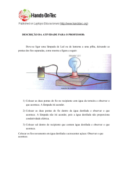
Fig. 29. Leptodactylus andreae. Distribuição corporal das glândulas granulosas: pele
total.
a) Vista da pele dorsal removida por inteiro. As glândulas granulosas concentram-se em duas
faixas longitudinais não-contínuas, desde a região pós-orbital da cabeça até a base dos
membros posteriores (seta). Na face dorsal da cabeça, as glândulas de veneno se limitam às
pálpebras e flancos dos tímpanos. Barra: 320 µm.
b) Vista da pele dorsal das coxas. Na face posterior, as coxas possuem acúmulos glandulares
circulares dispostos lado a lado, compondo uma faixa que se estende desde a cloaca até os
joelhos. Barra: 320 µm.
c) Detalhe das faixas glandulares dorsais, mostrando que as glândulas granulosas podem ser
distinguidas individualmente devido à sua baixa densidade. Barra: 80 µm.
d) Vista da pele ventral removida por inteiro. Na cabeça, uma faixa de glândulas de baixa
densidade margeia o lábio inferior e outra ocupa a região gular (seta preta). Há dois
agregados peitorais em forma de gota (seta vermelha) e um inguinal em forma de V, próximo
à base das coxas. Barra: 320 µm.
85
Fig. 30. Leptodactylus andreae. Distribuição corporal das glândulas cutâneas: dorso.
D – derme; E – epiderme; g – glândulas granulosas; m – glândulas mucosas.
a) Pele dorsal da cabeça. Notam-se glândulas granulosas e mucosas, essas com lúmen amplo.
Hematoxilina-eosina. Barra: 50 µm.
b) Pele dorsal. Nessa região, as glândulas granulosas não foram detectadas pela histologia, à
exceção de algumas na região das listas amarelas dorsais. As mucosas ocorrem em
quantidade. Azul de toluidina-fucsina. Barra: 50 µm.
c) Pele dorsal das coxas. Na face posterior das coxas, há concentração de glândulas
granulosas, compondo uma faixa de acúmulo. Menos freqüentes, as glândulas mucosas
ocupam posições mais superficiais na derme. Azul de toluidina-fucsina. Barra: 50 µm.
86
Fig. 31. Leptodactylus andreae. Distribuição corporal das glândulas cutâneas: ventre.
D – derme; E – epiderme; g – glândulas granulosas; m – glândulas mucosas.
a) Pele da margem do lábio inferior. Embora essa região apresente algum acúmulo de
glândulas granulosas, sua detecção histológica mostrou-se difícil. As glândulas mucosas, que
ocorrem em grande quantidade, possuem maior diâmetro do que no restante da superfície
corporal. Azul de toluidina-fucsina. Barra: 50 µm.
b) Pele da região ventral. A pele dessa região é rica em glândulas de muco, mas não foram
detectadas glândulas de veneno. Azul de toluidina-fucsina. Barra: 50 µm.
c) Pele ventral na região inguinal. Poucas glândulas granulosas foram encontradas nessa
região através da histologia. As glândulas mucosas, por outro lado, ocorrem em abundância.
Azul de toluidina-fucsina. Barra: 50 µm.
87
Fig. 32. Bioquímica das secreções cutâneas.
a) Análise por eletroforese (SDS-PAGE). A notável diversidade de proteínas nas secreções
cutâneas de Leptodactylus lineatus é indicada pelo grande número de bandas com diferentes
pesos moleculares nas duas amostras consideradas (L1, L2). Por outro lado, as secreções de
Ameerega picta possuem apenas uma fraca banca nas três amostras consideradas (A1, A2,
A3), indicando presença apenas pontual de proteínas. R é o padrão de pesos moleculares.
b) L. lineatus, análise de cromatografia por RP-HPLC. O grande pico na fração não-retida
(estrela) sugere grande quantidade de proteínas nas secreções dessa espécie. A presença de
picos entre os 12 e os 30 min aponta para a presença de peptídeos ou de compostos orgânicos
aromáticos.
c) A. picta, análise de cromatografia por RP-HPLC. A fração não-retida (estrela) é discreta no
cromatograma das secreções de A. picta. A presença de picos entre os 12 e os 30 min aponta
para a presença de peptídeos ou de compostos orgânicos aromáticos.
88
7. R EF ER ÊN C IAS B IB LIOGR ÁF IC AS
Alvarez B.B.; Delfino, G.; Nosi, D.; Terreni, A. 2005. Ultrastructure of Poison Glands of
South American frogs: A comparison Between Physalaemus albonotatus and
Leptodactylus chaquensis (Anura: Leptodactylidae). Journal of Morphology,
263:247–258.
Angel, R.; Delfino, G.; Parra, G.J. 2003. Ultrastructural patterns of secretory activity in
poison cutaneous glands of larval and juvenile Dendrobates auratus (Amphibia,
Anura). Toxicon, 41(1): 29-39.
Arifulova, I.; Delfino, G.; Dujsebayeva, T.; Fedotovskikh, G.; Nosi, D.; Terreni, A. 2007.
Serous Cutaneous Glands in the South American Horned Frog Ceratophrys ornata
(Leptodactylformes, Chthonobatrachia, Ceratophrydae): Ultrastructural Expression of
Poison Biosynthesis and Maturation. Journal of Morphology, 268:690-700.
Bancrof, J.D.; Stevens, A. 1996. Theory and Practice of Histological techniques. Churchill &
Livingstone, Edinburgh.
Bates H.W. 1861. Contributions to an insect fauna of the Amazon Valley. Lepidoptera:
Heliconidae. Transactions of the Entomological Society, 23(3): 495-566.
Bernarde, P. S. & Kokubum, M. N. C. 2009. Seasonality, age structure and reproduction of
Leptodactylus (Lithodytes) lineatus (Anura, Leptodactylidae) in Rondônia state,
southwestern Amazon, Brazil. Iheringia, 99(4):368-372.
Bevins, C.L.; Zasloff, M. 1990. Peptides from frog skin. Annual Review of Biochemistry,
59: 395-414
Bozzola, J.J.; Russell, L.D. 1999. Electron Microscopy: Principles and Techniques for
Biologists. Jones and Bartlett Publishers, Boston.
Brizzi, R.; Delfino, G.; Pellegrini, R. 2002. Specialized mucous glands and their possible
adaptive role in the males of some species of Rana (Amphibia, Anura). Journal of
Morphology, 254:328-341.
Brodie, E.D.; Howard, R.R. 1973. Experimental Study of Batesian Mimicry in the
Salamanders Plethodon jordani and Desmognathus ochrophaeus. American Midland
Naturalist, 90(1): 38-46.
Brodie, E.D. 1977. Salamander antipredator postures. Copeia, 1977:523-535.
Brodie, E.D.; Tumbarello, M.S. 1978. The antipredator functions of Dendrobates auratus
(Amphibia. Anura. Dendrobatidae): skin secretion in regard to a snake predator
(Thamnophis). Journal of Herpetology, 12: 264-265.
Brodie, E.D. 1993. Differential Avoidance of Coral Snake Banded Patterns by Free-Ranging
Avian Predators in Costa Rica. Evolution, 47(1): 227-235.
Brower, L.P.; Brower, J.V.Z.; Westcott, P.W. 1960. Experimental Studies of Mimicry. 5. The
Reactions of Toads (Bufo terrestris) to Bumblebees (Bombus americanorum) and
Their Robberfly Mimics (Mallophora bomboides), with a Discussion of Aggressive
Mimicry. The American Naturalist, 878: 343-355.
Chiari, Y.; Vences, M.; Vieites, D.R.; Rabemananjara, F.; Bora, P.; Ramilijaona, O.; Meyer,
A. 2004. New evidence for parallel evolution of colour patterns in Malagasy poison
frogs (Mantella). Molecular Ecology, 13: 3763- 3774.
Daly, J.W. 1998. Thirty years of discovering arthropod alkaloids in amphibian skin. Journal
of Natural Products, 61: 162–172.
89
Daly, J.W. 2003. Ernest Guenther award in chemistry of natural products. Amphibian skin: a
remarkable source of biologically active arthropod alkaloids. Journal of Medicinal
Chemistry, 46(4): 445-452.
Daly, J.W.; Brown, G.B.; Mensah-Dwumah, M.; Myers, C.W. 1978. Classification of skin
alkaloids from neotropical poisondart frogs (Dendrobatidae). Toxicon, 16: 163–188.
Daly, J. W.; Myers, C. W.; Warnick, J. E. and Albuquerque E. X. 1980. Levels of
batrachotoxin and lack of sensitivity to its action in poison-dart drogs (Phyllobates).
Science, 208: 1383-1385.
Daly, J.W.; Highet, R.J.; Myers, C.W. 1984. Occurrence of skin alkaloids in non-dendrobatid
frogs from Brazil (Bufonidae), Australia (Myobatrachidae) and Madagascar
(Mantellidae). Toxicon, 22: 905–919.
Daly, J.W.; Myers, C.W.; Whittaker, N. 1987. Further classification of skin alkaloids from
neotropical poison frogs (Dendrobatidae), with a general survey of toxic/noxious
substances in the amphibia. Toxicon, 25 (10): 1023–1095.
Daly, J.W.; Spande, T.F.; Garraffo, H.M. 2005. Alkaloids from amphibian skin: a tabulation
of over eight-hundred compounds. Journal of Natural Products, 68 (10): 1556–1575.
Daly, J.W.; Ware, N.; Saporito, R.A.; Spande, H.M.; Garraffo, H.M. 2009. NMethyldecahydroquinolines: An unexpected class of alkaloids from Amazonian
poison frogs (Dendrobatidae). Journal of Natural Products 72(6): 1110-1114.
Darst, C.R.; Cummings, M.E. 2006. Predator learning favours mimicry of a less-toxic model
in poison frogs. Nature, 440: 208-211.
Delfino, G.; Brizzi, R.; Calloni, C. 1990. A morphofunctional characterization of the serous
cutaneous glands in Bombina orientalis (Anura: Discoglossidae). Zoologischer
Anzeiger, 225: 295-310.
Delfino, G.; R. Brizzi, R.; Alvarez B.B.; Kracke-Berndorff, R. 1998. Serous cutaneous glands
in Phyllomedusa hypochondrialis (Anura, Hylidae) secretory patterns during
ontogenesis. Tissue & Cell, 30 (1): 30-40.
Delfino, G.; R. Brizzi, R.; Alvarez B.B.; Taddei, L. 1999. Secretory polymorphism and
serous cutaneous gland heterogeneity in Bufo granulosus (Amphibia, Anura).
Toxicon, 37: 1281-1296.
Delfino, G.; Nosi, D.; Brizzi, S.; Alvarez B.B. 2001. Serous cutaneous glands in the
paludiculine frog Physalaemus biligonigerus (Anura, Leptodactylidae): patterns of
cytodifferentiation and secretory activity in premetamorphic specimens. Acta
Zoologica, 82: 149–158.
Delfino, G.; R. Brizzi, R.; Nosi, D.; Terreni, A. 2002. Serous Cutaneous Glands in New
World Hylid Frogs: An Ultrastructural Study on Skin Poisons Confirms Phylogenetic
Relationships Between Osteopilus septentrionalis and Phrynohyas venulosa. Journal
of Morphology, 253:176–186.
Duellman, W.E. 1978. The biology of an equatorial herpetofauna in Amazonian Ecuador.
Miscelaneous Publications of the University of Kansas Museum of Natural History, 1352.
Duellman, W. E.; Trueb, L. 1986. Biology of the Amphibians. McGraw-Hill Book Co., New
York.
Dumbacher, J.P.; Wako, A.; Derrickson, S.R.; Samuelson, A.; Spande, T.F.; Daly, J.W. 2004.
Melyrid beetles (Choresine): a putative source for the batrachotoxin alkaloids found
in poison-dart frogs and passerine birds. Proceedings of the National Academy of
Sciences of the USA, 101 (45): 15857-15860.
90
Farquhar, M.G.; Palade, G.E. 1998. The Golgi apparatus: 100 years of progress and
controversy. Trends in Cell Biology, 8: 2–10.
Fox, H. 1986. Dermal glands. In: Biology of fhe Integument. Vol. , Vertebrates. BereiterHahn, J.; Matoltsy, A. G.; Richards, K. S. (eds), pp. 116-135. Springer, Berlin.
Fox H. 1994. The structure of the integument. In: Amphibian biology. Vol 1, The Integument.
Heatwhole, H. (ed.), p 1-32. Chipping Norton, NSW, Australia, Surrey Beatty &
Sons.
Fritz, G.; Rand, A.S.; dePamphilis, C.W. 1981. The aposematically colored frog,
Dendrobates pumilo, is distasteful to the large, predatory ant, Paraponera clavata.
Biotropica, 13(2): 158-159.
Frost, D. R. 2010. Amphibian Species of the World: an Online Reference. Version 5.1
Acessível em http://research.amnh.org/herpetology/amphibia/index.php. American
Museum of Natural History, New York.
Grant, T.; Frost, D.R.; Caldwell, J.P.; Gagliardo, R.; Haddad, C.F.B.; Kok, P.J.R.; Means,
D.B.; Noonan, B.P.; Schargel, W.E.; Wheeler, W.C. 2006. Phylogenetic systematics
of dart-poison frogs and their relatives (Amphibia: Athesphatanura: Dendrobatidae).
Bulletin of the American Museum of Natural History. 299: 1–262.
Greene, H. W., McDiarmid, R. W. 1981. Coral snake mimicry: Does it occur? Science,
213:1207-12.
Harper Jr, G.R.; Pfennig, D.W. 2007. Mimicry on the edge: why do mimics vary in
resemblance to their model in different parts of their geographical range? Proceedings
of the Royal Society, 274B: 1955-1961.
Jared, C.; Antoniazzi, M.M.; Silva, J.R.M.C.; Freymüller, E. 1999. Epidermal Glands in
Squamata: Microscopical Examination of Precloacal Glands in Amphisbaena alba
(Amphisbaenia, Amphisbaenidae). Journal of Morphology. 241: 197–206.
Jared, C; Antoniazzi, M.M.; Jordão, A.E.C.; Silva, J.R.M.C.; Greven, H.; Rodrigues, M.T.
2009. Parotoid macroglands in toad (Rhinella jimi): Their structure and functioning in
passive defence. Toxicon, 54(3):197-207.
Kiernan J.A. 1999. Histological and Histochemical Methods: Theory and Practice.
Butterworth-Heinemann, Oxford.
Lacombe, C.; Cifuentes-Diaz, C.; Dunia, I.; Auber-Thomay, M.; Nicolas, P.; Amiche, M.
2000. Peptide secretion in the cutaneous glands of South America tree frog
Phyllomedusa bicolor: an ultrastructural study. European Journal of Cell Biology,
79:631-341.
Laemmli, U. K. 1970. Cleavage of structural protein during assembly of bacteriphage T4.
Nature, 227:680-685.
Lamar, W.W.; Wild, E.R. 1995. Comments on the natural history of Lithodytes lineatus
(Anura: Leptodactylidae) with a description of the tadpole. Herpetological Natural
History, 3(2):135-142.
Lenzi-Mattos R.; Antoniazzi, M.M.; Haddad, C.F.B.; Tambourgi, D.V.; Rodrigues, M.T.;
Jared, C. 2005. The inguinal macroglands of the frog Physalaemus nattereri
(Leptodactylidae): structure, toxic secretion and relationship with deimatic behaviour.
Journal of Zoology, 266: 385–394.
Lillywhite, H.B. 2006. Water relations of tetrapod integument. Journal of Experimental
Biology, 209:202-226.
91
Lynch, J.D. 1985. Mimetic and Non-Mimetic Populations of Eleutherodactylus gaigeae
(Dunn) in lower Central America and Colombia (Amphibia: Anura, Leptodactylidae)
Studies on Neotropical Fauna and Environment, Volume 20:195 – 202.
Mallet, J.; Joron, M. 1999. Evolution of diversity in warning color and mimicry:
polymorphisms, shifting balance, and speciation. Annual Review of Ecology and
Systematics, 30: 201-233.
Mappes J & Alatalo R.V. 1997: Batesian mimicry and signal accuracy. Evolution 51: 20482051.
Marki, F.; Witkop, B. 1963. The venom of the Colombian narrow poison frog Phyllobates
bicolor. Experientia, 19: 329-376.
Müller, J. F. 1878. Über die Vortheile der Mimicry bei Schmetterlingen. Zoologischer
Anzeiger, 1: 54–55.
Myers, C. W., Daly, J. W., and Malkin, B. 1978. A dangerously toxic new frog (Phyllobates)
used by Emberá Indians of Western Colombia, with discussion of blowgun fabrication
and dart poisoning. Bulletin of the American Museum of Natural History, 161: 307366.
Myers, C. W.; Daly, J. W. 1983. Dart-poison frogs. Scientific American, 248 (2): 96- 105.
Navas, C.A.; C. Jared; M.M. Antoniazzi. 2002. Water economy in the casque-headed treefrog Corythomantis greeningi (Hylidae): role of behavior, skin and skull coossification. Journal of Zoology, Lond. 257: 525-532.
Nelson, C.E.; Miller, G.A. 1971. A possible case of mimicry in frogs. Herpetological Review,
3:109.
Paschinger, K.; Fabini, G.; Schuster, D.; Rendić, D.; Wilson, I.B. 2005. Definition of
immunogenic carbohydrate epitopes. Acta Biochimica Polonica, 52: 629–632.
Pasteur, G. 1982. A Classification Review of Mimicry Systems. Annual Review of Ecology
and Systematics, 13:169-199.
Randall, J.E. 2005. A review of mimicry in marine fishes. Zoological Studies, 44:299–328.
Reiskind, J. 1965. Behaviour of avian predator in an experiment simulating batesian mimicry.
Animal Behaviour,13(4): 466-469.
Regös, J.; Schlüter, A. 1984. Erste Ergebnisse zur Fortpflanzungsbiologie von Lithodytes
lineatus (Schneider, 1799) (Amphibia: Leptodactylidae). Salamandra, 20:252-261.
Rowe, C.; Lindström, L.; Lyytinen, A. 2004. The importance of pattern similarity between
Müllerian mimics in predator avoidance learning. Proceedings of the Royal Society of
London B, 271:407–413.
Santos, R.R.; Grant, T. 2010. Diel pattern of migration in a poisonous toad from Brazil and
the evolution of chemical defenses in diurnal amphibians. Evolutionary Ecology, no
prelo.
Saporito, R. A., M. A. Donnelly, R. L. Hoffman, H. M. Garraffo, and J. W. Daly. 2003. A
siphonotid millipede (Rhinotus) as the source of spiropyrrolizidine oximes of
dendrobatid frogs. Journal of Chemical Ecology, 29:2781–2786.
Saporito, R. A.; Garraffo, H.M.; Donnelly, M.A.; Edwards, A.L.; Longino, J.T.; Daly, J.W.
2004. Formicine ants: an arthropod source for the pumiliotoxin alkaloids of
dendrobatid poison frogs. Proceedings of the National Academy of Sciences of the
USA, 101: 8045–8050.
92
Saporito, R.A.; Donnelly, M.A.; Garraffo, H.M.; Spande, T.F.; Daly, J.W. 2006. Geographic
and seasonal variation in alkaloid-based chemical defenses of Dendrobates pumilio
from Bocas del Toro, Panama. Journal of Chemical Ecology, 32 (4): 795–814.
Saporito, R.A.; Donnelly, M.A.; Norton, R.A.; Garraffo, H.M.; Spande, T.F.; Daly, J.W.
2007. Oribatid mites: a major dietary source for alkaloids in poison frogs.
Proceedings of the National Academy of Sciences of the USA, 104 (21): 8885–8890.
Saporito, R.A; Zuercher, R.; Roberts, M.; Gerrow, K.G.; Donnelly, M.A. 2007. Experimental
evidence for aposematism in the poison frog Oophaga pumilio. Copeia, 4: 1006-1011.
Schlüter A.; Regös, J. 1981. Lithodytes lineatus (Schneider, 1799) (Amphibia:
Leptodactylidae) as a dweller in nests of the leaf cutting ant Atta cephalotes
(Linnaeus, 1758) (Hymenoptera: Attini). Amphibia-Reptilia, 2: 117-121.
Schwartz, E.F.; Stucchi-Zucchi, A.; Schwartz, C.A.; Salomão, L.C. 2003. Skin secretion of
Siphonops paulensis (Gymnophiona, Amphibia) forms voltage-dependent ionic
channels in lipid membranes. Brazilian Journal of Medical and Biological Research,
36:1279-1282.
Schwartz, C.A.; Castro, M.S.; Pires Jr., O.R.; Maciel, N.M.; Schwartz, E.N.F.; Sebben, A.
2007. Princípios ativos da pele de anfíbios: panorama atual e perspectivas. In:
Herpetologia no Brasil II. Nascimento, L.B.; Oliveira, M.E. (eds.), pp. 146-168.
Sociedade Brasileira de Herpetologia, Belo Horizonte.
Servedio, M.R. 2000. The Effects of Predator Learning, Forgetting, and Recognition Errors
on the Evolution of Warning Coloration. Evolution, 54(3): 751-763.
Sherratt, T.N. 2002. The evolution of imperfect mimicry. Behavioral Ecology, 13: 821–826.
Sherratt, T.N. 2008. The evolution of Müllerian mimicry. Naturwissenschaften, 95:681–695.
Smith, S. M. 1975. Innate recognition of coral snake pattern by a possible avian predator.
Science, 187: 759–760.
Stebbins, R. C.; Cohen, N. W. 1995. A Natural History of Amphibians. Princeton University
Press. New Jersey.
Stevens, M. 2007. Predator perception and the interrelation between different forms of
protective coloration. Proceedings of the Royal Society of London B, 274: 1457–
1464.
Symula, R.; Schulte, R.; Summers, K. 2001. Molecular phylogenetic evidence for a mimetic
radiation in Peruvian poison frogs supports a Müllerian mimicry hypothesis.
Proceedinf of the Royal Society of London B, 268: 2405-2421.
Szelistowski, W.A. 1985. Unpalatability of the Poison Arrow Frog Dendrobates pumilio to
the Ctenid Spider Cupiennius coccineus. Biotropica, 17(4):345-346.
Terreni, A.; Nosi, D.; Greven, H.; Delfino G. 2003. Development of serous cutaneous glands
in Scinax nasica (Anura, Hylidae): patterns of poison biosynthesis and maturation in
comparison with larval glands in specimens of other families. Tissue & Cell, 35: 274–
287.
Thomas EO, Tsang L, Licht P. 1993. Comparative histochemistrymof the sexually dimorphic
skin glands of anuran amphibians. Copeia, 1993:133–143
Toledo, R. C.; Jared, C. 1993. The calcified dermal layer in anurans. Journal of Comparative
Biochemistry and Physiology, 104A: 443-448.
Toledo, R.C.; Jared, C. 1995. Cutaneous granular glands and amphibian venoms.
Comparative Biochemistry and Physiology, 111(A): 1-29.
93
Toledo, L.F.; Guimarães, L.D.; Lima, L.P.; Bastos, R.P.; Haddad, C.F.B. 2004. Notes on
courtship, egg-laying site, and defensive behavior of Epipedobates flavopictus
(Anura: Dendrobatidae) from two mountain ranges of central and southeastern Brazil.
Phyllomedusa, 3(2):145-147.
Toledo, L. F.; Haddad, C.F.B. 2009. Colors and some morphological traits as defensive
mechanisms in anurans. International Journal of Zoology, 2009: 1-12
Vitt, L.J.; Caldwell, J.P. 2009. Herpetology: An Introductory Biology of Amphibians and
Reptiles, 3rd edition. Academic Press, San Diego. 697 pp.
Vitt, L.J.; Caldwell, J.P.; Shepard, D.B.; França, F. G. R.; Colli, G.R. 2010. Amphibians and
Reptiles of the Lower Cristalino River Region of the Southern Amazon. Banco de
dados eletrônico acessível em
http://www.omnh.ou.edu/personnel/herpetology/vitt/Cerrado/Cristalino/ assessado em
agosto de 2010.
Weygoldt, P. 1987. Evolution of parental care in dart poison frogs (Amphibia: Anura:
Dendrobatidae). Zeitschrift für zoologische Systematik und Evolutionsforschung,
25:51-67.
Zug, G. R. 1993. Herpetology. An Introductory Biology of Amphibians and Reptiles.
Academic Press Inc., San Diego.
94
8. A N E XOS
Encontra-se abaixo relacionada a composição dos corantes e soluções utilizados na seção
Material e Métodos:
Solução de Paraformaldeído Tamponado a 4% em tampão PBS 0,1M, pH 7,2
Paraformaldeído..................................................................................... 20 g
Água Destilada…………………………………………………………450 mL
Dissolver o paraformaldeído na água destilada, sob aquecimento, com agitador magnético.
Após dissolver completamente, acrescentar gotas de NaOH 1N até a solução ficar
transparente. Acrescentar o tampão PBS.
Tampão PBS: NaCl.................................................................................4,5 g
Tampão fosfato 0,1M pH7,2…….……………………...………..........50 mL
Preparo da solução de tampão fosfato 0,1M pH 7,2:
Solução estoque A:
Fosfato de sódio monobásico 0,2M.......................3,12 g
Água destilada.....................................................100 mL
Solução estoque B:
Fosfato de sódio dibásico 0,2M.............................2,83 g
Água destilada.....................................................100 mL
Misturar 14 mL de solução A com 36 mL de solução B e completar com água destilada para
100 mL.
1. Solução Karnovsky em tampão cacodilato de sódio 0,1M - pH 7,2
Solução A -
Glutaraldeído 10% em cacodilato de sódio 0,2 M pH 7,2:
Glutaraldeído 50% ..................................................10 ml
Cacodilato de sódio 0,2M pH 7,2 ............................ 40 ml
Solução B -
Paraformaldeído 8%:
Paraformaldeído ..........................................................4 g
Água destilada .........................................................50 ml
Acrescentar 2-4 gotas de NaOH para clarear. Misturar a solução A com a solução B a 1:1.
2. Solução A (pré-inclusão em historresina)
Peróxido de benzoíla....................................................................... 0,15 g
Hidroxietil metacrilato..................................................................... 15 ml
95
Dissolver o peróxido de benzoíla (1%) em hidroxietil metacrilato, agitando por 10-15
minutos.
3. Solução B (inclusão em historresina)
Solução A........................................................................................ 5 ml
Polimerizador.............................................................................. 0,33 ml
Misturar em agitador magnético por 4 minutos.
4. Albumina de Mayer
Clara de ovo "batida em neve"....................................................... 50 ml
Glicerina..........................................................................................50 ml
Misturar bem e filtrar em papel de filtro. Adicionar cristais de timol como conservante.
5. Azul de Toluidina 0,1%
Azul de Toluidina............................................................................ 0,1 g
Borato de sódio.....................................................................................1g
Água destilada.............................................................................. 100 ml
6. Fucsina Básica 0,05%
Fucsina básica.............................................................................. 0,05 g
Água destilada............................................................................. 100 ml
7. Hematoxilina de Harris
Hematoxilina...................................................................................... 1 g
Alúmen de potássio...........................................................................20 g
Dióxido de mercúrio........................................................................ 0,5 g
Água destilada............................................................................... 200 ml
Dissolver a hematoxilina em aproximadamente 10 ml de álcool absoluto e, separadamente,
dissolver o alúmen em água quente destilada. Após, misturar as duas soluções e ferver até
ebulição. Adicionar o dióxido de mercúrio.
8. Eosina Y (Gill)
Eosina Y......................................................................................... 0,5 g
Etanol 96%................................................................................... 100 ml
Ácido acético glacial.................................................................... 3 gotas
96
Dissolver a eosina no álcool e acrescentar o ácido acético.
9. Azul de Bromofenol
Bicloreto de mercúrio....................................................................................10 g
Azul de Bromofenol .................................................................................100 mg
Etanol 95% ...................................................................................……….100 ml
10. Solução tampão PBS pH 7,2
Solução stock
NaCl...................................................................................................................26,4g
Na2HPO4.12H2O………………………………………………………...….....7,95g
NaH2PO4.H2O…………………………………………………………………0,93g
Dissolver em 1 litro de água destilada. Essa solução está 3 vezes concentrada. Diluir na hora
do uso.
11. Alcian Blue pH2,5
Alcian Blue.........................................................................................1 g
Ácido Acético 3%......................................................................... 100 ml
12. Reativo de Schiff
Fucsina básica.................................................................................. 1 g
Água destilada............................................................................. 400 ml
Metabissulfito de potássio................................................................. 9 g
HCl.................................................................................................10 ml
Levar ao bico de Bunsen a fucsina com a água destilada. Retirar antes de ferver e filtrar.
Levar ao agitador magnético e colocar aos poucos o metabissulfito. Em seguida, com
agitador magnético adicionar o HCl puro. Adicionar 1 colher de café de carvão ativado.
Ativar por 3-5 minutos e filtrar novamente.
13. Água sulforosa
HCl 1N............................................................................................10 ml
Metabissulfito de sódio 10%........................................................... 10 ml
Água destilada............................................................................. 180 ml
97
14. Safranina 0,5%
Água destilada............................................................................. 50 ml
Álcool absoluto............................................................................ 50 ml
Safranina........................................................................................... 1g
Misturar e adicionar em 100 ml de água destilada, 0,5 ml da solução acima.
15. Tetróxido de ósmio 1% em tampão cacodilato 0,1M pH 7,2
OsO4 4%
Água destilada
Tampão cacodilato 0,2 M
Primeiramente misturar o tetróxido de ósmio e a água destilada em uma proporção de 1:1.
Misturar o volume desta solução de ósmio a um volume igual de tampão cacodilato 0,2 M.
16. Acetato de uranila 0,5% + Sacarose a 13,3%
Água destilada............................................................................. 100 ml
Acetato de uranila........................................................................... 0,5 g
Sacarose.........................................................................................13,3 g
17. Resina Epon
Polybed 812........................................................................………. 21 ml
DDSA...............................................................................……........13 ml
NMA................................................................................................ 11 ml
DMP-30......................................................................................... 0,7 ml
Misturar com agitador magnético os três primeiros componentes e por último adicionar o
catalisador (DMP-30).
18. Citrato de chumbo
Nitrato de chumbo......................................................................... 1,33 g
Citrato de sódio............................................................................. 1,76 g
Água destilada fervida (fria)........................................................... 30 ml
Misturar em balão volumétrico de 50 ml, agitando vigorosamente por 5 minutos. Continuar
agitando de vez em quando por mais 30 minutos, até a solução tornar-se leitosa. Acrescentar
8 ml de NaOH 1 N (4%), recentemente preparada com água destilada fervida. Completar o
volume até 50 ml com água destilada fervida.
98
Baixar