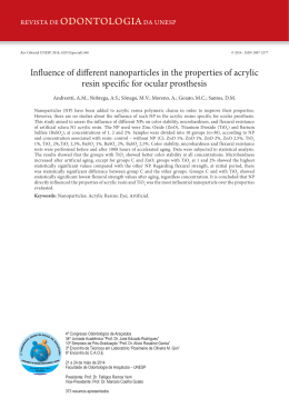
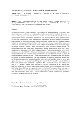
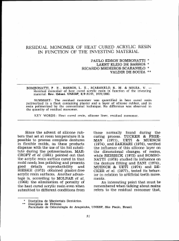
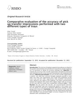
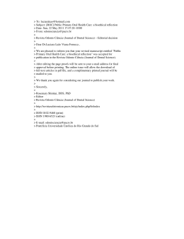
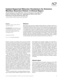
ACTA-1-2012-SEG:3-2011 11/07/2012 11:26 a.m. Página 96
96
INFLUENCE OF DIFFERENT MATERIALS AND TECHNIQUES
TO TRANSFER MOLDING IN MULTIPLE IMPLANTS
Júlio C.B. Faria1, Fernando L.G. Cruz1, Laís R. Silva-Concílio2, Ana C.C. Neves2
1
Department of Restorative Dentistry, Prosthodontics Area, Dental School,
Federal University of Juiz de Fora, Juiz de Fora, Minas Gerais, Brazil
2
Department of Restorative Dentistry, Prosthodontics Area, Dental School,
University of Taubaté, Taubaté, São Paulo, Brazil
ABSTRACT
The aim of this study was to compare different materials and techniques used in transfer molding of multiple implants, by evaluating the space between implants and superstructure. Four external
hexagon implants were fixed in a master template and the
same on a superstructure. Transfer molding of implants were done
using the direct and indirect techniques, with transfers united
or not, using the union chemically activated acrylic resin
(QA) and other groups polymerized acrylic resin (FT), and sectioned and not split. The casts were made with polyether and models divided into 8 groups (n = 5). The space between the
superstructure and the master implants was measured with
a microscope and the data was analyzed statistically by Student’s
t test (p <0.05). For the material of union there was no significant difference, except when the groups were compared with the
resin Duralay QA (G4) and the resin Duolay FT (G8) and groups
using resins Duolay QA (G5) and Duolay FT (G7) for the union
of the transfers. When comparing the groups who had the union
between the transfers and sectioned again united with those in
which the union was not severed there was no statistically significant difference. QA resin was superior to the FT with respect to
the union of transfers. Techniques with united transfers or not
were similar.
Keywords: Dental impression technique, dental Implants, dental materials.
INFLUENCIA DE DIFERENTES MATERIAIS E TÉCNICAS DE MOLDAGEM
DE TRANSFERÊNCIA EM MÚLTIPLOS IMPLANTES
RESUMO
O objetivo deste estudo foi comparar diferentes materiais e técnicas utilizados na moldagem de transferência em implantes
múltiplos, por meio da avaliação do espaço entre supra-estrutura e implantes. Quatro implantes de hexágono externo foram
fixados em um modelo mestre e sobre os mesmos uma supraestrutura. Moldagens de transferência dos implantes foram feitas
utilizando as técnicas indireta e direta, com transferentes unidos
ou não, utilizando para a união resina acrílica quimicamente ativada (QA) e em outros grupos resina acrílica fotoativada (FT),
sendo seccionadas e não seccionadas. As moldagens foram realizadas com poliéter e os modelos divididos em 8 grupos (n=5). O
espaço existente entre a supra-estrutura mestre e os implantes foi
mensurada com microscópio e os dados obtidos analisados esta-
tisticamente pelo teste t de Student (p<0.05). Para o material de
união não houve diferença significativa, exceto quando comparados os grupos com a resina Duralay QA (G4) e a resina Duolay
FT (G8) e as resinas Duolay QA (G5) com a Duolay FT (G7)
para união dos transferentes. Quando comparados os grupos que
tiveram a união entre os transferentes seccionada e novamente
unida com aqueles em que a união não foi seccionada, não foi
observada diferença estatística significativa. A resina QA se
mostrou superior à FT no que diz respeito à união dos transferentes. As técnicas com transferentes unidos ou não se mostraram
similares.
INTRODUCTION
Oral rehabilitation treatments with osseointegrated
implants have a high rate of success1. Yet, several
conditions may affect the predictability and the success rates of dental implants and of implant-supported prostheses. One of the main factors, among
other, is the biomechanical condition2,3.
The passive fit of the prosthetic supra-structure4,5
is extremely important for the success of implantsupported prostheses, thus enabling a consistent
distribution of the occlusal stresses generated by
them. Even though most of the components of
implant-supported prostheses are pre-fabricated
and, as such, they have a more accurate level of
Acta Odontol. Latinoam. 2012
Palavras-chave: Técnica de moldagem odontológica, implantes
dentários; materiais dentários.
ISSN 0326-4815
Vol. 25 Nº 1 / 2012 / 96-102
ACTA-1-2012-SEG:3-2011 11/07/2012 11:26 a.m. Página 97
Materials and techniques to transfer molding
fitting than conventional prostheses, the transfer
of the fitting of such components still remains a
problem for the development of a work model
according to the laboratory work. Such difficulties
increase according to the number of implants and
to their fitting6.
The techniques most widely used in transfers are
the indirect technique, with a closed mold, and the
direct technique, in which an open mold is used. In
the indirect technique, transfers are rounded or conical and are not fixed into the mold, but may be
refitted inside it before the mold is filled with the
cast to obtain the model. On the other hand, the
direct technique allows the normally retentive transfer to remain inside the mold, thus reducing the tilting effect of the implants and turning their refitting
unnecessary.
Obtaining an accurate mold is possible through the
union of the mold transfers with dental floss and
chemically-activated acrylic resin7. Yet, this technique requires a higher clinical timing8. An alternative for decreasing this clinical timing would be
using a photopolymerizable acrylic resin9,10
Based on these possibilities and on the need for the
passivity of the prostheses, the purpose of this study
was to assess the accuracy obtained with three different techniques of implant transfer molds, apart
from assessing how the activation of the resins used
in the union impact on the level of accuracy of the
technique.
MATERIALS AND METHODS
For the calculation of the samples in this study, a ttest for independent samples was used, assisted by
the BioEstat 5.0 software, with a 0.9-power and a
significance level equal to 0.05. Data used for this
calculation was obtained from studies available in
the literature, which used a methodology similar to
the one applied in this work9. As a result of the ttest, it was suggested that each one of the groups be
made up of three test specimens. Yet, by using a factor of safety, it was determined that five specimens
be used for each group.
A master model in self-polymerizable acrylic
resin was made (Duralay, Reliance Dental Mfg.
Co., Worth, Ill., USA). Length: 50 mm; Width: 20
mm, and Height: 15 mm. Four Neodent (Batch #
140207. Titamax, Neodent, Curitiba, Brazil),
external hexagonal implants, with a 3.75mmdiameter and a 13mm-length, were loaded into the
Vol. 25 Nº 1 / 2012 / 96-102
97
master model. For fitting the implants, a Neopronto System surgical bar, (Batch # 92327, Neodent,
Curitiba, Brazil) was used as a guide and four
drills were made on the model with a surgical drill
- 3.0mmx15mm - (Neodent, Curitiba, Brazil),
assembled on a Driller BLM 600 Plus (Driller,
São Paulo, Brazil). The diameter of the drills was
larger than that of the implants, thus allowing the
latter to be locked onto the master model by
adding condensed acrylic resin in that space. The
implants were fitted and numbered from 1 to 4,
clockwise (Fig. 1). The layout, parallelism and
distance between the implants were supplied by
means of the surgical bar.
A supra-structure was waxed onto UCLA-type calcinable cylinders, without anti-rotational and with
Tilite strip (Neodent, Batch # 666464, Curitiba,
Brazil), loaded into the implants previously fitted
into the master model (Fig. 2). The supra-structure
was cast with an Tilite – nickel-chrome-titanium
alloy (Talladium do Brasil, Curitiba, Paraná, Brazil)
by means of the lost wax casting method11 (Fig. 3).
The fitting and passivity of the settling were
checked by means of a probe (Exploratory Probe #
5, Duflex, SSWhite, Juiz de Fora, MG, Brazil) and
of the Sheffield single-screw test12,13. After these
Fig. 1: Master Model with Clockwise Numbering.
Fig. 2: Calcinable Cylinders (Front View).
ISSN 0326-4815
Acta Odontol. Latinoam. 2012
ACTA-1-2012-SEG:3-2011 11/07/2012 11:26 a.m. Página 98
98
Júlio C.B. Faria, Fernando L.G. Cruz, Laís R. Silva-Concílio, Ana C.C. Neves
checks, the supra-structure/master model set was
analyzed by a microscope (Olympus STM, Olympus, Tokyo, Japan) to check the level of accuracy
of the settling of the interface (Magnification: 30x
and Accuracy: 0.001mm.
This master model and supra-structure combination were named as Control Group, and became
the standard for fitting and measuring of the
specimens.
For producing the individual molds that were subsequently used for the molding, the relief of the
Control Group was made by means of a #7 Wax
Strip (Uraby, Rio de Janeiro, Brazil). Immediately after this, the molding process was performed
with Stern Tek (Stern Gold, Germany) and with
the mold filled with a special Type IV cast (Troquel/Quattro, Polidental, Brazil). The model that
was thus obtained was included in a similar way
to the one followed in the procedures of inclusion
and acrylization of total prostheses and the individual molds of acrylic resin were pressed7. This
procedure was repeated until forty individual
molds were obtained, and five molds were des-
Fig. 3: Master Supra-Structure (Vestibular View).
Fig. 4: Square Transfers (Union with RAAQ).
Acta Odontol. Latinoam. 2012
tined to each group in the study. The molds destined to the molding technique with square transfers were drilled so as to allow for access to the
screws of the transfers.
Rounded transfers were used, Batch # 700399
(Neodent, Curitiba, Brazil), as well as square
transfers, Batch # 679564 (Neodent, Curitiba,
Brazil). For techniques using united transfers, the
union was used with dental floss (Oral B, Gillette
do Brasil, Brazil) and Duralay acrylic resin
(Reliance Dental Mfg. Co., Worth, Ill., USA), or
Duolay acrylic resin (Vipi, São Paulo, Brazil) (Fig.
4). The latter, supplied with a liquid for chemical
activation and another liquid for photoactivation.
When using the Duralay chemical resin, after
flossing the dental floss through the four transfers,
the acrylic resin was applied by means of the
“brush technique” over the dental floss and around
the transfers. In the sectioning techniques, a section was performed between the transfers with a
diamond double-faced disk (Fava, São Paulo, SP,
Brazil) and then a new union was made with the
same resin. In the techniques in which Duolay
chemical acrylic resin was used, the procedures
followed were the same as above, for the techniques involving sectioning as well as for those
without sectioning. When using the Duolay photoactivated acrylic resin, after flossing the dental
floss through the transfers, the acrylic resin was
applied and photopolymerized during approximately four minutes; when the technique with no
sectioning was used, the application took approximately ten minutes (four minutes for pre-sectioning, two minutes for sectioning, and four
additional minutes for the new union). The photopolymerization equipment used was an Optilux
401 with halogenous lamp (Demetron, Kerr Corp,
Danbury, CT, USA).
In the mold made by means of closed molding, the
transfers were removed from the master model and
coupled to their corresponding replicas and refitted in the mold. In the open mold technique, as the
transfers were removed during the molding
process, only the coupling of the replicas to the
transfers was performed. All the eight experimental groups were molded with polyester (Impregum,
3M ESPE, Seefelf, Germany). After the pressing
of the material recommended by the manufacturer,
a model with special casting Type IV was made
(Troquel/Quatro, Polidental, Brazil). Once the
ISSN 0326-4815
Vol. 25 Nº 1 / 2012 / 96-102
ACTA-1-2012-SEG:3-2011 11/07/2012 11:26 a.m. Página 99
Materials and techniques to transfer molding
complete crystallization of the material was
obtained, the fitting of the master supra-structure
onto the implants was analyzed. The assessed
groups can be seen in Table 1.
Before the analysis, the supra-structure was
screwed (square screws, 4.1 mm Ø – Neodent,
Batch # 800015664, Curitiba, Brazil), Torque:
32Ncm (Dyna Torq, ITL Dental, Irvine, CA, USA,
Series # 222851), following the tightening
sequence of the implants. 2, 3, 4 and 1. Measurings were made at the central point, previously
marked in the master supra-structure by means of
a drill (# 3203, KG Sorensen, São Paulo, Brazil),
from the vestibular and lingual face with the aid of
a linear microscope (Measuring Microscope STM
- OLYMPUS®, Tokyo, Japan) (Fig. 5).
For the statistical analysis, when means were parametric, a t-test was used for independent samples
(p≤0,05) and when they were not parametric, the
Mann-Whitney test was used (p≤0,05).
Table 1: Specification of Assessed Groups.
Group
n=5
1
Transfer Technique
Rounded
Indirect
2
Square
Direct
------
No
3
Square
Direct
Duralay (chemical)
No
4
Square
Direct
Duralay (chemical)
Yes
5
Square
Direct
Duolay (chemical)
No
6
Square
Direct
Duolay (chemical)
Yes
7
Square
Direct
Duolay (photo)
No
8
Square
Direct
Duolay (photo)
Yes
Vol. 25 Nº 1 / 2012 / 96-102
Material
For Unions
------
Section/
Reunited
No
Table 2: Assessment of the Material for Unions in
the Groups.
Group Type of Technique
Transfer
Type of
Union
Mean
(dp)
3
Square
Direct
Dental Floss
+
Duralay Resin
Chemical
0,0079
(0,0065)
5
Square
Direct
Dental Floss
+
Duolay Resin
Chemical
0,0122
(0,0087)
3
Square
Direct
Dental Floss
+
Duralay Resin
Chemical
0,0079
(0,0065)
7
Square
Direct
Dental Floss
0,0545
+
(0,0711)
Duolay Resin
Photoactivated
4
Square
Direct
Dental Floss
+
Duralay Resin
Chemical
+
Sectioned and
Reunited
0,0027
(0,0027)
6
Square
Direct
Dental Floss
+
Duolay Resin
Chemical
+
Sectioned and
Reunited
0,0514
(0,1118)
4
Square
Direct
Dental Floss
+
Duralay Resin
Chemical
+
Sectioned and
Reunited
0,0027
(0,0027)
8
Square
Direct
Dental Floss
0,0116
+
(0,0039)
Duolay Resin
Photoactivated
+
Sectioned and
Reunited
5
Square
Direct
7
Square
Direct
Fig. 5: View of the Specimen being Assessed by means of Linear
Olympus STM Measuring Microscope.
RESULTS
For the material used for unions, there was no significant difference, except when the groups using
Duralay chemically activated resin (QA) (G4) and
Duolay photoactivated resin (FT) (G8), were compared with the groups using Duolay QA resins (G5)
and Duolay FT (G7) for unions of the transfers.
When both groups were compared, the group
whose union was sectioned and then reunited and
the group with no sectioned union, no significant
statistical difference was observed (Tables 2 and 3).
99
Dental Floss
+
Duolay Resin
Chemical
Value
of p
0,061
0,144
0,835*
0,004*
0,0122
(0,0087)
0,012*
Dental Floss
0,0545
+
(0,0711)
Duolay Resin
Photoactivated
* Significant Values
ISSN 0326-4815
Acta Odontol. Latinoam. 2012
ACTA-1-2012-SEG:3-2011 11/07/2012 11:26 a.m. Página 100
100
Júlio C.B. Faria, Fernando L.G. Cruz, Laís R. Silva-Concílio, Ana C.C. Neves
Table 3: Assessment of the Technique, With and
Without Sectioning in the Groups.
Group Type of Technique
Transfer
Type of
Union
Mean
(dp)
3
Square
Direct
Dental Floss
+
Duralay Resin
Chemical
0,0079
(0,0065)
4
Square
Direct
Dental Floss
+
Duralay Resin
Chemical
+
Sectioned and
Reunited
0,0027
(0,0027)
5
Square
Direct
Dental Floss
+
Duolay Resin
Chemical
0,0122
(0,0087)
6
Square
Direct
Dental Floss
+
Duolay Resin
Chemical
+
Sectioned and
Reunited
0,0514
(0,1118)
7
Square
Direct
Dental Floss
0,0545
+
(0,0711)
Duolay Acrylic
Resin
Photoactivated
8
Square
Direct
Dental Floss
0,0116
+
(0,0039)
Duolay Acrylic
Resin
Photoactivated
+
Sectioned and
Reunited
Value
of p
0,158
0,094
0,144*
DISCUSSION
The mold techniques most widely used are the
“closed or indirect mold technique”, that uses
rounded transfers, and the “open or direct mold
technique”, that uses square transfers, whether united or not14-16.
In general, distortions inherent with the indirect
technique are related to the refitting of transfers
within the molds14,17.
In the direct technique, used after being recommended by Bränemark et al.7, square transfers may
be united by means of an acrylic resin. Even though
several authors have been doing research on the
efficacy of this technique14,17,18, results have not
been conclusive so far as regards their superiority
in relation with the other transfer techniques.
In this research, apart from the techniques with nonunited transfers, other techniques were compared,
such as those that make the union of transfers with
acrylic resin and those in which, after the union of
transfers, the sectioning of the union takes place and
transfers are united once again with a lower amount
Acta Odontol. Latinoam. 2012
of resin so as to minimize distortions in the material19,20. The union of the transfers in the indirect technique can be made only with self-polymerizable or
photo-polymerizable acrylic resin9, with self-polymerizable acrylic resin associated with dental floss,
with orthodontic floss and even with the use of bars
or pre-machined pins21. Self-polymerizable acrylic
resins have been the most widely used materials, nevertheless, due to contraction of about 6.5% to 7.9%
occurring during polymerization20,22, distortions are
associated to the use of this material in the union of
transfers17,23. Besides, another disadvantage derived
from the use of self-polymerizable acrylic resins is
the long time necessary for this technique, increasing
the level of tiredness in patients and the costs of treatment due to the increase in clinical timing. Due to
these factors, in this research, the union made with
photopolymerizable acrylic resin was tested, and this
might decrease treatment costs because of the reduction of clinical timing, and would make this treatment
more comfortable for patients.
Polyether is the material chosen for moldings in
prostheses on implants due to its high linear stability, its low residual contraction during storage, its
great rigidity and resistance to the rotation of the
transfers.10,24.
The measuring procedures with the microscope was
used because of their scientifically proved level of
accuracy (0.001mm) and efficacy5,14,17,18. In other
surveys, accuracy levels were measured by means
of other measuring devices, for example, softwareassisted devices10.
As regards the assessment of the material used for
unions, the Duolay acrylic resin chosen was chemical polymerization or by means of light, as it would
decrease the time needed for union in approximately four minutes, when the technique without sectioning was used; and in ten minutes when the
technique with sectioning was used (four minutes
for pre-sectioning; two minutes for sectioning, and
four additional minutes for the new union), vis a vis
the twenty minutes used in the technique without
sectioning and forty minutes, in the technique with
sectioning elapsed when using the conventional
Duralay resin22. This showed to be possible in different tested groups as the statistically significant
difference of the adjustment was only verified when
groups 4 and 8 were compared (p=0,004) and 5 and
7 (p=0,012). In both cases this may have happened
due to the absence or to the excess of polymeriza-
ISSN 0326-4815
Vol. 25 Nº 1 / 2012 / 96-102
ACTA-1-2012-SEG:3-2011 11/07/2012 11:26 a.m. Página 101
Materials and techniques to transfer molding
tion of Duolay resin applied with light. Insufficient
or excessive time used for the application of the
light and the volatilization of the liquid of the resin
may bring about a greater contraction of the Duolay acrylic resin25.
In the assessment of the techniques with or without sectioning no significant difference was
observed between the assessed groups (Table 3).
This result is consistent with studies undertaken by
Herbst et al.5, Kim et al.25 and Del’Acqua et al.26.
Due to the contraction of polymerization in the
acrylic resins, several researchers have studied
ways of minimizing the distortion present in this
material20,27. According to Mojon et al.22 the reduction in the volume of the acrylic resin used in the
union of the transfers would reduce the contraction
of the material. Yet, this was not observed in this
study, probably due to the excellence of the prosthetic components or of the material of the molding used, or to the attention given to the timing of
101
polymerization of the resins and for that of the
pouring of the molds.
As this was an in vitro study, it did not have the
adverse conditions typical of clinical practices such
as the presence of saliva, adaptation difficulty of
components of molds and/or prosthetics, angle of
implants, accurate adaptation of moldings, among
other. Nevertheless, there was a high level of difficulty, irrespective of the technique that was adopted, in obtaining accurate models in the molds of
transfer in multiple implants.
CONCLUSION
From the results obtained, it was possible to conclude that the use of the techniques with transfers,
whether united or not, did not alter the accuracy of
the fitting of the supra-structure. Yet, the use of
chemically activated resin for the union of transfers
supplied a better fitting when compared with the
photoactivated resin.
CORRESPONDENCE
Dr. Julio César Brigolini de Faria
Avenida Barão do Rio Branco, 2406 sl 904 - Centro,
36016-904 / Juiz de Fora
Minas Gerais, Brazil.
E-mail: [email protected]
REFERENCES
1. Suito H, Tomotake Y, Watanabe M, Nagao D, Ishida Y,
Ichikawa T. Survival of immediate implant restoration: A
retrospective study through 9-year-observation. J Prosthod
Res 2011;55:141-145.
2. Miyata T, Kobayashi Y, Araki H, Ohto T, Shin K. The influence of controlled occlusal overload on peri-implant tissue.
Part 4: a histologic study in monkeys. Int J Oral Maxillofac
Implants 2002;17:384-390.
3. Esposito M, Grusovin MG, Kakisis I, Coulthard P, Worthington H.V. Interventions for replacing missing teeth: treatment
of perimplantitis. Cochrane Database Syst Rev. 2008 Apr
16;(2):CD004970.
4. Jemt T, Lekholm U. Measurements of bone and frame-work
deformations induced by misfit of implant superstructures.
A pilot study in rabbits. Clin Oral Implants Res 1998;9:
272-280.
5. Herbst D, Nel JC, Driessen CH, Becker PJ. Evaluation of
impression accuracy for osseointegrated implant supported
superstructures. J Prosthet Dent 2000;83:555-561.
6. Vigolo P, Majzoub Z, Cordioli G. Evaluation of the accuracy
of three techniques used for multiple implant abutment
impressions. J Prosthet Dent 2003;89:186-192.
7. Bränemark PI, Zarb GA, Albrektsson T. Tissue-integrated
prostheses. Chicago: Quintessence; 1985.
Vol. 25 Nº 1 / 2012 / 96-102
8. Barnabé W, Santos Júnior GC, Mendonça Neto T, Rubo JH.
Uso simplificado de moldeiras de estoque e barras de resina
acrílica nas moldagens em implantodontia. RBP Rev Bras
Implantodon Prótese Sobre Implant 2005;12:105-108.
9. Assif D, Nissan J, Varsano I, Singer A. Accuracy of implant
impression splinted techniques: effect of splinting material.
Int J Oral Maxillofac Implants 1999;14:885-888.
10. Assunção WG, Tabata LF, Cardoso A, Rocha EP, Gomes
EA. Prosthetic transfer impression accuracy evaluation for
osseointegrated implants. Implant Dent 2008;17:248-256.
11. De La Cruz JE, Funkenbusch PD, Ercoli C, Moss ME,
Graser GN, Tallents RH. Verification jig for implant-supported prostheses: A comparison of standard impressions
with verification jigs made of different materials. J Prosthet Dent 2002;88:329-336.
12. Henry PJ, Tan AE, Uzawa S. Fit discrimination of implantsupported fixed partial dentures fabricated from implant
level impressions made at stage I surgery. J Prosthet Dent
1997;77:265-270.
13. Kan JY, Rungcharassaeng K, Bohsali K, Goodacre CJ,
Lang BR. Clinical methods for evaluating implant framework fit. J Prosthet Dent 1999;81:7-13.
14. Carr AB. Comparison of impression techniques for a twoimplant 15-degree divergent model. Int J Oral Maxillofac
Implants 1992;7:468-475.
ISSN 0326-4815
Acta Odontol. Latinoam. 2012
ACTA-1-2012-SEG:3-2011 11/07/2012 11:26 a.m. Página 102
102
Júlio C.B. Faria, Fernando L.G. Cruz, Laís R. Silva-Concílio, Ana C.C. Neves
15. Assif D, Fenton A, Zarb G, Schmitt A. Comparative accuracy of implant impression procedures. Int J Periodontics
Restorative Dent 1992;12:112-121.
16. Hsu CC, Millstein PL, Stein RS. A comparative analysis of
the accuracy of implant transfer techniques. J Prosthet Dent
1993; 69:588-593.
17. Spector MR, Donovan TE, Nicholls JI. An evaluation of
impression techniques for osseointegrated implants. J Prosthet Dent 1990;63:444-447.
18. Goiato MC, Domitti SS, Consani S. Influência dos materiais de moldagem e técnicas de transferência em implante,
na precisão dimensional dos modelos de gesso. JBC J Bras
Odontol Clín 1998;2:45-50.
19. Ivanhoe JR, Adrian ED, Krantz WA, Edge MJ. An impression technique for osseointegrated implants. J Prosthet Dent
1991; 66:410-411.
20. Dumbrigue HB, Gurun DC, Javid NS. Prefabricated acrylic
resin bars for splinting implant transfer copings. J Prosthet
Dent 2000;84:108-110.
21. Naconecy MM, Teixeira ER, Shinkai RS, Frasca LC, Cervieri
A. Evaluation of the accuracy of 3 transfer techniques for
Acta Odontol. Latinoam. 2012
implant-supported prostheses with multiple abutments. Int J
Oral Maxillofac Implants 2004;19:192-198.
22. Mojon P, Oberholzer JP, Meyer JM, Belser UC. Polymerization shrinkage of index and pattern acrylic resins. J
Prosthet Dent 1990;64:684-688.
23. McDonnell T, Houston F, Byrne D, Gorman C, Claffey N.
The effect of time lapse on the accuracy of two acrylic
resins used to assemble an implant framework for soldering. J Prosthet Dent 2004;91:538-540.
24. Chee W, Jivraj S. Impression techniques for implant dentistry. Br Dent J 2006;201:429-432.
25. Kim S, Nicholls JI, Han CH, Lee KW. Displacement of
implant components from impressions to definitive casts.
Int J Oral Maxillofac Implants 2006;21:747-755.
26. Del’Acqua MA, Arioli-Filho JN, Compagnoni MA, Mollo
Jr F de A. Accuracy of impression and pouring techniques
for an implant-supported prosthesis. Int J Oral Maxillofac
Implants. 2008;23:226-36.
27. Hussaini S, Wong T. One clinical visit for a multiple
implant restoration master cast fabrication. J Prosthet Dent
1997; 78:550-553.
ISSN 0326-4815
Vol. 25 Nº 1 / 2012 / 96-102
Baixar