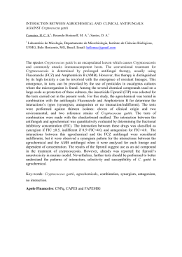
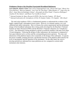
Silva et al., Nasal cryptococcosis in a sheep in Brazilian Semi-Arid. Braz J Vet Pathol, 2010, 3(2), 127-130.
127
Case Report
Nasal cryptococcosis in a sheep in Brazilian Semi-Arid
Saulo T. Gusmão da Silva1, José C. de Almeida Souza2, Carla L. de Mendonça3, Marisa A. Izael3,
Antônio F. Dantas4, Roseana Portela4, Franklin Riet-Correa4, José A. Bastos Afonso3
1
Graduate student of the Programa de Pós-Graduação em Sanidade e Reprodução de Ruminantes, Unidade Acadêmica de Garanhuns/Clínica de
Bovinos, Universidade Federal Rural de Pernambuco - UFRPE, Garanhuns-PE, Brasil;
2
Unidade Acadêmica de Garanhuns-PE, UFRPE, Garanhuns-PE, Brasil;
3
Clínica de Bovinos, Campus Garanhuns, UFRPE, Garanhuns-PE, Brasil,
4
Universidade Federal de Campina Grande, Campus Patos, Paraíba, Brasil.
Corresponding author: José A. Bastos Afonso, Clínica de Bovinos, Campus Garanhuns, UFRPE, Av.: Bom Pastor, s/n. Caixa postal 152. Bairro
Boa Vista. CEP 55292-901, Garanhuns - PE, Brazil.; Phone +55 87 3761-3233 Fax: +55 87 3762 2397 E-mail: [email protected]
Submitted June 10th 2010, Accepted September 14th 2010
Abstract
A case of nasal cryptococcosis is reported in a two years old male hair sheep in the Brazilian semi-arid. Severe
respiratory signs and a mass occupied a large portion of the right nasal cavity were observed. Cryptococcosis was
diagnosed by the typical histologic appearance of the fungus.
Key words: Crypococcus; nasal cavity; Brazilian Semi-Arid
Introduction
Cryptococcosis is a potentially fatal, systemic,
granulomatous fungal infection that affects mammals,
such as humans, dogs, cats and horses, as well as bird,
reptiles and some invertebrates. It is caused by two
fungal species: Cryptococcus neoformans, with the
varieties grubii (serotype A) and neoformans (serotype
D); and C. gattii (serotypes B and C) (3). Bird
excrement is a source of infection and pigeons play an
important role as a reservoir of C. neoformans in the
urban environment and C. gattii has been isolated from
several tree species (3). However, both species are
isolated from a large variety of biological and
environmental samples (19, 22). Infections by C.
neoformans are more common in immunocompromised
individuals and C. gattii has emerged as a primary
pathogen of healthy hosts (3). Both species cause
human infections all over Brazil, causing mainly
nervous system and respiratory infections (9, 16, 22).
Cryptococcosis in animals can be due to both
species C. gattii and C. neoformans (2, 5, 21, 33). It is
common in cats and less frequent in dogs causing
rhinitis, skin lesions mainly of the nose and head,
meningitis, ocular lesions, and systemic disease (5). In
horses, the infection by C. neoformans has been
associated to rhinitis (27, 34), meningitis, encephalitis,
nasal granuloma, pneumonia, abortion with mycotic
placentitis and pneumonia in the fetus (14, 24, 26). A
case of cryptococcosis of the skin and subcutaneous
tissue is reported in a donkey (13). In goats C. gattii
causes epidemic outbreaks of pneumonia and
encephalitis (28). Mastitis by Cryptococcus spp is
reported in goats (31), sheep (30), cattle (18), and
buffalo (21).
In Brazil, in domestic animals, cryptococcosis
was reported for the first time in a sheep with
pulmonary infection (8). Pulmonary lesions in a sheep
slaughtered for consumption (15) and a case of rhinitis
(23) are also reported. The disease is more frequent in
dogs (4, 11, 12, 20), cats (6, 17), and horses (14, 34). A
case of meningoencephalitis by C. neoformans was
recently reported in a bovine (10).
The aim of the present paper is to report a case
of nasal cryptococcosis in a sheep in Brazilian SemiArid.
Case Report
A ram two and a half-years-old hair sheep of
the Santa Inês breed, weighing 98 kg, was admitted at
the Bovine Clinic of the Federal Rural University of
Pernambuco, Campus of Garanhuns, state of
Pernambuco, Northeastern Brazil. Approximately two
weeks earlier, the sheep had suffered head trauma in a
fence and, five days later, an increased volume of the
Brazilian Journal of Veterinary Pathology. www.bjvp.org.br . All rights reserved 2007.
Silva et al., Nasal cryptococcosis in a sheep in Brazilian Semi-Arid. Braz J Vet Pathol, 2010, 3(2), 127-130.
right inner nasal cavity appeared. During the physical
exam, the animal was alert, presenting congested
conjunctiva and enlarged right pre-scapular and
submandibular lymphnodes. Percussion of the nasal
sinus produced a dull sound in the frontal region near
the nostril. The presence of hemorrhagic granulation
tissue was noted, inside the right nasal cavity (Figure
1). The animal exhibited tachypnea with polypnea, and
inspiratory dyspnea accompanied by audible expiratory
grunt. The animal had been vaccinated for clostridiosis
and treated with anti-helmintic.
128
nasal mucosa and infiltrated the adjacent structures,
extending throughout the entire nasal cavity from the
nasal vestibule to the ethmoid bone, causing facial
deformity and obstructed airflow. Resorption of the
right nasal bone and nasal turbinate bones (dorsal and
mid) was also noted. The remaining organs did not
exhibit any significant lesion.
Fragments from different regions of the mass
were collected, fixed in 10% buffered formalin,
embedded in paraffin, sliced to 5-6 µm and stained
with hematoxylin-eosin (HE), periodic acid-Schift
(PAS) and Alcian Blue. Microscopically, the lesion
was a locally extensive, necrotizing granulomatous
rhinitis, with infiltration by macrophages, lymphocytes
and plasma cells. Extensive areas of necrosis were also
observed. Myriads of round or oval levaduriform
structures, with a thin basophilic wall, measuring
approximately 5.0 to 20 µm in diameter were present
within the exudate. Surrounding the yeasts, there was a
light halo not stained by HE, giving a soap bubble
appearance, which was stained by Alcian blue and PAS
(Figure 2). These characteristics are typical of
Cryptococcus spp. There was also moderate
proliferation of the fibrous tissue, areas of hemorrhage,
and infiltration neutrophils in the periphery of the
lesion.
Figure 1: Nasal granulation tissue in sheep.
The examination of the nasal cavity revealed a
pendulous mass with a rough appearance that occupied
a large portion of the right nasal cavity, compressing
the septum and causing partial occlusion of the left
nasal cavity. The animal remained at the clinic for 31
days, during which time it had a reduced appetite, a
variable degree of dehydration, an area of alopecia in
the region of the nasal sinus, and softening of the nasal
bone structure. At that time it also exhibited an
abdominal breathing pattern with respiratory noises,
severe inspiratory dyspnea, sporadic cough, moments
of apnea, and fetid muco-purulent nasal secretion.
The animal was submitted to penicillin-based
antibiotic treatment (Pencivet Plus PPU/Intervet) at 24hour intervals and also received three applications of
dexamethasone
(Azium/Schering-Plough).
Daily
cleaning of the nostril was performed with a boric acid
solution, along with massage with dimethyl sulfoxide
in the sinus and daily nasal atomization with
physiologic solution but without satisfactory results.
Due to the unsuccessful therapy it was decided
to remove the mass surgically. The animal was
submitted to 24 hours of fasting and tranquilization
with 0.05 mg/kg of acepromazine. Due to the severity
of the clinical condition and compromised respiratory
function, the animal died during the procedure and was
necropsied.
The macroscopic exam of the nasal cavities
revealed
a
light-colored
mass,
measuring
approximately 25 cm in length, with an irregular shape,
gelatinous aspect, brittle consistency and surface with
polypoid nodules. This mass originated in the right
Figure 2 Sheep with nasal cryptococcosis. The
histologic section of the nose shows numerous yeast of
Cryptococcus neoformans and few inflammatory cells.
PAS. X40.
Discussion
Criptococcosis is a rare disease in sheep
causing mastitis (30) or pulmonary infections (8, 15).
In this case the lesion was localized in the upper
respiratory tract causing rhinitis. Similar lesions had
been reported in cat, dogs (5) and horses (27, 34).
Pulmonary and cerebromeningeal cryptococcosis are
the main form of the disease in humans and farm
animals (2, 8, 9, 10, 14, 15, 16, 22, 26, 28). The
infection of the nasal cavity seems to be by inhalation
of spores (3, 5). However pulmonary infections occur
more frequently following inhalation and deposition of
the spores into the pulmonary alveoli (3, 5).
Brazilian Journal of Veterinary Pathology. www.bjvp.org.br . All rights reserved 2007.
Silva et al., Nasal cryptococcosis in a sheep in Brazilian Semi-Arid. Braz J Vet Pathol, 2010, 3(2), 127-130.
Cryptococcus spp are neurotropic and can invade the
central nervous system from the respiratory tract either
hematogenously or via direct extension through the
cribiform plate (7). In this case the infection probably
occurred due to spore inhalation from the environment,
where the contamination was very probably due to the
presence of ground, decomposed vegetables, and bird
and bat feces, which are found both in urban and rural
areas of Brazil (22). Also the semi-intensive livestock
farming frequently used in Northeastern Brazil and the
Semi-Arid climate may increase the infection risk in
farm animals. It is also possible that the head trauma
that the animal suffered before the onset of the clinical
signs favored the infection by the fungus.
The clinical findings of inspiratory dyspnea
and difficulty in the airflow in the upper respiratory
tract observed in this case are similar than those
reported in other species (5, 34). However, these
findings are most often accompanied by chronic
pneumonia, emaciation and even neurological signs (5,
28), not observed in this case. The gelatinous aspect
and polypoid formations found in the mucosa of the
nasal cavity are similar to those observed in cases of
cryptococcosis in other species (7). The deformation of
the nasal cavity causing an obstruction to airflow was
also reported (7).
The frequency of cryptococcosis in the SemiArid is unknown, but a similar case of the disease, also
in a Santa Inês sheep, was reported in southeastern
Brazil (23). Most aspects of nasal cryptococcosis in
sheep are similar than those observed in
conidiobolomycosis (25, 32) and nasal phytiosis (25),
which are very common diseases in sheep in
northeastern Brazil, but also occur in other Brazilian
regions (1, 29). Both disease cause lesions in the nose,
but in conidioblomycosis the lesions affect the
rinopharingeal region (25, 32) and in pythyosis of the
rinofacial region (25, 29).
Histologic and
microbiologic examinations are important for the
differential diagnosis of these three diseases in sheep.
Sheep with cryptococcosis could contaminate
the environment. However, it is questionable to what
extent these animals represent a danger to public health
or other animals, as it is not clear whether
Cryptococcus spp can be transmitted between animals
or from animals to humans (5). The euthanasia of
animals infected with cryptococcosis is recommended,
due to the lack of effective medications and potential
public health risks. Few farm animals infected by the
disease are treated, as treatment is long, expensive, has
a large number of side effects and there is a frequent
occurrence of relapse (34).
References
1. BOABAID FM., FERREIRA EV., ARRUDA LP.,
GASPARETTO ND., SOUZA, RL., SILVA MC.,
DUTRA V., NAKAZATO L., COLODEL EM.
Conidiobolomicose em ovinos no Estado de Mato
Grosso. Pesq. Vet. Bras., 2008, 28, 77-81.
129
2. BARÓ T., TORRES-RODRÍGUEZ JM., DE
MENDOZA MH., MORERA Y., ALIA. C. First
identification of autochthonous Cryptococcus
neoformans var. gattii isolated from goats with
predominantly severe pulmonary disease in Spain. J.
Clin. Microbiol, 1998, 36, 458-461.
3. BOVERS M., HAGEN F., BOEKHOUT, T.
Diversity of the Cryptococcus neoformansCryptococcus gattii species complex. Rev. Iberoam.
Micol., 2008, 25, S4-12.
4. BROLO MDI., BARBOSA ALT., CAVALHEIRO
A., LOPES STA., SANTURIO JM., SCHOSSLER
JE., MAZZANTI A. Diagnóstico de criptococose
canina pela citologia aspirativa por agulha fina.
Ciência Rural, 2008, 38, 826-829.
5. CASTELLÁ G., LOURDES ML., CABAÑES FJ.
Criptococosis y animales de
compañía. Rev.
Iberoam. Micol., 2008, 25, S19-S24.
6. CORREA GLB. Criptococose em gatos. Ciência
Rural, 1994, 24, 431-437.
7. COSWELL JL., WILLIANS KJ.. The respitratory
system. In: MAXIE MG. Jubb, Kennedy, and
Palmer´s pathology of domestic animals. 4ª Ed. San
Diego: Academic Press, 2007, V2, p. 643-644.
8. DACORSO FP., CHAGAS WA. Criptococose
pulmonar em caprino. Anais. Col. Anat. Bras. 1957,
3, 55-69.
9. DARZÉ C., LUCENA R., GOMES I., MELO A.
Clinical and laboratory characteristics of 104
cryptococcus meningoencephalitis cases. Rev. Soc.
Brasil. Med. Trop., 2000, 33, 21-26.
10.GALIZA, GJN.; SILVA, MLCR.; DANTAS,
AFM.: SIMÕES, SVD.; RIET-CORREA F..
Doenças do sistema nervoso de bovinos no semiárido
nordestino. Pesq. Vet. Bras., 2010, 30, 267-276.
11. HONSHO, CS.. MINE, SY; ORIÁ, AP.; BENATO,
N.; CAMACHO, AA.; ALESSI, AC.; LAUS, JL..
Generalized systemic cryptococcosis in a dog after
immunosuppresive corticotherapy. Arq. Bras. Med.
Vet. Zootec., 2003, 55, 155-159.
12.JULIANO, RS.; SOUZA AI.; SCHEIDE R..
Criptococose no sistema nervoso de cães - relato de
três casos. Rev. Pat. Trop., 2006, 35, 65-70.
13.KHUDAKARAM-TAFTI, A.; DEHGHANI, S.
Cutaneous cryptococcosis in a donkey. Comp. Clin.
Pathol., 2006, 15, 271-273.
14. KOMMERS, GD.; SOUZA, TM.; SOUTO, MAM.;
DE LA CORTE, FD.; BARROS, CSL. Criptococose
pulmonar granulomatosa em um eqüino. Ciência
Rural, 2005, 35, 938-940.
15.LEMOS, LS.; SANTOS, ASO.; VIEIRA-DAMOTA, O.; TEXEIRA, GN.; CARVALHO, ECQ.
Pulmorary crytococosis in slaughtered sheep:
Anatomopathology and culture. Vet. Microbiol.,
2007, 125, 350-354.
Brazilian Journal of Veterinary Pathology. www.bjvp.org.br . All rights reserved 2007.
Silva et al., Nasal cryptococcosis in a sheep in Brazilian Semi-Arid. Braz J Vet Pathol, 2010, 3(2), 127-130.
130
16.LINDENBERG, ASC.; CHANG, MR.; PANIAGO,
AMM.; LAZÉRA, MS.; MONCADA, PMF.;
BONFIM, GF.; NOGUEIRA, SA.; WANKE, B.
Clinical and epidemiological features of 123 cases of
cryptococcosis in Mato Grosso do Sul, Brazil. Rev.
Inst. Med. Trop. S. Paulo, 2008, 50, 75-78.
25.RIET–CORREA,
F.;
DANTAS,
AFM.;
AZEVEDO, EO.; SIMÕES, SDV.; SILVA, SMS.;
VILELA, R.; Mendonza L. Outbreaks of rhinofacial
and rhinopahryngeal zygomycosis in sheep in
Paraíba, northeastern Brazil. Pesq.Vet. Bras., 2008,
28, 29-35.
17.MARCASSO, RA.; SIERRA, S.; VICKY, MB.;
BRACARENSE, APFRL.; YAMAMURA, AAM.;
BIASI, F.; LOPES, BA.; AMUDE, AM.; CORTÊZ,
DEA. Criptococose felina. Semina: Ciências
Agrárias, 2005, 26, 229-238.
26.RILEY, CB.; BOLTON, JR.; MILLS, JN.,
THOMAS, JB... Cryptococcosis in seven horses.
Aust. Vet. J., 1992, 69, 135-139.
18.MONGA, DP.; MOHAPATRA, LN.; KALRA, DS..
Bovine
mastitis
caused
by
Cryptococcus
neoformans. Indian J. Med. Res., 1970, 58, 12031205.
19.NISHIKAWA, MM.; LAZÉRA, MS.; BARBOSA,
GG.; TRILLES, L.; BALASSIANO, BR.;
MACEDO, RCL.; BEZERRA, CCF.; PÉREZ, MA.;
CARDARELLI, P.; WANKE, B. Serotyping of 467
Cryptococcus neoformans isolated from clinical and
environmental sources in Brazil: Analysis of host
and regional patterns. J. Clin. Microbiol., 2003, 41,
73-77.
20.OLIVEIRA, IA. NOBRE, MO.; FERREIRO, L..
Pesquisa de criptococose em cães atendidos no
Hospital de Clínicas Veterinárias da UFRGS, Porto
Alegre, Brasil. Acta Scientiae Veterinariae, 2005, 33,
253-258.
21.PAL, M.. Mastitis in a water buffalo (Bubalus
bubalis) due to Cryptococcus neoformans var.
neoformans. Rev. Iberoam. Micol., 1991, 8, 89-91.
22.PAPPALARDO, MCSM.;
MELHEM, MSC.
Cryptococcosis: a review of the Brazilian experience
for the disease. Rev. Inst. Med. Trop. S. Paulo, 2003,
45, 299-305.
23.PEREIRA, RN.; PEROTIA, JH.; DUNE, ACC.;
FERREIRA
LIMA,
IG.;
ALESSI,
AC.;
CARVALHO, AM.; CANOLA, PA.; CATTELAN,
JW.; CANOLA, JC. . Criptococose nasal em ovino:
relato de caso. Arq. Bras. Med. Vet. Zooct., 2005, 57,
18 (Abstract).
24.PETRITES-MURPHY, MB.; ROBBINS, LA.;
DONAHUE, BS.. Equine cryptococcal endometritis
and placentitis with neonatal cryptococcal
pneumonia. J. Vet. Diagn. Invest., 1966, 8, 383-386.
27.ROBERTS, MC.; SUTTON, RH.; LOVELL DKA..
A protracted case of cryptococcal nasal granuloma in
a stallion. Aust. Vet. J., 1981, 57, 287–291.
28.RODRÍGUEZ, JMT.; MENDOZA, M.; RAMÍREZ,
EA.; ROCA, GS.). Cryptococcosis by Cryptococcus
gattii in immunocompetent goats in Spain and
review of the literature. Acta Scientiae Veterinariae,
2006, 34, 245-253.
29.SANTURIO,
JM.;
ARGENTA,
JS.;
SCHWENDLER, SE.; CAVALHEIRO, AS.;
PEREIRA, DIB.; ZANETTE, RA.; ALVES, SH.;
DUTRA, V.; SILVA, MC.; ARRUDA, LP.;
NAKAZATO, L.; COLODEL, EM.. Granulomatous
rhinitis associated with Pythium insidiosum infection
in sheep. Vet. Rec., 2008, 163, 276-277.
30.SHNAWA, IMS.; NIGAM, JM. A note on
cryptococcal mastitis in sheep. Indian Vet. Med. J.
,1987, 7, 175-176.
31.SINGH, M.; GUPTA, PP.; RANA, JS.; JAND, SK..
Clinico-pathological studies on experimental
cryptococcal mastitis in goats. Mycopathologia,
1994, 126, 147-155.
32.SILVA, SMMS.; CASTRO, RS.; COSTA, FAL.;
VASCONCELOS, AC.; BATISTA, MCS.; RIETCORREA,
F.;
CARVALHO
EMS..
Conidiobolomycosis in sheep in Brazil. Vet. Pathol.,
2007, 44, 314-319.
33.STEPHEN, C.; LESTER, S.; BLACK, W.; FYFE,
M.; RAVERTY, S.. Multispecies outbreak of
cryptococcosis on southern Vancouver Island,
British Columbia. Can. Vet. J., 2002, 43, 792-794.
34.ZOPPA, ALV.; CRISPIM, R.; SINHORINI, IL.;
BENITES, NR.; SILVA, LCLC.; BACCARIN,
RYA. Obstrução nasal por granuloma fúngico em
eqüino: relato de caso. Arq. Bras. Med. Vet. Zootec.,
2008, 60, 315-321.
Brazilian Journal of Veterinary Pathology. www.bjvp.org.br . All rights reserved 2007.
Baixar