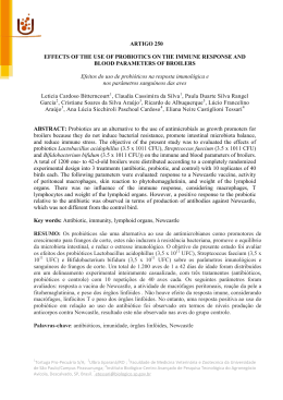
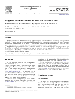
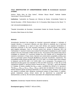
LUCIANA RODRIGUES DA CUNHA
GENOTYPIC AND PHENOTYPIC CHARACTERIZATION OF
Lactobacillus gasseri ISOLATED FROM A NEWBORN INFANT
Tese apresentada à Universidade
Federal de Viçosa, como parte das
exigências do Programa de PósGraduação em Ciência e Tecnologia
de Alimentos, para obtenção do
título de Doctor Scientiae.
VIÇOSA
MINAS GERAIS-BRASIL
2011
LUCIANA RODRIGUES DA CUNHA
GENOTYPIC AND PHENOTYPIC CHARACTERIZATION OF
Lactobacillus gasseri ISOLATED FROM A NEWBORN INFANT
Tese apresentada à Universidade
Federal de Viçosa, como parte das
exigências do Programa de PósGraduação em Ciência e Tecnologia
de Alimentos, para obtenção do
título de Doctor Scientiae.
APROVADA: 18 de março de 2011.
Pesq. Cláudia Lúcia de O. Pinto
(Coorientadora)
Prof. Paulo César Stringheta
(Coorientador)
Profa Elisabeth Neumann
Pesq. Marcelo Bonnet Alvarenga
Profa Célia Lúcia de Luces Fortes Ferreira
(Orientadora)
To my parents
Abel Fernandes da Cunha e Lúcia M. Rodrigues da Cunha
for their unconditional love and constant support
ii
ACKNOWLEDGMENTS
I would like to thank GOD for everything I have in my life.
I would like to thank my chair Célia L. L. F. Ferreira for her assistance,
guidance, opportunities and friendship over these years.
I would like to thank Dr. Todd R. Klaenhammer for his scientific support,
friendship and enormous assistance with my Doctoral Training in USA. Thank you so
much for believing me and giving me the opportunity to conduct part of my Doctoral
studies in your laboratory, and be part of your wonderful team. I really appreciate all
your reassurances and encouragement.
I would like to thank the Federal University of Viçosa and the Department of
Food Science and Technology for the opportunity.
I would like to thank National Council for Scientific and Technological
Development (CNPq) for providing me the fellowship during part of my Doctoral
Training in Brazil and also to Foundation Coordination for Improvement of Higher
Education Personnel (CAPES) for providing me the fellowship to conduct the
research experiment in North Carolina State University (NCSU), USA (Grant number,
BEX 4654-08-0).
I would like to thank my committee members, Dr. Marcelo Bonnet Alvarenga,
Dra Elisabeth Neumann, the EPAMIG`s researcher Dra. Cláudia Lúcia de O. Pinto
and Dr. Paulo César Stringheta for their receptivity and suggestions.
I would like to thank all the professors of the Department of Food Science and
Technology and Food Microbiology for their support and contributions to my
scientific background. Special thanks to Nélio José de Andrade, Nilda de Fátima
Ferreira Soares, Antônio Fernandes de Carvalho and Maria Cristina Dantas Vanetti
for believing me and my potential.
I would like to thank all my co-works of the Laboratory of Lactic Cultures,
specially Célio, Juliana, Milene, Carolina, Tatiane, Joice, Fabiana, Eder, Mônica,
Sara, Érika, for their friendship and for creating a great atmosphere in the lab. I very
much appreciate Karina da Silva Chaves and Juliana Nóbrega for their friendship and
always being there to listen and laugh with me.
I really would like to express my gratitude to Yong Jun Goh, Rosemary
Sanozky-Dawes, Evelyn Durmaz, Sara O`Flaherty, Akinobu Kajikawa, Grace
Douglas, Joakim Andersen, Erika Pfeiler, Jonathan Baugher and Joel Schroeter at the
iii
TRK lab for their receptivity, generous help in the lab and for making my experience
in USA so enjoyable. Jun Goh, Evelyn and Rosemary deserve a significant portion of
my gratitude for their technical assistance and precious help in my experiment. I can
not express how thankful I am to have those three angels in my life in the USA. They
are outstanding scientists and taught me to love the `` world of genetics``. They were
always there to help me as I needed and without them, none of this would have been
possible. From the botton of my heart, thank you so much!
I would like to thank all the collaborators of the Department of Food Science
and Technology, UFV specially, Vaninha, Osvaldo, Geralda, Perereca, Adão,
Sr. Manuel, Dimas, Pio, Fernando, Divino, Piu, Valério, Juarez, Sr Luis and José
Geraldo for their support and friendship over these years. I really would like to
express my gratitude to Geralda and Gilcemir that did not measure efforts to help me
with CAPES process. I also wish to extend my gratitude to all staff and Faculties at
the Department of Food, Bioprocessing and Nutrition Sciences of North Carolina
State University (NCSU), Raleigh, North Carolina, USA, in special Dr. Brian Farkas,
Sue Strong, Carol Reilly, Shirley Lyles, and Sabrina Whitley-Ferrell, for their
support, friendship and help during my training in the USA.
I deeply would like to thank Edith Ramos Neta, who is the best friend I could
have. I cannot express how thankful I am to have Edith around these years. She
greatly assisted me in obtaining my training at the Dr. Klaenhammer`s Lab and help
me a lot during my training in USA. She has always being a great friend and I am
very grateful for her being there to listen and advice me.
I would like to thank Marcelo Augusto Ferraz, who has also been a
wounderful friend over these years. Marcelo is a unique person that I love very much.
He has always being there to help and make me laugh.
I want to thank my friends in the Federal University of Viçosa, specially
Michele Bertoldi, Danilo Pereira, Rodrigo Resende, Roberta Careli, Geruza Dias,
Maria Patricia Milagres, João Paulo Rigueira, Wender Souza, Alexandre Resende,
Washington da Silva, Nathalia Ramos, Joesse, Rita Superbi, Vanessa de Castro, Junia
Capua, Rosineia de Paula, Maurício, José Carlos, Ana Andrea, Nathan Pimentel, for
their friendship and fun times we`ve had together.
I am very grateful to the friends Logan Buck, Dr. Farkas, Karen Coachman,
Luciano Silva, Fátima Roma for their friendship and help in the USA.
iv
I would like to give my special thank you to my boyfriend Joel Oliveira for the
support he has given me, for his unconditional love, and for all the wonderful
memories that we shared together in the USA and are still sharing in Brazil.
I would like to extend my special thank you to Denilce de Fátima da Cunha
(Pepete) and Chumbinho, who are not my blood, but the sister and brother I could
choose. Thank you for supporting and caring me.
I want to express my gratitude to my family, my grandmother Carlita, aunts, in
special Maria Arruda Rodrigues, Leonor Rodrigues da Silva, Carlita Helena
Rodrigues and Lia Cristina Rodrigues Brandi, uncles, cousins for providing a loving
environment for me.
Lastly, but not least, I deeply would like to express my gratitude to my parents
Abel e Lúcia, my sister Mariana and, my brother Leandro, for their active
participation and constant support. I am very fortunate to have such a loving family
and I would not be the person I am without their love and support.
Finally, I would like to thank all people that contributed to my personal
growth and to the development of this work.
v
BIOGRAPHY
LUCIANA RODRIGUES DA CUNHA was born on November 23, 1980 in Viçosa,
Minas Gerais, Brazil. She obtained a Bachelor’s degree in Food Engineering at
Federal University of Viçosa, Viçosa, Minas Gerais, Brazil, on January 2004. At the
same year, she was admitted by the Graduate Program in Food Science and
Technology at the Department of Food Technology of the Federal University of
Viçosa and received a fellowship of National Council of Technological and Scientific
Development (CNPq). Luciana completed her Master degree in Food Science and
Technology with a minor in Food Science and Food Microbiology, in 2006.
Following that, she was admitted by the same Graduate Program at the Department of
Food Technology of the Federal University of Viçosa, and her studies were funded by
the National Council of Technological and Scientific Development (CNPq). In 2009,
she was awarded a scholarship from Foundation Coordination for Improvement of
Higher Education Personnel (CAPES) to conduct part of her Doctoral studies at the
Department of Food, Bioprocessing and Nutrition Sciences of North Carolina State
University (NCSU), Raleigh, North Carolina, USA under the direction of Dr. Todd R.
Klaenhammer.
vi
LIST OF CONTENTS
LIST OF FIGURES ...................................................................................................... xi
LIST OF TABLE ......................................................................................................... xv
RESUMO ................................................................................................................... xix
ABSTRACT .............................................................................................................. xvii
1 - INTRODUCTION................................................................................................ 1
2 - LITERATURE REVIEW..................................................................................... 3
2.1 - Human Milk and Child Health ...................................................................... 3
2.2 - Human Milk Banks ....................................................................................... 4
2.3 - Probiotics - History and definitions .............................................................. 4
2.4 - Principal Probiotic Microorganisms ............................................................. 6
2.4.1 - Genus Lactobacillus ........................................................................... 6
2.4.1.1 - Lactobacillus gasseri .......................................................................... 7
2.5 - Probiotics and beneficial effects to health of children .................................. 9
2.5.1 - Modulation of the intestinal microbiota ............................................. 9
2.5.1.1 - Production of antimicrobial compounds ............................................. 9
2.5.1.2 - Competition for nutrients and adhesion sites .................................... 12
2.5.1.3 - Stimulation of the immune system .................................................... 14
2.5.2 - Maturation of the Intestinal Microbiota ........................................... 14
2.5.3 - Probiotics and Gastrointestinal Diseases ......................................... 18
2.5.3.1 - Diarrhea ............................................................................................. 18
2.5.3.1.1 - Antibiotic Associated Diarrhea (DAA) .................................. 19
2.5.3.1.2 - Diarrhea associated with Rotavirus infection ........................ 20
2.5.3.2 - Necrotizing Enterocolitis .................................................................. 22
2.5.3.3 - Probiotics and Alergies ..................................................................... 23
2.5.3.3.1 - Eczema and atopic dermatitis................................................. 24
2.5.3.4 - Probiotics and Respiratory infections ............................................... 25
2.5.3.5 - Probiotics and obesity ....................................................................... 26
2.5.4 - Selection of probiotic bacteria ......................................................... 27
2.5.4.1 - Aspects of functionality .................................................................... 28
2.5.4.1.1 - Survival of probiotic bacteria against gastrointestinal barrier 28
vii
2.5.4.1.2 - Antagonism to pathogens ....................................................... 29
2.5.4.1.3 - Adherence to intestinal tissues ............................................... 29
2.5.4.2 - Technological Aspects ...................................................................... 30
2.5.4.3 - Aspects of Safety............................................................................... 31
2.5.4.3.1 - Identification of probiotic strain............................................. 31
2.5.4.3.2 - Determination of virulence factors......................................... 32
2.5.4.3.3 - Resistance to antibiotics ......................................................... 32
2.5.5 - Legislation on probiotics .................................................................. 34
2.6 - REFERENCES ............................................................................................ 36
3 - EXPERIMENTAL HYPOTHESES................................................................... 57
CHAPTER I CHARACTERIZATION OF Lactobacillus gasseri ISOLATES
FROM A BREAST-FED INFANT .......................................................................... 60
ABSTRACT ................................................................................................................ 60
RESUMO .................................................................................................................... 61
1 - INTRODUCTION.............................................................................................. 62
2 - MATERIALS AND METHODS ....................................................................... 63
2.1 - Bacterial strains and growth conditions ...................................................... 63
2.2 - Bacterial identification ................................................................................ 64
2.3 - Pulsed-Field Gel Electrophoresis (PFGE) .................................................. 64
2.3.1 - Preparation of genomic DNA in agarose blocks .............................. 64
2.3.2 - Restriction enzyme digestion and PFGE.......................................... 65
2.4 - Testing of Hemolytic Activity and Bile Tolerance ..................................... 65
2.5 - Antagonistic activity against pathogenS ..................................................... 66
2.6 - Testing of Antibiotic Susceptibility ............................................................ 67
2.7 - Plasmid analysis .......................................................................................... 67
2.8 - Tolerance to simulated gastric and small intestinal juices .......................... 68
2.9 - Adherence assay .......................................................................................... 68
3 - RESULTS .......................................................................................................... 69
3.1 - Bacterial identification and Pulsed-Field Gel Electrophoresis (PFGE) ...... 69
3.2 - Testing of Hemolytic Activity and Bile Tolerance ..................................... 70
3.3 - Antagonistic activity against selected pathogens ........................................ 71
3.4 - Antibiotic Susceptibility.............................................................................. 72
3.5 - Plasmid content and selection of strains for further studies ........................ 73
viii
3.6 - Tolerance to simulated gastric and small intestinal juices .......................... 74
3.7 - Adherence assay .......................................................................................... 75
4 - DISCUSSION .................................................................................................... 76
5 - CONCLUSION .................................................................................................. 82
6 - REFERENCES ................................................................................................... 83
CHAPTER II SEQUENCE ANALYSIS OF FOUR PLASMIDS OF Lactobacillus
gasseri NCK2141 ISOLATED FROM A NEWBORN INFANT .......................... 90
ABSTRACT ................................................................................................................ 90
RESUMO .................................................................................................................... 91
1 - INTRODUCTION.............................................................................................. 92
2 - MATERIALS AND METHODS ....................................................................... 93
2.1 - Bacterial strain, media and growth condition ............................................. 93
2.2 - DNA manipulation, plasmid isolation and sequencing ............................... 94
2.3 - Sequence annotation.................................................................................... 94
2.4 - Blunt cloning ............................................................................................... 95
2.4.1 - Restriction enzyme digestion of pTRK989 ...................................... 95
2.4.2 - Ligation and transformation ............................................................. 97
2.5 - Phenotypic assay - Antibiotic susceptibility ............................................... 97
3 - RESULTS AND DISCUSSION ........................................................................ 99
3.1 - Plasmid DNA analysis and sequencing....................................................... 99
3.2 - General features of the plasmids ................................................................. 99
3.3 - Putative replication functions .................................................................... 104
3.4 - Transposable elements .............................................................................. 105
3.5 - Mobilization .............................................................................................. 106
3.6 - Partitioning ................................................................................................ 106
3.7 - Lantibiotic biosynthesis ............................................................................ 107
3.8 - Putative transport regions .......................................................................... 108
3.9 - Collagen adhesion ..................................................................................... 109
3.10 - Multidrug resistance transporter (lmrB) .................................................. 110
3.10.1 - Construction of lmrB recombinants ............................................. 111
3.10.2 - Phenotypic assay .......................................................................... 112
4 - CONCLUSIONS .............................................................................................. 113
5 - REFERENCES ................................................................................................. 114
ix
CHAPTER III FUNCTIONAL ANALYSIS OF FIBRONECTIN BINDING
PROTEIN IN Lactobacillus gasseri ........................................................................ 120
ABSTRACT .............................................................................................................. 120
RESUMO .................................................................................................................. 121
1 - INTRODUCTION............................................................................................ 122
MATERIALS AND METHODS .......................................................................... 124
1.1 - Bacterial strains, media and growth conditions ........................................ 124
1.2 - DNA manipulations and sequence analysis .............................................. 124
1.3 - Construction of L. gasseri fbp mutants ..................................................... 126
1.4 - Adherence assay ........................................................................................ 127
1.5 - Tolerance to simulated gastric juice .......................................................... 128
2 - RESULTS ........................................................................................................ 130
2.1 - Adherence to immobilized fibronectin and effect of the growth condition on
the adhesion ....................................................................................................... 130
2.2 - Characterization of Fibronectin-binding protein from L. gasseri NCK2141
........................................................................................................................... 131
2.3 - Construction of fbp mutants ...................................................................... 131
2.4 - Phenotypic analysis of the fbp insertion mutants ...................................... 132
2.4.1 - Survival to simulated gastric juice ................................................. 132
2.4.2 - Adherence ability of NCK 2147 and NCK 2148 ........................... 133
3 - DISCUSSION .................................................................................................. 134
4 - CONCLUSION ................................................................................................ 137
5 - REFERENCES ................................................................................................. 138
GENERAL CONCLUSION ................................................................................... 143
x
LIST OF FIGURES
Figure 1: Phylogenetic tree showing the relationships among the species of the Family
Lactobacillaceae, including genera Lactobacillus (abbreviated with ―L” in the
tree), Paralactobacillus and Pediococcus (abbreviated with ``P`` in the tree).
Extracted from Felis et al 2009. .......................................................................... 8
Figure 2: Changes in the intestinal microbiota in babies from birth to 7 days
(Mitsuoka, 1989). .............................................................................................. 15
Figure 3: Changes in the intestinal microbiota with age (Mitsuoka, 1989). .................. 16
CHAPTER I CHARACTERIZATION OF Lactobacillus gasseri ISOLATES
FROM A BREAST-FED INFANT ............................................................................. 60
Figure 1: PFGE of 30 Lactobacillus gasseri isolates digested with restriction enzyme
Sma I. The side bars denote the position of size markers of the molecular
weigh ladders. The pulsed time was ramped from 2s to 20s............................. 70
Figure 2: Percent of growth (A600nm) in the presence of oxgall compared to the control
(MRS medium without bile). Each column represents the mean of two
replicates. The bars represent the standard deviation. ....................................... 71
Figure 3: (a) Inhibition of E. coli by Lactobacillus gasseri (UFVCC1091). (b)
Inhibition of L. bulgaricus by NCFM (used as a control in this assay). The
numbers show the proteases (1mg/ml ) dropped (5 µl) around the colony: 1 Proteinase
K;
2-
Bacillus
protease;
3-
Bovine;
4-
Papain;
5- Pronase E type XIV; 6- Chymotrypsin; 7- Trypsin; 8- Fungal; 9- Pepsin ... 72
Figure 4: (a) Agarose gel electrophoresis plasmid profile of Lactobacillus gasseri
UFVCC1091. The side bars denote supercoiled size markers. (b) PCR
products using specific primer pairs (P1 [pTRK1023], P2 [pTRK1024], P3
[pTRK1025] and P4 [pTRK1026]) designed to amplify each of the four
plasmids of UFVCC1091. 1Kb Plus DNA Ladder. The arrows show P2
(pTRK1024) missing in strains UFVCC1083, 1092 and 1112. The
electrophoretic profile of strains UFVCC1083, 1091, 1092 and 1112 are
shown. This result was similar for all 29 strains. ............................................. 73
xi
Figure 5: Survival of stationary phase cells of Lactobacillus gasseri in simulated
small intestinal juice at pH 8.0 (Panel a) and gastric juice at pH 2.0 (Panel b).
Percent survival represents viable cells (CFU/mL) remaining after exposure at
the time points indicated versus pre-treatment (time 0). The data represent the
means of two independent replicates. ............................................................... 74
Figure 6: Adherence of Lactobacillus gasseri UFVCC1083, 1091 and 1112 to mucin
(Panel a), Caco-2 cells (Panel b) and HT-29 (Panel c) in vitro. The bacteria
grown in static MRS broth in ambient atmospheric (filled) and on MRS agar
under anaerobic conditions (open). The data represent the means ± standard
errors of the means for two independent replicates........................................... 75
CHAPTER II SEQUENCE ANALYSIS OF FOUR PLASMIDS OF Lactobacillus
gasseri NCK2141 ISOLATED FROM A NEWBORN INFANT ............................. 90
Figure 1: Construction strategy and restriction enzymes sites of pTRK1027 vector. .... 98
Figure 2: Physical and genetic map of plasmid pTRK1023 (a), pTRK1024 (b), pTRK
1025 (c) and pTRK1026 (d) of Lactobacillus gasseri NCK2141. Each ORF is
numbered, and arrows indicate positions and directions of predicted ORFs.
ORF numbers refer to Tables 2, 3, 4, and 5. ................................................... 100
Figure 3: Areas of significant similarity among RepA pSK118-44 (standard) and
RepA protein from pTRK1023, pTRK1025 and pTRK 1026. The green areas
show high matches at the same base position. ................................................ 104
Figure 4: Verification of recombinant plasmids by restriction enzyme digestion with
XhoI. Lane M, 1 kb plus DNA ladder marker. Lane 1, undigested pTRK1027
isolated from E. coli MC1061 (4.8 kb). Lane 2, digested pTRK1027 isolated
from E. coli MC1061 (control). Lanes 3 and 4, digested pTRK1027 isolated
from L. gasseri NCK2144 isolates. Lanes 5 and 6, digested pTRK1027
isolated from L. gasseri NCK2145 isolates. The black and red arrows indicate
the XhoI-digested plasmids. ............................................................................ 112
CHAPTER III FUNCTIONAL ANALYSIS OF FIBRONECTIN BINDING
PROTEIN IN Lactobacillus gasseri ........................................................................... 120
xii
Figure 1: Construction of the pTRK1028 integration vector. ori, origin of replication
of pWV01; erm, gene encoding Em resistance; MCS, multiple cloning sites
from pUC19. Only unique restriction sites are shown (Adapted from Goh et al
2009)................................................................................................................ 129
Figure 2: Adherence of Lactobacillus gasseri NCK2140, 2141, 2142 and the control
strain NCK99 to fibronectin in vitro. The bacteria grown in static MRS broth
in ambient atmosphere (black bars) and on MRS agar under anaerobic
condition (gray bars) were used for adherence testing at a concentration of 1.0
x 108 cells/ml. Bacterial cells were exposed to microwells coated with
fibronectin for 1 h at 37ºC, followed by plating on MRS agar medium for
enumeration of adhered bacterial cells. The data represent the means ±
standard errors of the means for four independent replicates. A Student`s ttest indicated that the results are significantly different (*) at a value of
P<0.05 for adherence of the strain grown in MRS broth compared to the same
strain grown in MRS agar. .............................................................................. 130
Figure 3: Colony PCR analysis of 16 selected Erm-sensitive double recombinants.
Lane M, DNA size marker (1 Kb plus); lane 1 to 8, selected NCK2148
isolated
Erm-sensitive
recombinants;
lanes
9 to 16, selected NCK2147 isolated Erm sensitive recombinants; Lane WT,
parent strain NCK 1041 (control). The expected amplicon sizes generated
from wild-type and ∆fbp genotypes are approximately 3.311 kb and 1.8 kb,
respectively...................................................................................................... 132
Figure 4: Survival of stationary phase cells of Lactobacillus gasseri NCK 2140 (a)
and NCK 2141 (b) (open square) compared with their respective fbp mutants
NCK
2148
(a)
and
NCK
2147
(b) (dark square) in simulated gastric juice at pH 2.0. Percentage of survival
represents viable cells (CFU/ml after treatment at various time points versus
before treatment (time 0). The data are the means ± standard errors of the
means for two independent replicates. ............................................................ 133
Figure 5: Adherence of Lactobacillus gasseri NCK 2148 (NCK 2140::pTRK2148)
and NCK 2147 (NCK 2141::pTRK2148) relative to their respective control
NCK 2140 and NCK 2141 to fibronectin (a) and HT-29 (b) in vitro. The
xiii
bacteria grown in static MRS broth in ambient atmosphere were used for
adherence test at a concentration of 1.0 x 10⁸ cells/ ml. Bacterial cells were
exposed to HT-29 monolayers, or microwells coated with fibronectin for 1 h
at 37ºC, washed, and plated on MRS agar medium for enumeration of
adhered bacterial cells. The data represent the means ± standard errors of the
means for two independent replicates. ............................................................ 134
xiv
LIST OF TABLE
CHAPTER I CHARACTERIZATION OF Lactobacillus gasseri ISOLATES
FROM A BREAST-FED INFANT ............................................................................. 60
Table 1: Primers tested in the study. .............................................................................. 68
Table 2: MIC values of L. gasseri to human therapeutic antibiotics.............................. 72
Table 3: Lactobacillus gasseri grouped according to their intrinsic characteristics. ..... 74
CHAPTER II SEQUENCE ANALYSIS OF FOUR PLASMIDS OF Lactobacillus
gasseri NCK2141 ISOLATED FROM A NEWBORN INFANT ............................. 90
Table 1: Bacterial strains, primers and plasmids used in this study. .............................. 96
Table 2: ORF analysis of the native plasmid pTRK1023 from L. gasseri NCK2141
with best matches to sequences in the public databases.................................. 101
Table 3: ORF analysis of the native plasmid pTRK1024 from L. gasseri NCK2141
with best matches to sequences in the public databases.................................. 102
Table 4: ORF analysis of the native plasmid pTRK1025 from L. gasseri NCK2141
with best matches to sequences in the public databases.................................. 102
Table 5: ORF analysis of the native plasmid pTRK1026 from L. gasseri NCK2141
with best matches to sequences in the public databases.................................. 103
CHAPTER III FUNCTIONAL ANALYSIS OF FIBRONECTIN BINDING
PROTEIN IN Lactobacillus gasseri ........................................................................... 120
Table 1: Bacterial strains, primers and plasmids used in this study. ............................ 125
xv
LIST OF ABBREVIATIONS
GRAS - Generally Recognized as Safe
LAB – Lactic Acid Bacteria
FAO/WHO – Food and Agriculture Organization and World Health Organization
HMB – Human Milk Bank
UFVCC – Universidade Federal de Viçosa Culture Collection
NCK – North Carolina Klaenhammer Culture Collection
MIC – Minimum Inhibitory Concentration
PFGE – Pulsed Field Gel Electrophoresis
CDS - Coding DNA Sequence
GAMOLA - Global Annotation of Multiplexed On-site bLasted DNA-sequences
BLAST - Basic Local Alignment Search Tool
TA System – Toxin-Antitoxin System
EPAMIG – Empresa de Pesquisa Agropecuária de Minas Gerais
xvi
RESUMO
CUNHA, Luciana Rodrigues da, D.Sc.,Universidade Federal de Viçosa, março de
2011. Caracterização genotípica e fenotípica de Lactobacillus gasseri
isolatedos de recém nascidos. Orientadora: Célia Lúcia de Luces Fortes Ferreira.
Coorientadores: Cláudia Lúcia de Oliveira Pinto and Paulo César Stringheta.
O objetivo deste estudo foi caracterizar novas estirpes de Lactobacillus gasseri
isoladas de recém-nascidos saudáveis para potencial uso como probiótico em bancos
de leite humano no Brasil. Este estudo foi dividido em três fases. Na primeira,
realizou-se a caracterização genotípica das estirpes com a identificação dos isolados
ao nivel de espécie e avaliação da diversidade genética entre eles). Caracterização
fenotípica também foi realizada considerando os aspectos funcionais e de segurança
recomendados pela FAO/OMS para a validação de novas culturas probióticas.
Avaliou-se trinta estirpes isoladas de fezes de recém-nascidos quanto à resistência a
antibióticos, atividade hemolítica, tolerância a sais biliares, presença de plasmídeos e
antagonismo a patógenos Gram-positivos e Gram-negativos. Com base na diversidade
genética (PFGE), resistência a antibióticos e perfil plasmidial, três estirpes
(NCK2140, 2141 e 2142) foram selecionadas e avaliadas quanto à resistência ao suco
gástrico e pancreático, capacidade de adesão à mucina, fibronectina, e às linhagens de
células intestinais humanas, Caco-2 e HT-29. Na segunda fase, os quatro plasmídeos
identificados na estirpe NCK2141 foram sequenciados pela técnica de shotgun,
anotados por meio do GAMOLA® software e os genes resultantes foram identificados
por meio do software Artemis®. Posteriormente, análises BLAST foram realizadas
para confirmar as anotações in silico. Na terceira fase, avaliou-se a capacidade das
estirpes selecionadas aderirem à fibronectina imobilizada e a influência dessa proteina
sobre as caracteristicas probióticas da célula como, resposta ao estresse digestivo e
habilidade de adesão. Os trinta isolados foram identificados como Lactobacillus
gasseri por meio do sequenciamento do 16S rDNA, e vinte nove deles foram
idênticos pela técnica de Eletroforese em Campo Pulsado (PFGE) utilizando-se as
enzimas de restrição Sma I e Apa I. O isolado NCK2142 apresentou-se estritamente
relacionado aos outros, diferindo-se apenas por uma única banda no gel. Todas as 30
estirpes não apresentaram atividade hemolítica e carreavam três plasmídeos, exceto a
estirpe NCK2141, que apresentou um quarto plasmídeo, codificando um sistema de
transporte que confere resistência à múltiplas drogas (lmrB). Essa mesma estirpe
xvii
mostrou maior tolerância a bile (0.5%) e resistência a eritromicina, cefalotina e
oxacilina. Todas as estirpes apresentaram antagonismo frente aos patógenos avaliados
sendo a inibição assossiada apenas à produção de ácidos orgânicos. Os três isolados
selecionados NCK2140, 2141 e 2142 foram resistentes ao suco gástrico e ao suco
pancreático e apresentaram boa capacidade de adesão in vitro a mucina, fibronectina e
às linhagens de células humanas do câncer de cólon, Caco-2 e HT-29. Os quatro
plasmídeos identificados, pTRK1023, 1024, 1025 e 1026 apresentaram morfologia
circular, replicação por mecanismo teta e 36, 7, 20 e 46 open reading frames (ORF),
respectivamente. Os conteúdos de G+C foram consistentes com aqueles encontrados
em outras estirpes de Lactobacillus (37 - 40%), exceto o pTRK1025 que foi de
44,57%. Algumas propriedades funcionais como, proteínas de ligação ao colágeno
(pTRK1026), biossíntese de lantibióticos (pTRK1023) e transporte de carboidratos
(pTRK1023 e pTRK1026) foram identificados nos plasmídeos, as quais podem
promover vantagens competitivas para as células hospedeiras. Mecanismo de
manutenção celular como, Sistema de Separação e Toxina-Antitoxina também foi
observado, explicando a dificuldade de cura dos plasmídeos. O gene lmrB
identificado no pTRK1024, é associado à resistência a alguns antibióticos, no entanto,
o experimento de clonagem não confirmou a relação do mesmo com a maior
resistencia à eritromicina, cefalotina e oxacilina apresentada pelo isolado NCK2141
carreador desse plasmideo. Por questões de segurança, essa estirpe não pode ser
utilizada como um probiótico. Maior adesão in vitro foi observada à fibronectina
quando as estirpes NCK2140, 2141 e 2142 foram cultivadas em ágar MRS e
incubadas em condições de anaerobiose. Observou-se também que os mutantes fbp
(NCK2147 e NCK2148) tiveram menor aderência à fibronectina imobilizada e à
linhagem celular humana HT-29. A inativação do gene fbp não influenciou a
susceptibilidade das estirpes recombinates ao suco gástrico.
xviii
ABSTRACT
CUNHA, Luciana Rodrigues da, D.Sc.,Universidade Federal de Viçosa, March, 2011.
Genotypic and phenotypic characterization of Lactobacillus gasseri isolated
from a newborn infant. Adviser: Célia Lúcia de Luces Fortes Ferreira. Coadvisers: Cláudia Lúcia de Oliveira Pinto and Paulo César Stringheta.
The objective of this study was to characterize Lactobacillus gasseri strains
isolated from a healthy newborn infant for potential use as a probiotic in human milk
banks in Brazil. This study was divided into three phases. The first examines the
genotypic (identification at the species level and evaluation of genetic diversity
among the isolates) and phenotypic characterization of the strains with regard to
safety and functional properties currently recommended by FAO/WHO for validation
of new probiotic strains. Thirty strains were isolated from breast fed newborn stools
and were evaluated for resistance to antibiotics, hemolytic activity, bile tolerance,
antagonism toward selected Gram-positive and Gram-negative pathogens and
presence of plasmids. Based on the results from PFGE, antibiotic resistance and
plasmid profiles, three strains (NCK2140, 2141 and 2142) were selected and
evaluated for resistance to small intestine and gastric juices, ability to adhere to
mucin, fibronectin, Caco-2 and HT-29 cell lines. In the second phase, the four
plasmids identified in the strain NCK2141 were sequenced by the shotgun sequencing
technique and were annotated by GAMOLA® software. The resulting genes and CDS
designations were viewed using Artemis® software. BLAST was carried out for each
designated gene to confirm and to refine the in silico annotations. In the third phase,
the ability of the selected strains to adhere to fibronectin binding protein and the effect
of growth conditions on the adhesion were examined. It was also investigated the
functional role of this protein in the adhesion and stress response of the cell. In this
study, all thirty isolates were identified as Lactobacillus gasseri through 16S rDNA
sequencing. Pulsed-Field Gel Electrophoresis (PFGE) using restriction enzymes Sma
I and Apa I revealed that 29 of the L. gasseri were identical; one isolate (NCK2142)
was closely related, but exhibited a distinctive DNA fingerprint. All 30 strains
harbored 3 plasmids, with one strain (NCK2141) that showed strong tolerance to
0.5% of bile and harbored a unique fourth plasmid encoding a putative multidrug
resistance transporter protein (lmrB). No hemolytic activity or antagonism, beyond
acid inhibition was observed. The three selected strains NCK2140, 2141 and 2142
xix
showed strong resistance to small intestinal and gastric juices, and adhered in vitro to
mucin, fibronectin and two intestinal epithelial cell lines, Caco-2 and HT-29. The
complete nucleotide sequence of the plasmids (pTRK1023, 1024, 1025, and 1026)
revealed that they are circular and contain 36, 7, 20 and 46 open reading frames,
respectively. All four plasmids are predicted to replicate by the theta mechanism, and
have G+C contents consistent with those found in other lactobacilli strains (37-40%),
with the exception of pTRK1025 (44.57%). The plasmids appear to encode functional
properties, such as collagen binding (pTRK1026), lantibiotic biosynthesis
(pTRK1023) and carbohydrate transport (pTRK1023 and pTRK1026) that may
provide competitive advantages to the plasmid-carrying cells. Cell maintenance
mechanisms (partitioning and TA system) were also observed, thus explaining the
failure to cure these plasmids. Plasmid pTRK1024 harbored a putative lmrB gene, an
ATP-binding cassette-type multidrug resistance transporter protein, which has been
associated with clindamycin and lincomycin resistance. Cloning experiments did not
correlate the erythromycin, cephalothin and oxacillin resistance of NCK2141 to
LmrB. However, for safety reasons, strain NCK2141 should not be used as a
probiotic. Significantly higher adherence to fibronectin was observed when the strains
NCK2140, 2141 and 2142 were grown on MRS agar under anaerobic condition. It
was also observed a reduction of in vitro adherence of fbp mutants (NCK2147 and
NCK2148) to immobilized fibronectin and HT-29 intestinal epithelial cells. The
inactivation of the fbp locus did not influence the gastric juice susceptibility of the
mutant strains.
xx
1 - INTRODUCTION
Breastfeeding is recognized worldwide as nurturing and protective to the
developing infant. Human milk supplies the nutrients and energy needs of the
newborn and protects them
against
infections. Despite
the
numerous
benefits
conferred by this human milk (HM), many children are deprived from its
consumption because their mothers cannot breastfeed them. Therefore, human milk
banks have a fundamental role as an important food source for such newborns.
Brazil is a world reference in the Human Milk Banking with more than 290
banks currently under operation. In 2010, the 152,813 thousand liters of HM collected
provided protection to more than 139,000 newborns. Almost all of these children were
prematurely born and were not able to breast feed. Furthermore, it is estimated that
7,000 Brazilian children would die every year if they were not breastfed properly,
which emphasizes the importance of the human milk banks (HMB). Pasteurization
(65oC x 30 min-1) insures the microbial safety of the milk deposited in the HMB.
However, studies from Borba et al (2003) indicated that this heat treatment reduces,
inactivates or eliminates some prebiotic constituents, which are essential for
establishing Bifidobacterium and other desirable lactic acid bacteria (LAB) in the
intestinal tract of the newborn, therefore compromising the formation of a beneficial
intestinal microbiota for them.
The
development of a beneficial microbiota contributes
to the
health of
the host. The gut microbiota acts as an important intestinal immune-modulator, not
only educating the naive infant immune system but also serving as an important
source of non inflammatory immune stimulators throughout life in healthy
individuals. In addition, the enteric microbiota can secrete molecules that inhibit host
pathogens, metabolize compounds that harm the host to less toxic substances and
produce a range of bioactive compounds such as conjugated linoleic acid, and short
chain fatty acids that may play a role in protection from lifestyle illnesses such cancer,
obesity and cardiovascular disease.
Given the importance of probiotic bacteria in the formation of a beneficial
microbiota and, since the heat treatment is an indispensable practice in HMB, a
possible alternative to overcome any undesirable effects of this heat treatment, could
be through the addition of well selected endogenous intestinal LAB strains into the
1
pasteurized milk provided in the milk banks. With this in mind, 30 LAB strains were
isolated in previous work in our laboratory of Lactic Acid Bacteria, from the target
population, healthy newborn fully breast fed.
In order to claim that a bacterial strain is a potential probiotic, in 2002, the
Food and Agriculture Organization (FAO) and World Health Organization (WHO)
established guidelines with some safety and functional criteria. According to this
document, all probiotic strains need to be properly identified at the genus and species
level using current and internationally scientific practices. In addition, they also need
to be evaluated for resistance to bile, antagonism toward pathogens, ability to adhere
to intestinal epithelial cells, tolerance to small intestine and gastric juices, hemolytic
activity and resistance to antibiotics. The importance of assessing the antibiotic
resistance profile pattern of new isolates is to limit the use of probiotic cultures
harboring plasmids containing transferable antibiotic-resistance genes.
Given the above background and the possible implementation of lactobacilli
strains in human milk banks, the objective of this study was to characterize recently
isolated lactobacilli from the stool of a newborn infant to select suitable strains for
potential use as a probiotic culture in human milk banks in Brazil. To this end, the
study was divided in three phases. In the first one the objective was to evaluate some
safety and functional aspects currently recommended by FAO/WHO and select those
strains with desirable probiotic characteristics for being further evaluated in vivo
studies. In the second phase, the goal was to sequence all those plasmids harbored by
the selected strains in the first phase to verify if they contained transferable antibioticresistance genes. Finally, the third phase aimed to investigate the effect the growth
condition on the adhesion of three selected Lactobacillus gasseri strains to
immobilized fibronectin binding protein and the functional role of this protein in the
adhesion and stress response of the cell.
2
2 - LITERATURE REVIEW
2.1 - Human Milk and Child Health
Human
milk is
balanced nutritional
considered the ideal
food
composition, containing the
for the newborn. It
right
amount of fatty
has
a
acids,
lactose, water and amino acids for newborn digestion, brain development and growth
(Willians and Stehlin, 2005), and it is enough to supply all the nutritional needs of
newborns during the first six month of life (Workgroup on Breastfeeding, 1997). In
addition, it has more than 45 bioactive compounds, which contribute to the maturation
of the newborn gastrointestinal tract (Kunz, 1999).
Besides providing optimal nutrition to infants, human milk contains a
multitude of immunological components, which are transferred from the mothers to
infant via breastfeeding. About 80 percent of the cells in breast milk are macrophages,
cells that kill bacteria, fungi and viruses (Willians and Stehlin, 2005). In addition to
macrophages, human milk contains proteins such as lactoferrin, lysozyme and casein,
lipids, oligosaccharides, enzymes, prostaglandins, growth factors, hormones, and cells
that work in many different ways to prevent infections and modulate the immune
system. This natural immune protection is not available to artificially fed infants
(Hanson, 2007). Studies have been shown that breastfed babies are less susceptible to
certain diseases, such as bacterial meningitis, bacteremia, diarrhea, respiratory tract
infections, necrotizing enterocolitis, otitis, urinary tract infections (Singhal et al
2002), hypercholesterolemia, and asthma (Chulada et al 2003) than not breastfed
infants. These special properties of human milk also provide long-term protection
from many diseases seen at higher rates in artificially fed infants, including an
increased risk of obesity, type 1 and 2 diabetes, and childhood leukemia (Hamosh,
2001). In addition, the protective effect of human milk against HIV postnatal
transmission in humans (Walter et al 2009) and in the reduction of the progression of
the disease (Coutsoudis et al 2003; Tozzi et al 1990) also has been reported.
Despite the numerous benefits conferred by human milk, many children are
deprived of it because their mothers cannot breastfeed them. Therefore, human milk
banks have a fundamental role as an important food source for such newborns
(Giugliani, 2002).
3
2.2 - Human Milk Banks
Human milk banks (HMB) are specialized centers established for encouraging
breastfeeding
and for collecting, screening, processing, storing and distributing
donated human milk (colostrum, transition milk and mature human milk). They must
be linked to a maternity or hospital and are responsible for quality control of the milk.
Brazil is a world reference in the Human Milk Banking with 283 banks under
current operation (Fiocruz, 2010). In 2007, 1,350 million liters of human milk were
collected through the Nacional Net of Human Milk providing protection to more than
107,000 newborns. Almost all these children were prematurely born and were not able
to breast feed. Furthermore, it is estimated that 7,000 Brazilian children would die
every year if they were not breastfed properly (Bom dia Brazil, 2008) which
emphasizes the importance of the human milk banks (HMB).
As evidenced in literature, human milk can be easily contaminated and vehicle
of various infectious diseases, such as AIDS, herpes, rubella, hepatitis B and C
(Goldin, 1997). Thus, for safety reasons, all milk is pasteurized (65oC x 30 min-1).
However, studies from Borba et al (2003) indicated that this heat treatment reduces,
inactivates or eliminates some prebiotic constituents. The prebiotic character of
human milk is important to establishing Bifidobacterium and other desirable probiotic
lactic acid bacteria (LAB) in the newborn´s intestinal tract. A possible way to
augment any undesirable effects of heat treatment could be through the addition of
well selected endogenous intestinal probiotic strains into the pasteurized milk
provided in the milk banks.
2.3 - Probiotics - History and definitions
The concept of probiotics was founded in 1908 when Elie Metchnikoff
suggested the benefits of lactic bacteria in the prolongation of life (Metchnikoff,
1908). The term ―probiotic‖ was introduced for the first time in 1965 by Lilly and
Stillwell to nominate substances of microbiological origin which stimulated the
growth of other microorganisms. This term was introduced to differentiate probiotics
from antibiotics. In 1989, Fuller emphasized the need for viability and defined
probiotics as live organisms which beneficially affect the host by improving its
intestinal balance. In 1999, Dunne et al brought up the question of security and
defined probiotics as non-pathogenic microorganisms, which when ingested exerted
positive influences on health and physiology of the host. With the understanding of
4
physiological and therapeutical properties of these microorganisms, the definition has
been suffering alterations until today and there is still no consensus, however the
definition most utilized internationally (Grupta and Garg, 2009; Petrof, 2009;
Sanders, 2009; Reid, 2008; Pineiro and Stanton, 2007) is that they are live
microorganisms which when administered in adequate concentrations benefit the
health of the host (World Health Organization [WHO] and Food and Agriculture
Organization [FAO], 2002). In Brazil, the department which regulates probiotic
products is the ANVISA (National Agency for Sanitary Vigilance), and according to
this Agency probiotics are live microorganisms capable of improving intestinal
microbial equilibrium, producing beneficial effects to the health of the individual.
According to the last two definitions, microorganisms must be alive to exert
beneficial effects. However, the scientific basis of this affirmation has been
questioned by some authors, since there are studies showing that non-viable
microorganisms (inactivated by heat or UV radiation), products or even bacterial
DNA may exert heath benefits to the host in specific situations (Kataria et al 2009).
Zhang et al (2005) reported that thermally inactivated Lactobacillus rhammnosus GG
promoted a reduction in IL-8 (interleukin-8) production induced by TNFα (tumor
necrosis factor alpha) in Caco-2 cells. Xiao et al (2003) administered capsules of
active or thermal inactivated Lactobacillus acidophilus LB to 64 and 59 patients with
chronic diarrhea, respectively. After four weeks of treatment, it was verified that the
inactive probiotic form was significantly more effective in the combat of diarrhea.
Children (3-24 months old) with acute diarrhea and moderate degrees of dehydration
also showed clinical improvement after receiving thermally inactivated L. acidophilus
(Simakachorn et al 2000). Besides the ability to modulate the immune system and
combat diarrhea, it has also been reported that inactivated probiotic bacteria have
potential to adhere to intestinal cells (Chauviere et al 1992).
Another aspect to be considered in the definition proposed by the FAO/WHO
and ANVISA is the specificity of the host. The definition is broad and does not
mention the origin of the strain. For many authors (Staton et al 2003; Dunne et al
2001, Klaenhammer and Kullen, 1999, Charteris et al, 1998) this is an important
criterion and if the probiotic was destined for human consumption it must be of
human origin. Thus, these microorganisms would have a greater chance of competing
with resident microbiota and establish themselves in the intestinal tract of the host
(Morelli, 2000). However, there are reports showing the beneficial effects promoted
5
by Saccharomyces boulardii, isolated from lychee (Litchi chinensis Soon) in the
treatment of diarrheas associated to antibiotics and Clostridium difficile (Kotowska et
al 2005; Vanderhoof & Young, 2002), ulcerative colitis (Guslandi et al 2000),
irritated bowel syndrome (Maupas et al 1983) and Crohn’s disease (Guslandi et al
2000).
2.4 - Principal Probiotic Microorganisms
The microorganisms most commonly used as a probiotics are producers of
lactic acid such as lactobacilli and bifidobacteria (Ferreira, 2003; Kopp-Hoolihan,
2001). Enterococcus faecium and Enterococcus faecalis have also been employed as
functional supplements in foods and their utilization is permitted in Brazil by the
ANVISA. However, there exists a concern on the part of the scientific community in
relation to utilization of these bacteria as probiotics due to the evidences relating these
microorganisms to cases of bacteremia (Gasser et al 1994), septicemia (Poh et al
2006; Schaberg et al 1991), urinary infections (Poh et al 2006; Lopes et al 2005;
Schaberg et al 1991) and endocarditis (Adam et al 1999). Moreover, virulence factors
such as hemolysins, gelatinases and surface proteins (Jahangiri et al 2010; Mundy et
al 2000), as well as conjugative plasmids encoding antibiotic resistance genes (Eaton
& Gasson, 2001) have also been encountered in these bacteria.
2.4.1 - Genus Lactobacillus
Lactobacillus are Gram positive microorganisms, nonspore-forming rods or
coccobacilli, deprived of flagella and are facultative anaerobic. They possess optimal
pH and temperature of growth in the range of 5.5 – 6.3 and 30 – 40ºC respectively
(Gomes and Malcata, 1999), and lower G+C content (Carr et al 2002). This genus is
amply distributed in the environment, especially in the intestinal tract (Wall et al
2007; Reuter 2001), mouth (Smith et al 2001), vagina (Martin et al 2008) and stomach
(Ryan et al 2008) of humans and other animals, as well as in plants, organic material,
sewage and fermented or deteriorated foods (Bernardeau et al 2006; Axelsson, 1989).
Taxonomically, the genus Lactobacillus belongs to the phylum Firmicutes,
class of the Bacillus, the order Lactobacillales and family Lactobacillaceae (Garrity
et al 2004). The genus was described for the first time by Beijerinck in 1901, where
the species were grouped based on phenotypic characteristics (optimal growth
temperature and fermentation of hexoses). Later, they were reclassified as obligatory
6
homofermentative, facultative heterofermentative and obligatory heterofermentative
(Kandller and Weiss, 1986). Obligatory homofermentative lactobacilli include those
which exclusively ferment glucose in lactic acid and do not ferment pentoses or
gliconate. The obrigatory heterofermentatives include lactobacilli which ferment
hexoses in lactic acid, acetic acid and/or ethanol and carbon dioxide, being that the
production of gas from glucose is a distinctive characteristic of these bacteria. The
facultative heterofermentatives include Lactobacillus which ferment hexoses in lactic
acid and may produce gas from gliconate, but not from glucose. These
microorganisms also ferment pentoses by activity of an induced phosphoketolase to
produce lactic and acetic acid (Vásques et al, 2005).
The genus Lactobacillus is phylogenetically diverse (Figure 1) and with
development of molecular techniques and genetic characterization, this was
reorganized and new groups were identified, based on the 16S rRNA region (Collins
et al 1991). Currently, in accordance with data obtained from the NCBI (National
Center of Biotechnology Information, USA), there are 195 different species of
Lactobacilli, including subspecies.
2.4.1.1 - Lactobacillus gasseri
Lactobacillus
gasseri
are
obligate
saccharoclastic,
homofermentative
organism, with optimum growth at 35 to 38ºC, and forms small rods with rounded
ends from 0.6 to 0.8 by 3 to 5 µm in size. L. gasseri is considered one of the true
autochthonous species of the human intestinal ―probiome,‖ defined as commensal
intestinal bacteria considered to have a beneficial influence on human health
(Azcarate-Peril et al 2008). Depending on consumption habits and geographic
location, L. gasseri has been determined to be one of the Lactobacillus species native
to the human gastrointestinal tract (GIT) of neonates (Wall et al 2007) and adults
(Reuter, 2001). L. gasseri has also been described as a common member of the
vaginal (Martin et al 2008) and oral Lactobacillus biota (Smith et al 2001), and it has
been proposed that the oral cavity acts as a reservoir and source of intestinal
lactobacilli (Dal Bello et al 2006).
L. gasseri was routinely classified as ―L. acidophilus‖ since morphologically it
differs only slightly from L.acidophilus and cannot be distinguished from L.
acidophilus by the classical taxonomic characteristics, such as carbohydrate
utilization, lactic acid isomer produced, etc. (Lauer and Kandler, 1980). In 1980, L.
7
gasseri was differentiated by DNA/DNA hybridization patterns from L. acidophilus
and named after Francis Gasser, who studied lactate dehydrogenases of Lactobacillus
species (Gasser and Mandel, 1968). Today, the complete genome of L. gasseri ATCC
33323 are deposited in the Bacterial Genome Database at the National Center for
Biotechnology Information (NCBI) and was sequenced by the Department of EnergyJoint Genome Institute in collaboration with the Lactic Acid Bacteria Genomics
Consortium (LABGC) (Klaenhammer et al 2002).
Figure 1: Phylogenetic tree showing the relationships among the species of the
Family Lactobacillaceae, including genera Lactobacillus (abbreviated with ―L” in
the tree), Paralactobacillus and Pediococcus (abbreviated with ``P`` in the tree).
Extracted from Felis et al 2009.
8
2.5 - Probiotics and beneficial effects to health of children
2.5.1 - Modulation of the intestinal microbiota
The relation between a balanced intestinal microbiota and health of the host
has been documented in literature. The enteric microbiota not only functions as a
barrier, but is involved in the immune response, inhibition of pathogen growth and
synthesis of bioactive compounds, such as conjugated linoleic acid, γ-aminobutíric
acid and short chain fatty acids, which exert an important role in the protection
against diseases including cancer, obesity and cardiovascular diseases (Wall et al
2009). Analogously, when there is a breakdown of intestinal homeostasis, and
pathogenic and/or opportunistic microorganisms are excessive, the enteric microbiota
may also contribute to the development of diseases (Kelly et al 2007). Therefore, this
suggests that predominance of a beneficial microbiota is associated with
the promotion of metabolism, nutrition and health of the host (Mackie et al 1999).
Probiotics may promote the modulation of the intestinal microbiota by
competition for epithelium adhesion sites and nutrients, production of antimicrobial
compounds and stimulation of the immune system (Sobko et al 2006).
2.5.1.1 - Production of antimicrobial compounds
Organic acids
Lactobacilli and bifidobacteria produce metabolites, such as acetic and lactic
acid, which possess an inhibitory effect on growth of pathogenic microorganisms
(Servin, 2004). This effect is related to the non-dissociated form of the acid (Podolak
et al 1996), which being lipophilic and apolar passively diffuse through the membrane
(Kashket, 1987) and promote acidification of the cellular cytoplasm. Therefore, the
photoelectrochemical gradient is collapsed promoting interruption of the substrate
transport system to the cell (Snijders et al 1985) and impairment of the metabolic
function of the microorganism. Acetic acid has a greater inhibitory effect than lactic
acids for the same pH value due to its pKa (pKa=5.76) being greater than that of lactic
acid (pKa=3.86), resulting in greater concentrations of the non-disassociated form in
the bacterial cytoplasm (Vasseur et al 1999).
Not only does it exert its activity by means of reducing the pH and its nondisassociated form, but lactic acid also acts in permeabilization of the membrane in
9
Gram-negative bacteria (Alakomi et al 2000) and as a chelation agent, capturing
elements such as iron, essential for bacterial growth (Presser et al 1997).
Studies in vitro have shown organic acids to be the principal inhibiting agent
to growth of pathogenic bacteria, as Escherichia coli (Maragkoudakis et al 2006;
Forestier et al 2001; Ogawa et al 2001; Dembeté et al 1998), Listeria monocytogenes
(Dembeté et al 1998), Salmonella typhimurium (Maragkoudakis et al 2006;
Keersmaecker et al 2006; Forestier et al 2001), Clostridium difficile (Forestier et al
2001), Helicobacter pylori (Maragkoudakis et al 2006; Midolo 1995) and
Staphylococcus aureus (Dembeté et al 1998).
Bacteriocin and/or molecules similar to bacteriocin
Bacteriocin are antimicrobial compounds of proteic origin, ribosomally
synthesized and produced by bacteria, and are generally active against species
intimately related with the producing strain (Klaenhammer, 1988; Tagg et al 1976).
However, some bacteriocin are capable of inhibiting growth of phylogenetically
distant microorganisms, as for example, Clostridium botulinum, Bacillus sp,
Enterococcus
faecalis,
Listeria
monocytogenes
and
Staphylococcus
aureus
(Tichaczek et al 1992). These compounds were principally characterized in gramnegative bacteria, in which colicins (Escherichia coli) are the most studied
(Lazdunski, 1988). Posteriorly, it was found that Gram-positive bacteria also produce
these compounds and within this class, lactic acid bacteria have been amply explored
for utilization of their bacteriocin in the conservation of foods (Cleveland et al 2001).
These molecules act promoting collapse of the membrane potential by means
of electrostatic bonds with the phospholipids (negatively charged). These bonds are
favored since the majority of bacteriocins are amplified and cationic. After bonding,
the hydrophobic portion of the bacteriocin is inserted in the membrane forming pores.
Formation of these pores allow for the exit of ions, principally potassium and
magnesium, promoting dissipation of the promoter force, compromising synthesis of
macromolecules and production of energy, resulting in cell death (Montville et al
1995).
Lactobacillus and Bifidobacterium are microorganisms capable of producing
bacteriocins and their substances have been cited as responsible for growth inhibition
of various pathogenic bacteria. Lozo et al (2004) identified bacteriocin (Bac217)
produced by Lactobacillus paracasei subsp. paracasei BGBUK2-16, which showed
10
inhibitory activity on growth of Staphylococcus aureus and Bacillus cereus. Yildirim
and Johnson (1998) identified bacteriocin (bifidocina B) produced by Bifidobacterium
bifidum with inhibitory effects on growth of some species of the genera Listeria,
Bacillus, Enterococcus, Lactobacillus, Leuconostoc and Pediococcus. Gassericin A, a
bacteriocin produced by Lactobacillus gasseri LA39 isolated from feces of newborns,
inhibited growth of Listeria monocytogenes, Bacillus cereus and Staphylococcus
aureus (Itoh et al 1995). Ten Brink et al (1994) identified bacteriocin produced by
Lactobacillus salivarus M17 (salivaricin B), which present inhibitory activity on
growth of Listeria monocytogenes, Bacillus cereus and Enterococcus ssp. The authors
identified another bacteriocin produced by Lactobacillus acidophilus M46 (acidocin
B), which has an inhibitory effect on Clostridium speorogenes.
Lactobacillus and Bifidobacterium may also antagonize growth of pathogens
by the production of lipid or protein compounds of low molecular weight. Collado et
al (2005) encountered inhibition of Helicobacter pylori by protein compounds,
thermostable and sensitive to proteases, produced by Bifidobacterium. The growth of
Giardia intestinalis and its bonding to Caco-2 human colon adenocarcinoma cells
lines was significantly reduced by antimicrobial substances of low molecular weight
(La1) produced by Lactobacillus johnsonii (Perez et al 2001). Liévin et al (2000)
attributed the antagonism of bifidobacteria on growth of Salmonella typhimurium to
production of lipophilic molecules of low molecular weight (<3500Da).
Production of H2O2
Some lactic acid bacteria produce hydrogen peroxide in the presence of
oxygen as a result of the action of flavoprotein oxidase or peroxides (NADH). Its
antimicrobial effect on various pathogens is the result of the oxidation of sulfhydryl
groups, promoting inactivation of various enzymes. Moreover, this substance may
promote alteration in permeability of the cell membrane by peroxidation of lipids in
the membrane and damages to DNA by formation of free radicals such as (O2-) and
hydroxyl (OH-) (Byezkowski and Gessner, 1988). (Equation 1).
11
O2 + 1e-
O2- (superoxide radical)
O2 + 1e- + 2 H+
H2O2 + 1eOH. + 1e- + H+
H2O2 (hydrogen peroxide)
Eq. 1
OH- + OH. (hydroxyl radical)
H2O
In the membrane peroxidation process, the free hydroxyl radical captures one
hydrogen atom from the unsaturated fatty acid of the phospholipid membrane and is
transformed in energy. The fatty acid, in the presence of oxygen, generates a free
radical and initiates a chain reaction which will destroy the cell membrane. The
damaged membrane loses its flexibility and its barrier functions allowing the entrance
of calcium into the cell and activating the phospholipases. These enzymes continue to
damage the cell since they attack the lysosomal membrane and liberate its enzymes,
accelerating degradation. In the DNA, the peroxide radical reacts with the iron ions
present in the molecule, producing the hydroxyl radical. This principally attacks the
pyrimidine in the bond with deoxyribose, rupturing the sugar-phosphate bond and
liberating the free bases of the nucleotides (Silva, 2003).
Lactobacillus plays an important role in maintaining vaginal health and
preventing the growth of pathogens including Escherichia coli and Gardnerella
vaginalis by the production of acid and H2O2. Lactobacillus crispatus and
Lactobacillus jensenii inhibit growth of gonococci in vitro by the production of H2O2
(Amant et al 2002). Aroutcheva et al (2001), while examining the antimicrobial
activity of twenty-one strains of Lactobacillus spp. of vaginal origin, observed that
approximately 80% of these produced H2O2, lactic acid and bacteriocins, and were
effective in inhibiting the growth of Gardnerella vaginalis, Lactobacillus brevis,
Lactobacillus salivarius and Lactobacillus gasseri in vitro.
2.5.1.2 - Competition for nutrients and adhesion sites
The adhesion of pathogens to the intestinal mucus is considered the first step
in the infective processes (Finlay and Falkon, 1997). It is mediated by bacterial
adhesins which recognize specific receptors of the mucus. Studies have shown that
some probiotic bacterial possess adhesins similar to those of the pathogens (Neesser et
al 2000), allowing for adhesion to intestinal sites, making them unavailable to the
pathogenic microorganisms (Reid and Hammond, 2005).
12
Collado et al (2007b) verified inhibition of adhesion of pathogenic bacteria, as
Bacteroides vulgatus DSM 1447, Clostridium histolyticum DSM 627, Clostridium
difficile DSM 1296, Escherichia coli K2, Enterobacter aerogenes DSM 30053,
Listeria monocytogenes ATCC 15313, Salmonella enterica serovar Typhimurium
ATCC 12028, Staphylococcus aureus DSM 20231 to human intestinal mucus by
probiotic bacteria (Lactobacillus rhamnosus GG [ATCC 53103], L. rhamnosus
LC705, L. casei, Shirota [Yakult], L. fermentum ME3, L. acidophilus NCFM, L.
plantarum Lp-115, L. salivarius Ls-33, Bifidobacterium longum 46, B. lactis Bb12, B.
lactis 420, B. breve) isolated from commercial milk products. The authors also
reported that these not only inhibited adhesion, but the probiotic bacteria were capable
of removing the pathogens already adhered to the intestinal mucus.
In another study performed by the same group, the authors verified inhibition
of pathogenic bacteria adhesion (Bacteroides vulgatus DSM 1447, Clostridium
histolyticum DSM 627, C. difficile DSM 1296, Escherichia coli K2, Listeria
monocytogenes ATCC 15313, Salmonella enterica serovar Typhimurium ATCC
12028, Staphylococcus aureus DSM 20231) to the human intestinal mucus by the
combination of probiotic bacteria Lactobacillus rhamnosus GG (ATCC 53103), L.
rhamnosus LC705 (DSM 7061), B. breve 99 (DSM 13692) and B. lactis Bb12 (DSM
10140). The authors also found that the probiotic bacteria in the form of a pool
promote greater inhibition to adhesion that when alone (Collado et al 2006). However,
the researchers, suggested the individual evaluation of each probiotic strain for later
selection of the pool since adhesion level of these bacteria to the intestinal cells are
dependent on genus, species and even the strain (Collado et al 2007b; Collado et al
2006).
Lee et al (2003) verified a reduction in the adhesion levels of E. coli and
Salmonella sp. to the intestinal mucus (isolated from the feces of healthy humans) and
in Caco-2 human colon adenocarcinoma cell line when incubated together with
Lactobacillus rhamnosus GG and Lactobacillus casei Shirota. Lactobacillus johnsonii
LJ1 and Lactobacillus casei Shirota significantly inhibited adhesion of Salmonella
typhimurium to the human intestinal mucus (Tuomola et al 1999).
The probiotic bacterial also inhibited growth of pathogens by competitive
utilization of nutrients in the gastrointestinal tract. These microorganisms metabolize
substrates, such as sugars, vitamins, minerals, proteins, as well as ingredients partially
13
degraded by the digestive enzymes of the host, making them unavailable to the
pathogen (Gibson et al 2005).
2.5.1.3 - Stimulation of the immune system
The intestine represents the largest lymphoid organ of the human body, being
responsible for 80% of the immunological response. This action is due to the presence
of antibodies and various immunocompetent cells dispersed on the inner wall and
epithelium, or organized in structures, which play fundamental role in the antigenic
presentation and elaboration of the immune response to the microorganisms and
proteins of the diet (Morais and Jacob, 2006).
In vitro studies and animal and human models suggest that probiotics may
stimulate the immune system. This effect is attributed to the capacity of these strains
to interact with the Peyer’s patches and the intestinal epithelial cells, stimulating the
Ig A producing B cells and the migration of intestinal T cells (Perdigon and Holgado,
2000). It has also been shown that probiotics favor unexpected phagocytic activity of
the alveolar macrophages, suggesting systemic action by secretion of mediators which
stimulate the immune system (Cross, 2002). Increase in the secretory IgA serum
levels and production of macrophages are extremely important in the immune
response of the intestine since this is the first line of non-specific defense of the
organism (Vanderhoof, 2001). Schiffrin et al (1997) in a randomized double blind
study administered of supplemented or non-supplemented (control) fermented milk
with Bifidobacterium bifidum Bb12 (1 x 10¹⁰ CFU/day) and Lactobacillus acidophilus
LA1 (7 x 10¹⁰ CFU/day) to 28 adults (23 – 62 years old) for 3 weeks and reported a
significant increase in phagocytic activity and in the serum levels of granulocytes and
monocytes, but not in lymphocytes or T cell activity. Fukushima et al (1998)
administered a formula supplemented with Bifidobacterium bifidum Bb12 (109
CFU/day) to Japanese children (15-31 months). After 21 days of treatment a
significant increase was verified in levels of Ig A in the feces of the infants fed with
the probiotic strain. The authors concluded that this increase in Ig A may increase the
resistance of mucus to intestinal infections.
2.5.2 - Maturation of the Intestinal Microbiota
The gastrointestinal tract includes a complex microbiota with more than 500
bacterial species. This is sterile when we are born, but soon becomes colonized by
14
microorganisms present in the environment and in the mother (Mitsuoka, 1982).
Roughly 24 hours after birth, the feces of most newborns already contain bacteria,
such as coliforms, Enterococcus, Clostridium and Sthaphylococcus in various
proportions (Figure 2), whose growth is favored at the beginning of colonization in
function of the positive oxido-reduction potential in the intestine.
Figure 2: Changes in the intestinal microbiota in babies from birth to 7 days
(Mitsuoka, 1989).
Although some of these microorganisms belong to species with pathogenic
potential, they do not cause harm to the host (Morelli, 2008). The first colonizers
consume the oxygen and produce new metabolics, preparing the intestinal
environment for the establishment of strictly anaerobic bacteria, as for example
Bifidobacterium, Clostridium and Bacteroides, which play an important role in
maturation of the intestinal tract of the new-born (Adlerberth and Wold 2009; Morelli,
2008).
Three to four days after birth, bacteria of the genus Bifidobacterium begin to
proliferate and predominate. In response to the increase of these bacteria, coliforms,
Enterococcus and putrefactive bacteria are restricted and diminish gradually, probably
by the reduction of nutrients and low oxido-reduction potential of the environment
(Mitsuoka, 1978). The bacterial microbiota of the intestinal tract stabilizes in the first
week of the newborns, with predominance of bifidobacteria, believing that this
predominance has an effect on prevention of intestinal infections (Mitsuoka, 1989).
With the improvement of molecular techniques, an additional group of
anaerobic bacteria was identified: Ruminococcus, which has been encountered in
large numbers in the intestinal tract of children fed with maternal milk (Favier et al
2003). The role of these microorganisms in the health of infants is still little
15
understood, but it is known that they are stimulated by prebiotics (Konstantinov et al
2003) and that they produce ruminocin A, a bacteriocin effective in inhibiting growth
of different species of Clostridium.
During weaning, when an adult diet is introduced to the child, a bacillary
microbiota, Gram negative, similar to adults becomes predominant. The number of
Bifidobacterium diminishes, being surpassed by Bacteroides, eubacterium and
peptostreptococci. As time passes, in the third age, the concentration of
Bifidobacterium diminished even further and some undesirable groups such as
clostridia, including Clostridium perfringens increase significantly, as well as
lactobacilli, streptococci and enterobacteriaceae (Figure 3) (Mitsuoka, 1996).
Figure 3: Changes in the intestinal microbiota with age (Mitsuoka, 1989).
The bifidobacteria species also suffer alterations with the advancing of age,
where the species of Bifidobacterium infantis and Bifidobacterium breve, which are
typical of newborns, are succeeded by Bifidobacterium adolescentis. Bifidobacterium
longum endures throughout the life of the host, and for this reason, is one of the most
sought for integration in functional foods (Mitsuoka, 1990).
These alterations are amply influenced by the composition of the diet of the
child (maternal milk x formulas), type of birth (cesarean section x normal birth),
gestational age, antibiotic therapy, state of health of the mother and hospitalization
(Adlerberth and Wold 2009; Morelli, 2008).
Of the factors mentioned above, the type of birth and feeding have the greatest
influence on development of microbiota of the newborn (Wall, et al 2009; Morelli,
2008). In a normal birth, infants are colonized first by the fecal and vaginal
16
microbiota of the mother, however, in those born by cesarean section, the maternal
microbiota does not have a strong influence in the initial contamination (Neu and
Douglas-Escobar, 2008), where the infants are first exposed to bacteria of the hospital
environment (doctors, nurses, equipment and other newborns) [Reinhardt et al, 2009,
Morelli, 2008]. According to Morelli (2008), infants born by cesarean section possess
a smaller concentration of bifidobacteria and Bacteroides fragilis, and greater
numbers of Clostridium difficile than those of vaginal birth. Grounlund et al (1999)
observed a lower concentration of Bacteroides fragilis and later colonization by
Bifidobacterium and Lactobacillus in newborns of cesarean section birth.
In relation to the effect of the diet, traditionally, the microbiota of the
newborns feed with breast milk has been considered to possess a greater number of
Bifidobacterium and lower concentration of facultative anaerobes, including
Streptococci, Staphylococci, Enterococci, Lactobacilli and Enterobacteria, while the
microbiota of newborns fed with formulas is more diverse and includes bacteria of the
group Bacteroides, Clostridium and Enterobacteriaceae (Wall, et al 2009). Some
authors attributed this difference to the presence of secretory IgA, lysozyme,
lactoferrin, nucleotides and bifidogenic factors in breast milk which stimulate the
growth of bifidobacteria and inhibit the pathogenic microbiota (Coppa et al 2004).
Additionally, breast milk appears to be a continuous source of bacteria for the
intestinal tract of the infant (Martin et al 2008). Bacteria with probiotic potential have
been isolated in breast milk, for example, L. gasseri, L. rhamnosus, L. plantarum, L.
fermentum, B. Breve, B. Adolescentis and B. Bifidum (Martin et al 2008). However,
the effect of the diet on intestinal microbiota composition, more specifically, on the
predominance of bifidobacteria in breastfeed newborns still controversial. Some
authors encountered no difference between the two types of feeding, and suggested
that modern formulas supplemented with prebiotics would increase the number of
bifidobacteria in the intestine to values similar to those encountered in breastfeed
babies (Adlerberth, et al 2009).
The gestation time also has an influence on enteric microbiota composition.
Due to the immaturity of the intestinal tract, premature newborns remain for long time
in Neonatal Intensive Care Units and under frequent use of broad spectrum
antibiotics, limiting the establishment of the beneficial micriobiota and allowing the
development of pathogens (Mshvildadze, et al 2008). Antibiotics negatively affect the
17
composition of intestinal microbiota of the infant by decreasing the number of
anaerobic bacteria [bifidobacteria and Bacteroides] (Martin et al 2008).
The environment is another factor which infers on intestinal colonization of
newborns. In developed countries, it is believed that the rigorous aseptic practices at
birth and the immediate care, resulting in low environmental contamination, may
affect intestinal colonization pattern (Martin and Walker, 2006). The hygiene
hypothesis, which is based on the practices of good sanitation conditions in developed
countries, diminishes exposure of newborns to microorganisms, resulting in reduced
microbial immune stimulation. On the other hand, infants born in poorer countries are
exposes to successive contaminations with pathogenic bacteria, thus increasing the
risk of infectious diseases (Kalliomaki and Walker, 2005). One study performed in
Pakistan accompanied the microbiota of infants delivered by vaginal birth at home,
and a premature and intense colonization of E. coli of the environment was observed
(Alderth et al 1998).
Besides the maturation and modulation of enteric microbiota, probiotics has
also shown an important role in the prevention and treatment of gastrointestinal
disturbances.
2.5.3 - Probiotics and Gastrointestinal Diseases
2.5.3.1 - Diarrhea
In Brazil and other regions of the world, infant mortality due to acute and
persistent diarrhea, malnutrition and dehydration among children younger than 5 has
decreased in the last decades. A study performed by the Brazilian Department of
Heath revealed a 93.9% decrease in infant death caused by diarrhea in the last 25
years. With this reduction, the problem is no longer the second leading cause of infant
mortality (24.3% in 1980) in the country and is now in fourth (4.1% in 2005), of a
total of six principal causes (Brazil, 2009).
Although evidence has shown a decline in the mortality rate, diarrhea
continues to be the second leading cause of death in the world among children (WHO,
2009). In 2008, nine million infants died and approximate 40% of these deaths were
caused by pneumonia and diarrhea (Wardlaw et al 2010). Annually, roughly 1.5
million children die from diarrhea, which kills more than AIDS, malaria and measles
together (WHO, 2009). Despite the high mortality rate, in developed countries as in
18
Brazil, its importance is also related to the impact of the diseases of the population,
brought by its harm to human health and affecting infant development as well as the
society due to the costs generated by the demand for medical care services and
hospitalizations. Therefore, prevention of diarrhea is a great challenge to public
health.
Therapies with probiotics have shown to be effective both in prevention as
well as cure of acute diarrhea of different etiologies.
2.5.3.1.1 - Antibiotic Associated Diarrhea (DAA)
Antibiotic Associated Diarrhea (DAA) is an intestinal disturbance which
affects 5-15% of patients subjected to antibiotic therapy and results from
disequilibrium in the enteric microbiota causes by action of the antibiotics (Bartlett,
2002).
Treatment with probiotics has shown to reduce the incidence and severity of
DAA as indicated in various studies. One of the pioneering studies performed in this
area was conducted by Vanderhoof et al 1999, who administered Lactobacillus GG to
210 children between 6 months and 10 years old who had received antibiotics for
treatment of respiratory infections. The use of probiotics resulted in a significant
reduction (26% to 8%) in the frequency of diarrhea and in the duration of the disease.
In a similar study, Arvola et al 1999 administered Lactobacillus rhamnosus GG (2 x
1010 CFU) in capsule two times a day to 167 children with respiratory infections.
Again, the use of the probiotic resulted in a reduction (16% to 5%) of diarrhea
occurrence. Corrêa et al (2005) verified the effect of formulas supplemented with
Bifidobacterium lactis and S. thermophilus in the prevention of diarrhea associated to
antibiotics in 157 children between 6 and 36 months old. The percentage of children
who developed the disease in the next 30 days after implementation of the antibiotic
for respiratory disturbances was significantly lower in the group receiving the
probiotic strain (16.3%) than in the control (31.2%).
However, not all studies support these results. Thomas et al 2001 administered
Lactobacillus GG (2 x 10⁹ CFU/day) or placebo during 14 days to 167 patients
subjected to antibiotic therapy. The authors observed no difference in the rate of
diarrhea occurrence between the placebo (29.9%) and the group receiving the
probiotic strain (29.3%). Similar results were also obtained in study performed by
Tankanow, 1999.
19
2.5.3.1.2 - Diarrhea associated with Rotavirus infection
Since its discovery in 1970, rotavirus has been considered one of the greatest
causers of infections associated to diarrhea in children throughout the world
(Maldonado et al 1990). In developing countries, this microorganism is responsible
for severe cases of dehydration associated to infectious diarrheas, causing the death of
more than 440 thousand people per year. In 2003, it was estimated that 1205 children
die daily from diarrhea caused by the rotavirus, where 80% of these are infants in
undeveloped countries (Parashar et al 2003).
Studies performed with probiotic microorganisms have shown promising
results in the treatment and prevention of acute diarrhea, as well as reducing the
severity and duration of infections promoted by the rotavirus in children (Isolauri,
2003; Szajewska and Mrukowict, 2001).
- Guandalini et al (2001) administered oral rehydration supplemented or not
(control) with Lactobacillus GG (1x 10¹⁰ CFU/ 250 mL) to children (1-36 months
old) and reported a reduction in the duration of diarrhea in the group which received
the probiotic strain. The authors also verified that in children with rotaviral infection,
the duration of diarrhea was 76.6 ± 41.6h in the control group and 56.2 ± 16.9 h in the
group given L. rhamnosus GG.
- Guarino et al (1997) administered oral rehydration supplemented or not
(control) with Lactobacillus GG for 6 days to 100 children (3 to 36 months old). The
authors observed a reduction in the duration of diarrhea from 6 to 3 days in children
who received the probiotic strain.
- Majamma et al (1995) administered 2.8 x 10⁹ CFU of Lactobacillus GG and
109 CFU of Lactobacillus casei in capsule to 49 children (4 – 35 months old) with
acute gastroenteritis promoted by the rotavirus, showing symptoms for more than 7
days. After 5 days of treatment, a reduction in diarrhea was observed along with
increase in rotavirus specific IgA, IgG, Ig M and secretory IgA.
- Saaverdra et al (1994) reported significant reduction in diarrhea promoted by
rotavirus in children (5 – 24 months old) administered formula supplemented with
Bifidobacterium lactis (1.9 x 10⁸ UFC/g) and Streptococcus thermophilus (0.14 x 10⁸
UFC/g).
- Isolauri et al (1991) administered lyophilized Lactobacillus GG (single dose
of 1010-11 CFU) or added in fermented milk (two times per day 1010-11 CFU) to
20
children (4 to 45 months old) for 5 days. The authors reported a reduction in the
duration of diarrhea caused by rotavirus in the groups which received the probiotic
strain (1.4 days) compared to the control (2.4 days) which received only pasteurized
milk.
It has been demonstrated that the promoting effect of L. rhamnosus is dose
dependent. Fang, et al (2009) randomly administered three different concentrations (0
CFU/day, 2 x 10⁸ CFU/day and 6 x 10⁸ CFU/day) of L. rhamnosus 35 (Lcr35) to 23
children with rotaviral gastroenteritis. After three days of treatment, a significant
reduction (86%) was verified in the count of rotavirus in the feces only in the group
which received the greatest dose of the probiotic (6 x 10⁸ CFU/day). The authors
suggest a minimal dose of de 6 x 10⁸ CFU/day for 3 days in order to obtain the
protective effect caused by this probiotic strain.
Clinical studies have also shown that probiotics are as effective in the control
of rotaviral gastroenteritis as conventional medicines. Teran et al (2009) evaluated in
vivo the effectiveness of the drug nitazoxanide, utilized in the treatment of infections
caused by protozoa and worms, and probiotics for combating diarrhea caused by
rotavirus. The children (28 days to 24 months old) were divided randomly into 3
groups. The first group received 15 mg/Kg/day nitazoxanide two times per day for 3
days. The second group received 1.25 x 10⁹ CFU/g of probiotics (L. acidophilus, L.
rhamnosus, B. longum and Sacharomyces boulardii) two times per day for 5 days.
The third group (control) received only an oral or systemic rehydration solution. The
authors reported a significant reduction (p=0.017) in the average hospitalization
period of the patients who received nitazoxanide (81h) and probiotics (72h) compared
to the control group (108h). Similarly, the average duration of diarrhea was reduced
(p=0.009) in children who received the medication (54h) and probiotics (48h)
compared to the control group (79h), demonstrating that the probiotics were as
effective as nitazoxanide in the treatment of diarrhea caused by rotavirus. The
probiotic effect has been attributed to the reduction in propagation of the rotavirus,
modulation of the intestinal microbiota, normalization of intestinal permeability and
increase in the expression of mucins and IgA secretory cells (Isolauri, 2004).
Although various studies have demonstrated the efficiency of probiotic strains
in the control of rotaviral gastroenteritis, more studies are necessary to confirm this
protective effect since the same results were not verified by other authors (Misra et al
2009; Sarker et al 2005; Costa-Ribeiro, et al 2003).
21
2.5.3.2 - Necrotizing Enterocolitis
Necrotizing enterocolitis (NEC) is a serious intestinal infection which attacks
premature newborns admitted in Neonatal Intensive Care Units (ICU). It is
characterized by abdominal distensions, bilious vomiting, diarrhea with the presence
of blood, lethargy, respiratory apnea and thermal instability (Caplan and Jilling,
2000). This disease has been reported in 10 – 25% of premature newborns admitted to
the Neonatal ICU and may affect 33 to 50% of all low birth weight babies. The
mortality rate is high (20 – 30%) among all that developed the disease, and those that
survive, may continue with sequels, such as short bowel syndrome and intestinal
obstruction (Glass et al 1991).
Necrotizing enterocolitis has a multifactorial origin and is caused by intestinal
and immunological immaturity, infection, hypoxia, hyperosmolar enteral feeding and
by the altered composition of intestinal microbiota. The contribution of each of these
factors is still unknown, however, it is known that retarding the installation of normal
microbiota favors enteritis by the lack of protective bacteria and reduced development
of the local and systemic immune system (Kliegman and Walsh, 1987). Studies have
shown that low birth weight premature babies delivered by caesarean section always
require intensive care and only receive breast milk after several days. The process by
which microorganisms, for example lactobacilli, are ingested during the vaginal birth
and propagated by breast milk does not occur in these children (Gewolb et al 1999).
Thus, they are exposed to pathogenic bacteria, such as Salmonella, Clostridium,
Escherichia, Streptococcus and Staphylococcus which colonize the intestine and
increase the risk of necrosis. Considering the association of the development of
predominantly beneficial intestinal microbiota with impediment of NEC (Hoy et al
1990), the administration of probiotics has been suggested by some authors with the
intent of optimizing development of these microbiota and consequently protecting
newborns from infectious diseases.
Hoyos, 1999 was the first to propose the utilization of probiotics in a study
performed in Columbia in 1999. The author administered 2.5 x 10⁸ CFU/day of
Bifidobacterium infantis to 1237 newborns and verified a reduction of 60% in the
indices of mortality and occurrence of NEC.
In the study conducted by Bin-Num et al (2005) in Israel, newborns weighing
less than 1500g received 109 UFC/day of Bifidobacterium infantis, Bifidobacterium
22
bifidum and Streptococcus thermophilus. The authors verified that the probiotic
reduced both the incidence (14 to 1%) and severity of NEC in newborns.
In India, in a randomized controlled double blind study, 186 newborns with a
gestational age of less than 32 weeks and weighing less than 1500g were feeding (two
times per day) with breast milk supplemented or not (control, n=95) with 2.5 x 10⁹
CFU/dose of Bifidobacterium infantis, Bifidobacterium bifidum, Bifidobacterium
longum
and
Lactobacillus
acidophilus. The authors verified that enteral
administration of probiotics significantly reduce the time of hospitalization and death
caused by NEC in very low birth weight newborns (Samanta et al 2009).
In a recent randomized controlled and double blind study performed at the
Institute of Medicine Fernandes Figueira in Brazil, 231 premature newborns with
body weights from 750 to 1499g received pasteurized breast milk supplemented or
not (control, n-112) with Lactobacillus casei and Bifidobacterium breve (3.5 x 10⁷ to
3.5 x 10⁹ CFU) for a period of 30 days, or until improvement or death, considering
that which occurred first. The authors reported (4 cases) of NEC only in the control
group and concluded that oral supplementation with B. breve and L. casei reduced the
occurrence of NEC in newborns (Braga et al 2011).
Although the majority of publications suggest a protective effect of probiotic
bacteria in the prevention of NEC and their secure use, some authors have reported
the onset of sepsis in children who received the probiotic strain (Lin et al 2008,
Wagner et al 1997), thus suggesting caution in the use of probiotics in
immunocompromised children.
2.5.3.3 - Probiotics and Alergies
The allergy, manifested in the form of topical eczema, allergic rhinitis and
asthma, is an excessive immune response to substances foreign to the organism.
Currently, it has been considered one of the chronic disorders most common in
children of developed countries (Taylor, 2007). It is believed that this increase is
related, in part, to good sanitary conditions and infrastructure of these sites, reducing
exposure of the children to commensal microorganisms and other organisms closely
associated with humans throughout their evolutionary history (Rook, 2007; Guarner,
et al 2006). Such microorganisms, including helminthes and saprophytic bacteria,
may be recognized by the immune system of children as harmless commensals, thus
triggering immune responses against themselves and other foreign substances in the
23
air or food (Rook, 2007). Thus, exposure to certain microorganisms in early life may
prevent the subsequent development of immune-mediated diseases, such as allergies,
asthma and atopic dermatitis (Hsieh et al 2008). One evidence of this fact, which is
about children whose parents adopt an anthroposophic lifestyle, with restricted use of
antibiotics, antipyretics and vaccinations, and make use of vegetables spontaneously
fermented by Lactobacillus have lower incidence of allergies and microbiota rich in
bifidobacteria and lactobacilli (Alm et al 2002).
It is believed that the composition of colonizing microorganisms is also critical
for proper development of a balanced immune response. Studies conducted with
children from countries with high and low incidences of allergic diseases have shown
differences in the colonizing species, with a reduction of lactobacilli, bifidobacteria
and enterococci and increase in clostridia and Staphylococcus aureus in children from
countries with high incidences of allergic disorders (Bottcher et al 2000). Studies have
also shown that breastfeeding infants have a greater concentration of bifidobacteria
and lower incidence of allergies than those given formulas (Watanabe et al 2003).
These findings suggest that certain microorganisms can direct the allergenic potential
of an individual, and that changes in the profile of bacteria in early life may possibly
change the potential for allergies (Furries et al 2005). Therefore, probiotics have been
considered an important alternative to modify the intestinal microbiota early in life,
thus preventing the development of allergic disorders in childhood (Huurre, et al
2008).
2.5.3.3.1 - Eczema and atopic dermatitis
Abrahamsson et al (2007), in a randomized double-blind, placebo-controlled
study administered Lactobacillus reuteri ATCC 55730 daily to 188 mothers during 36
gestational weeks and to their respective babies for a year. The authors found
decreased levels of IgE-associated eczema during the second year of life.
In a randomized, double-blind, placebo-controlled, study conducted by
Kalliomäki et al (2007) in Finland, Lactobacillus rhamnosus GG was administered to
pregnant women with a history of atopic eczema, rhinitis or asthma and to their babies
for six months. The authors verified a significant reduction (50%) in the incidence of
atopic eczema in children who received the probiotic strain.
Kalliomäki et al (2003 and 2001) administered L. rhamnosus GG to 159
mothers with atopic diseases during the entire gestational period and their respective
24
babies for 6 months after birth. The infants were accompanied for 2 years (Kalliomäki
et al 2001) and 4 years (Kalliomäki et al 2003). The authors found that the incidence
of atopic dermatitis was 23% in children who received the probiotic and 46% in the
placebo group in both experiments (2 and 4 years).
Isolauri et al (2000) administered whey hydrolysate supplemented or not
(control) with Bifidobacterium lactis and Lactobacillus rhamnosus GG to 27 breastfed
newborns with atopic eczema. After two months of treatment, the authors found that
the SCORAD (Scoring Atopic Dermatitis - index used to evaluate the severity of
dermatitis) was significantly lower in the groups receiving the probiotic strains
compared to the control group.
Majamaa and Isolauri (1997) showed that children with atopic dermatitis and
cow’s milk allergy, when fed with a whey hydrolysate formula supplemented with
Lactobacillus GG for a period of 30 days, showed a reduction of more than 50% in
the indices of atopic dermatitis compared with the placebo group.
2.5.3.4 - Probiotics and Respiratory infections
In a study conducted by Hatakka et al (2001), milk supplemented or not
(control) with Lactobacillus rhamnosus GG was administered to 571 healthy children
(1-6 years old) for 18 days. The authors found a 17% relative reduction in the number
of children with respiratory and respiratory tract infections. The authors also observed
a 19% reduction in antibiotic use for treatment of respiratory infections.
It is believed that the protective effect of probiotic bacteria is related to
mechanisms of the intestinal mucosal barrier and the imbalance Th1 and Th2 cells to
several antigens (Vaarala, 2002). The cytokines of Th2 cells (IL-4, IL-5, IL10 and IL13) predominate in allergic responses and are responsible for the production of IgE
antibodies and activation of eosinophils (Heyman and Menard, 2002). Lactobacillus
inhibits the production of Th2 cytokines, and thereby controls the allergic response.
However, the role of probiotics in allergy prevention remains controversial
since other authors have not confirmed this effect. In an Australian study conducted
by Abrahamson et al (2007), 178 newborns from mothers with a history of allergy
received Lactobacillus acidophilus LAVRI-A1 or placebo for 6 months. After the
treatment period there was no difference in the incidence of atopic dermatitis between
groups receiving the probiotic (23/89) and placebo (20/88). The authors found that
after 12 months, the rate of sensitization was significantly higher in the probiotic
25
group (P = 0.03), suggesting that treatment with L. acidophilus increased the risk of
cow's milk allergy (P = 0.012).
Additionally, it is not clear whether all probiotic strains are able to prevent the
development of allergies and if this effect could be extended to all age groups. Studies
have shown that some strains of L. reuteri, isolated from breast milk, induced the
production of tumor necrosis factor (TNF) by the macrophages; however, other strains
inhibited the production of the same compound (Lin et al 2008; Rook, 2007). Helin et
al (2002) administered L. rhamnosus GG to adults and adolescents and found that the
probiotic strain did not prevent pollen allergy in these individuals.
2.5.3.5 - Probiotics and obesity
Childhood obesity is among the most important problems nowadays (Owen et
al 2005) due to its health implications, including diabetes mellitus type 2,
cardiovascular disease, pulmonary hypertension, apnea, reflux, cancer, among others
(Ogden and Leibel, 2007). This disease is the result of changes in energy balance, i.e.,
the form in which the body regulates absorption, use and storage of energy. Genetic
factors combined with social behavior, cultural and environmental factors directly
influence the energy balance and consequently body weight (Dibaise, et al 2008).
Studies have suggested that the intestinal microbiota also exerts an influence on the
weight of the individual, and thus it is a factor for the development of obesity, since it
directly affects nutrient acquisition and energy regulation. By means of energetic
economization, the undigested substrates that reach the colon are metabolized by
indigenous bacteria to form monosaccharaides and short chain fatty acids. These
compounds are more easily absorbable by the human body and transported to the liver
where they are converted into more complex lipids. Concomitantly, the microbiota
regulates the expression of host genes that promote the storage of energy absorbed by
the adipocytes (Backhed et al 2004). Evidence of this has been shown by some
authors. Cani et al (2009) found that consumption of oligofructose (prebiotics) by rats
resulted in increased concentration of bifidobacteria and reduced levels of
lipopolysaccharides (LPS) in the plasma. This compound has been linked to the
development of obesity, and increase in its concentration in the plasma has been
reported in rats fed with high-fat diets (Cani et al 2007). Turnbaugh et al (2008)
transferred the microbial communities of obese and lean rats to germ-free rats and
verified a greater accumulation of fat in those who received the intestinal microbiota
26
originating from the obese rats. Kalliomäki et al (2008) found that obese children had
lower concentrations of Bifidobacterium and higher numbers of Staphylococcus than
infants of normal weight. Backhed et al (2004) found that germ-free rats possessed
40% less body fat than those with a normal microbiota. The authors also found that by
transferring the intestinal microbiota from normal rats to germ-free rats, the body fat
content increased by 60% in two weeks.
These studies indicate that manipulation of the intestinal microbiota by
administration of probiotics may reduce susceptibility to obesity and alter the
metabolism of the body in positive directions in pediatric practice (Hsieh and
Versalovic, 2008). Kondo et al (2010) administered Bifidobacterium breve B-3 (108
to 109 CFU/day) for 8 weeks to rats with obesity induced by a high fat diet. After
treatment, the authors found that the probiotic suppressed the accumulation of weight
and epididymal fat and promoted improvement in the levels of serum total
cholesterol, glucose and insulin. Increase in the concentration of Bifidobacterium
breve in the feces of the evaluated animals was also reported.
However, the role of probiotics in the prevention of obesity is still
controversial since there are studies showing weight gain in the groups which were
administered with probiotic (Chouraqui et al 2008; Guandalini et al 2000).
2.5.4 - Selection of probiotic bacteria
The benefits conferred by probiotic bacteria have led to the search for new
strains to be added to infant formulas, with the intent of stimulating intestinal
colonization similar to that found in healthy newborns, and thus prevent development
of chronic gastrointestinal diseases and disorders. As reported above, the potential
beneficial effects promoted by probiotic bacteria are species and even strain specific.
Moreover, the benefits provided by traditional probiotic bacteria cannot be
extrapolated to the new isolates and, therefore a careful and individualized evaluation
of these microorganisms should be performed.
In 2002, the World Health Organization [WHO] and Food and Agriculture
Organization [FAO], provided a guide for evaluation of probiotics in foods. In this
document, items of safety and functionality that need to be evaluated before the
incorporation of new strains in the human food chain are listed.
27
2.5.4.1 - Aspects of functionality
The organism should present: i) resistance to gastrointestinal barriers ii) ability
to adhere to cells and intestinal tissues, iii) antagonism to pathogens.
2.5.4.1.1 - Survival of probiotic bacteria against gastrointestinal barrier
Probiotic bacteria, when ingested, must be resistant to enzymes of the oral
cavity, such as lysozyme (Fuller, 1992), and the gastric acidity of the stomach, which
secretes hydrochloric acid and enzymes. More than two liters of gastric juices are
secreted per day, promoting a pH of 1.5. This low pH, combined with the
antimicrobial action of pepsin, represents an effective barrier to the entrance of
bacteria in the intestinal tract (Morelli, 2000). Evidence of the destructive action of
this environment was shown by Conway et al 1987. The authors found that lactic acid
bacteria used in the production of yoghurt were significantly more sensitive to human
gastric juices than enteric lactobacilli.
After resisting acidic stress in the stomach, bacteria must have the ability to
resist the toxicity of bile salts and pancreatin in the intestine. Bile salts, major
components of bile, consist of a ring structure of cholesterol linked to amino acids
(glycine or taurine) by an amide bond. The amphiphilic nature of these conjugated
compounds promotes their ability to act as emulsifiers, thus facilitating the absorption
of dietary lipids. Likewise, bile salts may act on cell membrane lipids of bacteria,
destroying them (Begley et al 2005). Some microorganisms in the intestinal tract are
capable of hydrolyzing the amide bond, creating unconjugated bile salts which can
also damage the cell membrane of the bacteria, causing their death (Kurdi et al 2006).
Some studies have shown that these compounds can also cross the cell membrane, and
in cytoplasm damage DNA and protein (Prieto et al 2004; Leverrier et al 2003).
Lactobacillus and Bifidobacterium are generally resistant to bile salts (Kroll et
al 2010; Cukrowska et al 2009; Charteris et al 1998) and the level of resistance varies
between species (Delgado et al 2007; Du Toit, et al 1998; Chateau et al 1994;
Gilliland, 1984). According to Du Toit, et al (1998), this difference is due to the
presence of hydrolases, which act to reduce the toxic effects of conjugated bile salts.
Membrane transport systems related to the efflux of bile have also been
reported as defense mechanisms. Pfeiler and Klaenhammer (2009) found induced
expression of ATPase subunits in the ABC transporter system (BAL 1679-1680) in
28
Lactobacillus acidophilus NCFM growing in MRS supplemented with 0.5% bile.
These genes were annotated as being of the multidrug resistance transporter (MDR), a
class of transport membranes able to reverse the toxic effects of bile and certain
antibiotics by pumping these compounds out of the cell.
Given all these barriers present in the gastrointestinal tract, it is essential that
the probiotic bacteria develop resistance mechanisms which enable them to reach
their site of action viable and in sufficient numbers to promote beneficial effects to the
host.
2.5.4.1.2 - Antagonism to pathogens
In order to have an impact on the intestinal microbiota, probiotics should
create an environment unfavorable to the growth of pathogens, protecting the host
from diseases (Saarela et al 2000). As already reviewed in this article, Lactobacilli
and bifidobacteria produce metabolites such as organic acids, hydrogen peroxide and
bacteriocins, which inhibits the growth of pathogens (Servin, 2004).
2.5.4.1.3 - Adherence to intestinal tissues
The gastrointestinal tract, especially the small intestine, is a dynamic
environment with peristaltic movements and high rate of cell renewal, a fact which
hinders the persistence of non-adhered bacteria in this environment. Thus, the ability
to adhere is an important characteristic of probiotic bacteria, since not only does it
allow the permanence in the intestinal tract, studies have shown that it is also related
to inhibition of pathogens (competition for adhesion sites) and modulation of the
immune response (Collado et al 2007; Vesterlund et al 2006; Coconnier and Servin
2003; Moreli, 2000).
In vitro cell models, involving human intestinal cells (HT-29, Caco-2), mucins
and extracellular matrices (laminin and fibronectin) have been used extensively to
study mechanisms of adhesion of enteropathogenic bacteria (Coconier et al 1992) and
the ability of probiotic microorganisms to adhere to the intestinal tissues (Cammarota
et al 2009; Provencio Munoz et al 2009; Horie et al 2005; Styriak et al 2003; Blum
and Renieri, 1999).
HT-29 and Caco-2 are human colon adenocarcinoma cell line which express
morphological and physiological characteristics of normal human enterocytes
(Brassart et al 1998). Although they are of colonic origin, the use of these cellular
29
models also promote a good approximation of adhesion in the small intestine due to
formation of a monolayer of polarized cells expressing intestinal microvilli on the
apical surface and with enzymes and transport proteins common to that environment
(Lenaerts et al 2007, Quarona and Hochman, 1996).
Fibronectin is a dimeric glycosylated protein (454 kDa) present in soluble
form in blood plasma and in the immobilized form on the surfaces and extracellular
matrices (ECM) of the host cells. It is found inserted in the mucus, potentially
exposed to the intestinal lumen in cases of trauma, infection or inflammation (Styriak
et al 2003; Lorca et al 2002). Adherence of pathogenic bacteria to ECM has been
associated with several infections (Ljungh and Wadstrom, 1995). Thus, probiotics
must be able of competing with pathogens for the same receptors, making them
unavailable in the intestine (Neesser et al 2000).
Mucins are high molecular weight glycoproteins, synthesized and secreted by
epithelial cells of various organs, including the intestine. They are characterized by
large molecular size, high carbohydrates concentrations and O-glycosidic bonds
between N-acetylgalactosamine and serine/threonine molecules in the peptide
backbone (Forstner et al 1994). Different genes related to the mucin have been
identified, and among those found in humans, MUC2 and MUC3 appear to
predominate in ileocolonic mucins. MC2 contains 23 amino acid repeated peptides
rich in threonine and proline residues, highly glycosylated and flanked by cysteinerich domains (Gum et al 1997). This is expressed in goblet cells in the small and large
intestines (Chang et al 1994), where the main mucin is expressed in the colon (Van
Klinken et al 1995). MC3 has periodic tandem repetitions of 17 amino acids rich in
threonine and serine residues and is expressed in goblet and enterocyte cells of the
small intestine (Chang et al 1994).
2.5.4.2 - Technological Aspects
The technological aspects are important criteria to be evaluated since probiotic
bacteria in the carrier food must withstand the processing and storage conditions, not
promoting changes in texture and sensory characteristics of the product, as well as
remain active and viable to exert their beneficial effects on the host (Vasiljevic and
Shah, 2008; Ferreira, 2003; Huis't Veld; Shortt, 1996). The viability of these strains is
dependent on multiple factors such as pH, presence of competitive microorganisms,
storage temperature and presence of inhibitory bacteria compounds in the food matrix
30
(Kurmann and Rasic, 1991). Some probiotic strains, especially bifidobacteria, are
more sensitive to pH and oxygen. Thus, microencapsulation has been considered a
good alternative (Kim et al 1996). This technique consists of encapsulating the solid,
liquid or gas materials in extremely small capsules, which can release the contents in
a controlled manner and under specific conditions (Ubbink and Krüger, 2006).
2.5.4.3 - Aspects of Safety
Bifidobacteria and lactobacilli have a long history of safe use in human and
animal diets (Shortt, 1999). However, in recent years, some reports have associated
probiotic lactic acid bacteria with clinical pathology (Ishibashi and Yamazaki, 2001).
Pathological situations in which the presence of these strains were identified are rare
and estimated at 0.05% - 0.4% of cases of endocarditis and bacteremia. However, it is
unlikely that these microorganisms are the causative agents of these diseases (Gasser,
1994). These occurrences have increased interest and questioning of the safety of
probiotics, especially with regard to new strains being introduced into the human food
chain. Security aspects such as genetic identification of the probiotic strain, presence
of virulence factors, antibiotic resistance, toxicity and bacterial translocation must all
be evaluated (FAO / WHO, 2001; Saarela, 2000).
2.5.4.3.1 - Identification of probiotic strain
According to a document established by FAO/WHO, 2001, all probiotic
strains must be properly identified to the species level using current and
internationally accepted methods. Generally, these are based on deoxyribonucleic
acid. Furthermore, probiotic bacteria must be properly named in accordance with
current nomenclature. The genetic diversity among strains of the same species should
be evaluated by pulsed field gel electrophoresis (PFGE). Randomly Amplified
Polymorphic DNA (RAPD) may also be used; however, the results are less
reproducible.
The specificity of the probiotic is another factor that must be taken into
consideration. Probiotics that are intended for humans must be isolated from the
microbiota of healthy individuals and those intended for animals should therefore be
isolated from the respective animal species (Borriello et al 2003).
31
2.5.4.3.2 - Determination of virulence factors
Virulence factors, such as haemolytic and gelatinase activities, are common
mechanisms among various pathogens. Hemolysin production enables them to acquire
iron ions, thus causing anemia and edema in the host (Vesterlund et al 2007). Iron is a
micronutrient that acts as a cofactor in various reactions, and it is thus necessary for
the growth of these microorganisms (Husain, 2008). Lactobacilli are reported as an
exception among living organisms, because there was no need for iron to grow; this is
considered an ecological advantage in the natural environment where they compete
with pathogenic bacteria (Elli et al 2000). However, some studies have shown
hemolysin producing lactobacillus strains (Maragkoudaki et al 2006; Baumgartner et
al 1998). According to Elli et al (2000), this behavior may be related to the demand
for iron ions by some probiotic strains for the metabolism of purine and pyrimidine in
an environment with a limited source of specific nucleotides. Therefore, it is essential
to examine the hemolytic activity of new potential probiotic isolates.
Gelatinase is an extracellular zinc metalloprotease capable of hydrolyzing
gelatin, collagen, casein, hemoglobin and other bioactive peptides. These proteases
are normally found associated with inflammation and virulence in humans and
animals (Kanemitsu et al 2001).
2.5.4.3.3 - Resistance to antibiotics
The assessment of antimicrobial susceptibility of new isolates aims to prevent
the indication of strains resistant to antibiotics, and the possibility of spreading this
resistance to pathogenic bacteria in the intestinal tract of host (Robredo et al 2000;
Austin et al 1999). The indiscriminate use of antimicrobials for human beings and
animals, as well as the use of these agents as growth promoters has promoted the
selection of resistant strains, causing serious problems in the treatment of bacterial
infections (Smith, 2001).
The food chain has been indicated as one of the principal routes of
transmission of resistance genes to the human commensal microbiota (Witte, 2000;
Teuber et al 1999), and although lactic acid bacteria are GRAS (Generally
Recognized as Safe) these bacteria may contribute to the transfer of resistance genes
(Ammore et al 2007). Huys et al (2004) reported resistance to tetracycline in
Enterococcus isolated from cheeses in Europe. According to the author, this
resistance may be linked to tet (M) genes found in all strains, which also carry
32
conjugative transposons. Cataloluk and Gogebaken (2004) reported the existence of
tet (M) and erm (B) genes in the majority (61.9%) of lactobacilli (L. acidophilus, L.
gasseri, L. plantarum and L. crispatus) isolated from human and dairy product, in
Turkey. Perreten et al (2001) identified plasmid (pK214) carrying antibiotic resistance
genes (tet [S], str, and cat) in Lactobacillus lactis ssp. lactis isolated from cheese
made with raw milk. The authors also found that this plasmid possesses a typical
lactococcica replication region.
Among lactic acid bacteria, special attention has been given to the probiotics,
since they being used in the large-scale for integration with food products (Ammore et
al 2008), as well as ingested daily in large numbers.
Probiotic bacteria naturally exhibit a wide variation in antimicrobial resistance
(Klein, 2010; Charteris et al 1998), which may be intrinsic or acquired. Intrinsic
resistance is based on the natural characteristics of the microorganism, such as
physiology and structural peculiarities of the strain (characteristics of the cell wall or
loss of active function of the antibiotic) and unlikely to be transmitted to other
microorganisms (Wright, 2005). Heterofermentative Lactobacilli, for example, are
intrinsically resistant to vancomycin (Ammore, et al 2008; Hamilton-Miller and Shah,
1998) due to the presence of D-Ala-D-lactate in their peptidoglycan rather than the
dipeptide D-Ala-D -Ala (Klein et al 2000). In contrast, acquired resistance may be
transferred horizontally between bacteria and results of mutations in the bacterial
genome or acquisition of resistance genes by means of conjugation processes (gene
transfer requires cell-cell contact, differing from other processes), transduction
(genetic transfer assisted by bacteriophages) and transformation (uptake of soluble
DNA from the medium by donor cells) [Mathur and Singh, 2005]. Transposons may
also contribute to gene transfer. In transposition, the segments can "jump" or transfer
themselves from one DNA molecule (plasmid, chromosome) to another (plasmid,
chromosome, phage) within a single cell (Tortora et al 2000).
The contribution of these different mechanisms is unknown, but it is believed
that conjugation is the main pathways for the spread of resistance (Salyers, 1995),
since many of these genes have been found in conjugative plasmids and transposons.
Furthermore, conjugation allows the transfer of DNA between phylogenetically
distant bacteria, while transformation and transduction typically occur between
organisms of the same species (Matheur and Singh, 2005).
33
In the conjugation process, the plasmids (DNA fragments often times circular
and double stranded, that can replicate independently of the genetic material of the
host cell) can self transfer from one cell to another. In this process, a plasmid DNA
strand is transferred from the receptor to donor cell. This DNA serves as a template
for synthesis of the complementary strand and at the end of the process; both the
donor and recipient organism have the plasmid, thus contributing to the spread of
resistance (Snyder and Chapness, 2003, Brooks et al 2000).
Plasmids encoding resistance to tetracycline, erythromycin, chloramphenicol,
streptomycin and lincomycin have been reported in L. reuteri (Lin et al 1996),
Lactobacillus fermentum (Fons et al 1997), Lactobacillus acidophilus (Vescovo et al
1982) and L. plantarum (Daniels, 2002), isolated from meats, silage and feces.
However, they are rarely found in Bifidobacterium and other species of Lactobacillus
(Ammore et al 2007).
The determination of resistance to antibiotics is verified in vitro by tests of
agar dilution, disk diffusion, E-test and microdilution (through which the MIC
[minimum inhibitory concentration] is normally calculated). The different methods
employed, combined with the different experimental conditions (incubation time and
temperature, amount of inoculum, culture medium) complicate the direct comparison
of results with those available in literature (Ammor et al 2007).
2.5.5 - Legislation on probiotics
The ANVISA (National Agency for Sanitary Vigilance) is an agency under the
Brazilian Ministry of Health and is responsible for establishing standards, proposing,
monitoring and implementing policies, guidelines and actions of Sanitary Vigilance,
and also grants registration of probiotic products in Brazil. This agency is advised by
CTCAF (Techno-scientific Advisory Commission on Foods with Allegations of
Functional Properties and/or Health), which has the function of supporting the
management of foods and toxicology in decisions related to that subject. Currently,
the following regulations related to probiotics are in effect:
Resolution RDC n.º 2, of January 7, 2002 - Technical Regulation of Bioactive
substances and Probiotic isolates with allegations of functional and/or health
properties. This resolution was created in order to standardize the procedures to be
adapted for safety assessment, registration and commercialization of Bioactive
Substances and Probiotic Isolates with the allegation of functional and/or health
34
properties. Under this regulation, the probiotic product should have no medicinal or
therapeutic purpose, and should be safe for human consumption without the need for
guidance and/or medical care, unless directed towards specific population groups.
Resolution RDC n.º 17, January 30, 1999 - Technical Regulations for Risk
Assessment and Food Safety. According to this resolution, product safety should be
proven by means of clinical studies, biochemical, nutritional and/or physiological
and/or toxicological studies in experimental animals.
Resolution RDC n.º 18, April 30, 1999 - Technical Regulations for Analysis and
Verification of Functional Properties and/or Health Claims in Food Labeling.
According to the ANVISA, functional property claims are those related to the
metabolic or physiological roles that the nutrient or non-nutrient has on growth,
development, maintenance and other normal functions of the human body. Allegation
of health properties is that which states, suggests or implies the existence of a
relationship between food or ingredient with the disease or health-related condition.
The allegation may refer to the maintenance of general health, the physiological role
of nutrients and non-nutrients, and disease risk reduction. Health allegations referring
to cure or disease prevention are not permitted.
Currently, ANVISA has 1 claim approved for probiotics products "The (state
species of microorganism) (probiotics) contributes to the balance of intestinal flora.
Its consumption should be associated with a balanced diet and healthy lifestyle‖.
There are still no approved health claims.
Resolution RDC n.º 19, April 30, 1999 – Technical Regulation of Procedures for
Registration of Foods with Allegations of Functional and/or Health Properties on their
Labels.
Resolution RDC n.º 323, November 10, 2003 – Technical Regulation for Registration,
Alteration and Revalidation of Probiotic Medical Registrations. In this case, the
probiotic is no longer treated as a food but as a medicine. The probiotic strains are
concentrated and available in the form of capsules. According to the regulation, the
applicant must present a report on the resistance to the gastrointestinal tract and
viability of microorganisms in conditions of the intestine, as well as demonstrate the
probiotic potential and safe use of the microorganism for a period of no less than three
years.
35
2.6 - REFERENCES
Abrahamsson, T.R., Jakobsson, T., Bottcher, M.F., Fredrikson, M., Jenmalm, M.C.,
Bjorksten, B. et al (2007). Probiotics in prevention of IgE-associated eczema: a
double-blind, randomized, placebo-controlled trial. Journal of Allergy and Clinical
Immunology. 119, 1174–1180.
Adams, M.R. (1999). Safety of Industrial Lactic Acid Bacteria. Journal of
Biotechnology. 68, 171-178.
Adlerberth, I., Wold, A. (2009). Establishment of the gut microbiota in Western
infants. Acta paediatrica. 98,229-238.
Alakomi, H.L., Skytta, E., Saarela, M., Mattila-Sandholm, T., Latva-Kala, K.,
Helander, I.M. (2000). Lactic acid permeabilizes gram-nagative bacteria by disrupting
the outer membrane. Applied Environmental Microbiol. 66, 2001-2005.
Alderth I, Jalil F, Carlsson B, Mellander L, Hanson LA, Larsson P, Khalil, K., Wold,
A.E. (1998). High turnover rate of Escherichia coli strains in the intestinal flora of
infants in Pakistan. Epidemiol infect. 121, 587-598.
Alm, J.S., Swartz, J., Bjorksten, B., Engstrand, L., Engstrom, J., Kuhn, I., Lilja, G.,
Mollby, R., Norin, E., Pershagen, G., Reinders, C., Wreiber, K., and Scheynius, A.
(2002). An anthroposophic lifestyle and intestinal microflora in infancy. Pediatric
Allergy and Immunology. 13, 402–411.
Amant, D.C., Valentin-Bon, I.E. And Jerse, A.E. (2002). Inhibition of Neisseria
gonorrhoeae by Lactobacillus species that are commonly isolated from the female
genital tract. Infection and Immunity. 70, 7169–7171.
Ammor, M.S., Florez, A.B., Van Hoek, A.H., Reyes-Gavilan, C.G., Aarts, H.J.,
Margolles, A., Mayo, B. (2008). Molecular characterization of intrinsic and acquired
antibiotic resistance in lactic acid bacteria and bifidobacteria. Journal of Molecular
Microbiol and Biotechnology. 14, 6-15.
Ammor, M.S., Florez, A.B., Mayo, B. (2007). Antibiotic resistance in nonenterococcal lactic acid bacteria and bifidobacteria. Food Microbiology. 24, 559-570.
ANVISA. Resolução RDC n.º 17, de 30 de janeiro de 1999. Available at
http://elegis.anvisa.gov.br/leisref/public/showact.php?id=108. Acessed 10/11/2010.
ANVISA. Resolução RDC n.º 18, de 30 de abril de 1999. Available at
http://elegis.anvisa.gov.br/leisref/public/showact.php?id=109. Acessed 10/11/2010.
ANVISA. Resolução RDC n.º 19, de 30 de abril de 1999. Available at
http://elegis.anvisa.gov.br/leisref/public/showact.php?id=110. Acessed 10/11/2010.
ANVISA. Resolução RDC n.º 2, de 7 de janeiro de 2002. Available at
http://elegis.anvisa.gov.br/leisref/public/showact.php?id=1567. Acessed 10/11/2010.
ANVISA. Resolução RDC n.º 323, de 10 de novembro de 2003. Available at
http://www.anvisa.gov.br/legis/resol/2003/rdc/323_03rdc.htm. Acessed 10/11/2010
36
Aroutcheva, A., Gariti, D., Simon, M., Shott, S., Faro, J., Simoes, J.A., Gurguis, A.
and Faro, S. (2001). Defense factors of vaginal lactobacilli. American Journal of
Obstetrics and Gynecology. 185, 375–379.
Arvola T, Laiho K, Torkkeli S, et al (1999). Prophylactic Lactobacillus GG reduces
antibiotic-associated diarrhea in children with respiratory infections: A randomized
study. Pediatrics. 104(5): e64.
Austin, D. J., Kristinsson, K. G., Anderson, R. M. (1999). The relationship between
the volume of antimicrobial consumption in human communities and the frequency of
resistance. Proceedings of the National Academy of Sciences of the United States
of America. 96, 1152–1156.
Axelson LT, Chung TC, Dobrogosz WJ, Lindgren SE. (1989). Production of a broad
spectrum antimicrobial substance by Lactobacillus reuteri. Microbial Ecology in
Health and Disease. 8, 131–136.
Azcarate-Peril, M.A., Altermann, E., Goh, Y.J., Tallon, R., Sanozky-Dawes, R.B.,
Pfeiler, E.A., O’Flaherty,S., Buck, L. Dobson,A., Duong,T., Miller, M.J., Barrangou,
R., Klaenhammer, T.R (2008). Analysis of the Genome Sequence of Lactobacillus
gasseri ATCC 33323 Reveals the Molecular Basis of an Autochthonous Intestinal
Organism. Applied and Environmental Microbiology. 4610–4625.
Backhed, F., Ding, H., Wang, T.,Hooper, L.V., Gou, Y.K., Nagv, A., Semenkovich,
C.F., Gordon, J.I. (2004). The gut microbiota as an environmental factor that regulates
fat storage. Proceedings of the National Academy of Sciences of the United States
of America. 101(44), 15718-15723.
Bartlett JG. (2002). Clinical practice. Antibiotic-associated diarrhea. New England
Journal of Medicine. 346, 334–339.
Baumgartner, A., Kueffer, M., Simmen, A. and Grand, M. (1998). Relatedness of
Lactobacillus rhamnosus strains isolated from clinical specimens and such from foodstuffs, humans and technology. Lebensmittel-Wissenschaft und-Technologie. 31,
489–494.
Begley, M., Gahan, C.G.M. and Hill, C. (2005). The interaction between bacteria and
bile. FEMS Microbiology Reviews. 29, 625-651.
Beijerinck, M.W., (1901). Sur les ferments lactiques de l'industrie. Archives
Néerlandaises des Sciences Exactes et Naturelles (Section 2). 6, 212–243.
Berg, R. (1980). Inhibition of Escherichia coli translocation from the gastrointestinal
tract by normal cecal flora in gnotobiotic or antibiotic decontaminated mice. Infection
and Immunity. 29, 1073-1081.
Berg, R.D. (1992). Translocation and the indigenous gut flora. Scientific basis of
probiotic concept. 55-85.
Berg, R.D. (1995). Bacterial Translocation from Gastrointestinal Tract. Trends in
Microbiology, vol.3 nº 4, 149-154.
37
Berg, R.D., Garlington, A.W. (1979). Translocation of certain indigenous bacteria
from the gastrointestinal tract to mesenteric lymph nodes and other organs in a
gnotobiotic mouse model. Infection and Immunity. 23, 403-411.
Bernardeau M., Guguen M. and Vernoux J.P. (2006). Beneficial lactobacilli in food
and feed: long-term use, biodiversity and proposals for specific and realistic safety
assessments. FEMS Microbiology Reviews. 30, 487–513.
Bin-Num et al (2005). Oral probiotics prevent necrotizing enterocolitis in very low
birth weight neonates. Journal of Pediatrics. 147, 192-196.
Blum, S., Reniero, R., Schiffrin, E.J., Crittenden, R., Mattila-Sandholm, T.,
Ouwehand, A.C., Salminen, S., Von Wright, A., Saarela, M., Saxelin. M., Collin, K.,
Morelli, L. (1999). Adhesion studies for probiotic: need for validation and refinement.
Trends and Food Science Technology. 10, 405-410.
Bom
dia
Brasil.
Available
at
WWW.
https://www.medplan.com.br/materias/2/7059.html. Last update 07/29/2008.
URL:
Borriello, S.P., Hammes, W.P., Holzapfel, W., Marteau, P., Schrenmeir, J., Vaara, M.,
Valtonen, V. (2003). Safety of Probiotics That Contain Lactobacilli or Bifidobacteria.
Clinical Infectious Diseases. 36, 775-780.
Bottcher M.F., Nordin E.K., Sandin A, Midtvedt T, Bjorksten B. (2000). Microfloraassociated characteristics in faeces from allergic and nonallergic infants. Clinical and
Experimental Allergy. 30,1590–1596.
Braga, T.D., da Silva, G.A., de Lira, P.I., de Carvalho, L.M. (2011). Efficacy of
Bifidobacterium breve and Lactobacillus casei oral supplementation on necrotizing
enterocolitis in very-low-birth-weight preterm infants: a double-blind, randomized,
controlled trial. The American Journal of Clinical Nutrition. 93, 81-86.
Brassart D, Schiffrin E, Rochat F, Offord E.A., Macé C. & Neeser J.R., (1998). The
future of functional foods: Scientific basis and future requirements. Lebensmittel
Technol. 7–8, 258–266.
Brook, I., Macvittie, T.J., and Walker, R.I., (1984). Recovery of aerobic and
anaerobic bacteria from irradiated mice. Infection and Immunity. 46, 270–271.
Brooks, G.F., Butel, J.S. & Morse, S.A. (2000). Microbiologia Médica, 21 ed., Rio
de Janeiro: Guanabara Koogan S.A., p. 117-141.
Byezkowski, J., Gessner, T. (1988). Biological role of superoxide íon-radical.
International Journal of Biochemistry. 20, 569-580.
Cammarota, M., Rosa, M., Stellavato, A., Lamberti, M., Marzaioli, I., Giulianoa, M.
(2009). In vitro evaluation of Lactobacillus plantarum DSMZ 12028 as a probiotic:
Emphasis on innate immunity. International Journal of Food Microbiology. 135,
90-98.
Cani, P.D, Amar, J., Iglesias, M.A., et al (2007). Metabolic endotoxemia initiates
obesity and insulin resistance. Diabetes. 56(7),1761-1772.
38
Cani, P.D., Possemiers, S., Van de Wiele, T., Guiot, Y., Everard, A., et al (2009). O
Changes in gut microbiota control inflammation in obese mice through a mechanism
involving GLP-2-driven improvement of gut permeability. Gut. 58,1091-1103.
Caplan, M. S., & Jilling, T. (2000). Neonatal necrotizing enterocolitis: Possible role
of probiotic supplementation. Journal of Pediatric Gastroenterology and
Nutrition, 30(Suppl 2), S18–S22.
Carr, F.J., Chill, D., Maida, N. (2002). The lactic acid bacteria: a literature survey.
Critical Reviews in Microbiology. 28, 281-310.
Cataloluk O. & Gogebakan B. (2004). Presence of drug resistance in intestinal
lactobacilli of dairy and human origin in Turkey. FEMS Microbiology Letters. 236,
7–12.
Chang, S.K., Dohrman, A.F., Basbaum, C.B., Ho, S.B., T. Tsuda, T., Toribara, N.W.,
Gum, J.R. and Kim, Y.S. (1994). Localization of mucin (MUC2 and MUC3)
messenger RNA and peptide expression in human normal intestine and colon cancer.
Gastroenterology. 107,28–36.
Charteris, W.P., Kelly, P.M., Morelli, L., Collins, JK. (1998). Ingredient selection
criteria for probiotic microorganisms in functional dairy foods. International
Journal of Dairy Technology. 51(4), 123-36.
Chateau, N., Deschamps, A.M., Hadh-Sassi, A. (1994). Heterogeneity of bile salts
resistance in the Lactobacillus isolates from a probiotic consortium. Letters Applied
Microbiology. 18, 42-44.
Chauvière, G., Coconnier, M.-H., Kernéis, S., Fourniat, J. and Servin, A.L., (1992).
Adhesion of Lactobacillus acidophilus strain LB to human enterocyte-like Caco-2
cells. Journal of General Microbiology. 138, pp. 1689–1696.
Chouraqui, J.P., Grathwohl, D., Labaune, JM., Hascoet, J.M., Montgolfier, I.,
Leclaire, M., Giarre, M., Steenhout, P. (2008). Assessment of the safety, tolerance,
and protective effect against diarrhea of infant formulas containing mixtures of
probiotics or probiotics and prebiotics in a randomized controlled trial. American
Journal of Clinical Nutrition 87, 1365–1373.
Chulada, P.C., Arabes S.J. Jr, Dunson, D., Zeldin, D.C. (2003). Breast-Feeding and
the prevalence of asthma and wheeze in children: analyses from the third National
Healt and Nutrition Examination Survery. Allergy and Clinical Immunology. v.
111, p.328-336.
Cleveland, J., Monteville, T.J., Nes, I.F., Chikindas, M.L. (2001). International
Journal of Food Microbiology. 71, 1-20.
Coconnier, M.H., Klaenhammer, T.R., Kerneis, S., Bernet, M.F., and Servin, A.L
(1992). Protein-mediated adhesion of Lactobacillus acidophilus BGF04 on human
enterocyte and mucus secreting cell lines in culture. Applied environmental
Microbiology. 58, 2034-2039.
Collado, M.C., González, A., González, R., Hernández, M., Ferrús, M.A., Saz, Y.
(2005). Antimicrobial peptides are among the antagonistic metabolites produced by
39
Bifidobacterium against Helicobacter
Antimicrobial Agents. 25, 385-391.
pylori.
International
Journal
of
Collado, M.C., Jalonen L., Meriluoto, J., Salminen, S. (2006). Protection mechanism
of probiotic combination against human pathogens: in vitro adhesion to human
intestinal mucus. Asia Pacific Journal of Clinical Nutrition 15(4), 570-575.
Collado, M.C., Meriluoto, J., Salminen, S. (2007). Role of commercial probiotic
strains against human pathogen adhesion to intestinal mucus. Letters in Applied
Microbiology 45, 454–460.
Collado, M.C., Surono, I.S., Meriluoto, J. and Salminen, S. (2007b) Potential
probiotic characteristics of Lactobacillus and Enterococcus strains isolated from
traditional dadih fermented milk against pathogen intestinal colonization. Journal of
Food Protection. 70, 700–705.
Collins, M.D., Rodrigues, U.M., Ash, C., Aguirre, M., Farrow, J.A.E., MartinezMurcia, A., Phillips, B.A., Williams, A.M. And Wallbanks, S. (1991). Phylogenetic
analysis of the genus Lactobacillus and related lactic acid bacteria as determined by
reverse transcriptase sequencing of the 16S rRNA. FEMS Microbiology Letters. 77,
5–12.
Conway, B.R., Gorbash, S.L., Goldin, B.R. (1987). Survival of lactic acid bacteria in
the human stomach and adhesion to intestinal cells. Journal of Dairy Science.70, 112.
Coppa, G.V., Stefano, B., Morelli, L., Soldi, S., Orazio, G. (2004). The first prebiotics
in humans: human milk oligosaccharides. Journal Clinical Gastroenterology. 38,
80-82.
Correa N.B.O., Filho, L.A.P, Penna, F.J.M., Lima, F.M.L.S., Nicoli, J. (2005). A
randomized formula controlled trial of Bifidobacterium lactis and Streptococcus
thermophilus for prevention of antibiotic-associated diarrhea in infants. Journal
Clinical Gastroenterology. 39,385–389.
Costa-Ribeiro, H., Ribeiro, T.C.M., Mattos, A.P., Valois, S.S., Neri, D.A., Almeida,
P. Cerqueira, C.M., Ramos, E., Young, R.J., Vanderhoof, J.A. (2003). Limitations of
probiotic therapy in acute, severe dehydrating diarrhea. Journal of Pediatric
Gastroenterology and Nutrition. 36, 112–115.
Coutsoudis, A., Pillary, K., Spooner, E., Kuhn, L., Coovadia, H.M. (2005). Influence
of infant-feeding patterns on early mother-to-chil transmission of HIV-1, 2003. In:
American Academic of Pediatrics. Breastfeeding and the use of human milk.
Pediatrics, v. 115, nº2.
Cross, M.L. (2002). Microbes versus microbes: immune signals generated by
probiotic lactobacilli and their role in protection against microbial pathogens. FEMS
Immunology and Medical Microbiology. 34, 245–253.
Cukrowska, B., Motyl, I., Kozakova, H., Schwarzer, R.K., Gorecki, R.K., Klewicka,
E. (2009). Probiotic Lactobacillus Strains: in vivo and in vitro Studies. Folia
Microbiology. 54, 533-537.
40
Dal Bello, F., and C. Hertel. (2006). Oral cavity as natural reservoir for intestinal
lactobacilli. Systematic and Applied Microbiology. 29:69–76
Danielsen, M. (2002). Characterization of the tetracycline resistance plasmid
pMD5057 from Lactobacillus plantarum 5057 reveals a composite structure.
Plasmid. 48, 98–103.
Delgado, S., O`Sullivan, E., Fitzgerald, G., Mayo, B. (2007). Subtractive Screening
for Probiotic Properties of Lactobacillus Species from the Human Gastrointestinal
Tract in the Search for New Probiotics. Food Microbiology and Safety, 72, Nº 8.
M310-M315.
Dembélé, T., Obdrzálek, V., Votava M. (1998). Inhibition of bacterial pathogens by
lactobacilli. Zentralbl Bakteriol. 288(3), 395-401.
DiBaise, J.K., Zhang, H., Crowell, M.D., Krajmalnik-Brown, R., Decker, G.A.,
Rittmann, B.E. (2008). Gut Microbiota and Its Possible Relationship With Obesity.
Mayo Clinical Proceedings. vol. 83 Nº 4, 460-469.
Dunne, C., Murphy, L., Flyin, S., O’Mahony, L., O’Halloran, S.,Feeney, M.,
Morrisey, D., Thorton, G., Fitzgerald, G., Daly, C.,Kiely, B., Quigley, E. M. M.,
O’Sullivan, G. C., Shanahan, F., & Collins, K. (1999). Probiotics: from myth to
reality. Demonstration of functionality in animal models of disease and in human
clinical trials. Antonie van Leeuwenhoek, 76, 279–292.
Dunne, C., O`Mahony, L., Murphy, L., Thornton, G., Morrissey, D., O`Halloran, S.,
Feeney, M., Flynn, S., Fitzgerald, G., Kiely, B.D.C., O`Sullivan, G.C., Shanahan, F.,
Collins, J.K. (2001). In vitro selection criteria for probiotic bacteria of human origin:
correlation with in vivo findings. The American Journal of Clinical Nutrition. 73,
386-392.
Du-Toit, M., Franz, C., Schillinger, U., Warles, B., Holzappfel, W. (1998).
Characterization and selection of probiotic lactobacilli for a preliminary minipigfeeding trail and their effect on serum cholesterol level and faeces moisture contents.
International Journal of Food Microbiology. 40, 93-104.
Eaton, T.J., Gasson, M.J (2001). Molecular Screening of Enterococcus Virulence
Determinants and Potential for Genetic Exchange between Food and Medical Isolates.
Applied Environmental Microbiology. 67(4), 1628–1635.
Elli, M., Zink, R., Rytz, A., Reniero, R., Morelli, L. (2000). Iron requirement of
Lactobacillus spp. in completely chemically defined growth media. Journal of
Applied Microbiology. 88, 695-703.
Fang, SB., Lee, HC., Hu, JJ., Hou, SY., Liu, HL., Fang, HW. (2009). Dose-dependent
effect of Lactobacillus rhamnosus on quantitative reduction of faecal rotavirus
shedding in children. Journal of Tropical Pediatrics. Vol. 55, No. 5. 297-301.
Favier, C.F., De Vos, W.M., Akkermans, A.D. (2003). Development of bacterial and
bifidobacterial communities in feces of newborn babies. Anaerobe. 9, 219-229.
41
Felis, G.E., Dellaglio, F., Torriani, S. (2009). Genomics of probiotic bacteria, p. 681723.In D. Charalampopoulos and R. A. Rastall, Robert (ed.), Prebiotics and
Probiotics Science and Technology, vol. 2. Springer, LLC., New York.
Ferreira, C.L.L.F. (2003). Prebioticos e Probióticos: Atualização e Prospecção Ed.
Suprema Gráfica e Editora, 1ªed, Viçosa-MG.
Finlay, B.B. & Falkow, 5. (1997). Common themes in microbial pathogenicity
revisited. Microbiology and Molecular Biology Reviews. 61, 136-169.
Fiocruz.
Fundação
Oswaldo
Cruz.
RedeBLH.
www.fiocruz.br//redeBLH/cgi/cgilua.exe/sys/start/htm?tpl=home&id=ce.
(2010).
Fons, M., Hege, T., Ladire, M., Raibaud, P., Ducluzeau R., Maguin, E. (1997).
Isolation and characterization of a plasmid from Lactobacillus fermentum conferring
erythromycin resistance. Plasmid. 37, 199–203.
Food and Agriculture Organization of the United Nations And World Health
Organization. (2002). Guidelines For The Evaluation of Probiotics. In: Food And
Agriculture Organization For The United Nations And World Health Organization
Working Group Report, On Line.
Forestier, C., De Champs, C., Vatoux, C. and Joly, B.(2001). Probiotic activities of
Lactobacillus casei rhamnosus: in vitro adherence to intestinal cells and antimicrobial
properties. Research in Microbiology. 152, 167–173.
Forstner, J.F., and Forstner, G.G. (1994). Gastrointestinal mucus. In: Physiology of
the Gastrointestinal Tract (3rd ed.), edited by L. R. Johnson. New York: Raven, p.
1255–1283.
Fukushima, Y., Kawata, Y., Hiroyoshi, H., Terada, A., Mitsuoka, T. (1998) Effect of
a probiotic formula on intestinal immunoglobulin A production in healthy children
International Journal of Food Microbiology. 42, 39–44
Fuller R. (1992). History and development of probiotics. In Probiotics. The Scientific
Basis, pp. 1-8. Chapman & Hall, London.
Fuller, R. (1989). Probiotic in man and animals. Journal of Applied Bacteriology.
Oxford, 6, 365-378.
Furrie, E. (2005). Probiotic and Allergy. Proceedings of the Nutrition Society. 64,
465–469.
Garrity, G.M., Bell, J.A., and Lilburn, T.G. (2004). Taxonomic Outline of the
Procaryotes. Bergey’s Manual of Systematic Bacteriology, 2nd edition, SpringerVerlag, New York.
Gasser, F. (1994). Safety of lactic-acid bacteria and their occurrence in human clinical
infections. Bulletin de L’Istitut Pasteur, 92, 45-67.
Gasser, F., Mandel, M. (1968). Deoxyribonucleic acid base composition of the genus
Lactobacillus. Journal of Bacteriology. 96, 580–588.
42
Gibson, G.R., McCartney, A.L., Rastall, R.A., (2005). Prebiotics and resistance to
gastrointestinal infections. British Journal of Nutrition. 93, S31–S34.
Gilliland, S.E., Staley, T.E. and Bush, L.J. (1984). Importance of bile tolerance of
Lactobacillus acidophilus used as dietary adjunct. Journal of Dairy Science. 67,
3045–3051.
Giugliani, E. R. J. (2002). Rede Nacional de Bancos de Leite Humano do Brasil:
Tecnologia para Exportar. Jornal de Pediatria v. 78, nº3.
Glass R.I., Lew J.F., Gangarosa R.E., LeBaron C.W., Ho M.S. (1991). Estimates of
morbidity and mortality rates for diarrheal diseases in American children. Journal of
Pediatrics. 118, S27–S33.
Goldin J. (1997). Unnatural constituints of breast milk: medication, lifestyle,
pollutants, viruses. Early Human Development, v. 49: S29-S43. (Suppl.).
Gomes, A.M.P., Malcata, F.X. (1999). Bifidobacterium spp. and Lactobacillus
acidophilus biological, biochemical, technological and therapeutical properties
relevant for use as probiotics. Trends in Food Science & Technology. 10, 139-157.
Gronlund, M.M et al (1999). Fecal microflora in health infants born by different
methods of delivery: permanent changes in intestinal flora after cesarean delivery.
Journal of Pediatric Gastroenterology and Nutrition. V.28, N0 1, 19-25.
Grupta, V., Garg, R. (2009). Probiotic. Indian Journal of Medical Microbiology.
27, 202-209.
Guandalini, S. et al (2000). Lactobacillus GG administered in oral rehydration
solution to children with acute diarrhea: a multicenter European trial. Journal of
Pediatric Gastroenterology and Nutrition. 30, 54–60.
Guandalini, S., Pensabene, L., Zikri, A., Duas, A.J. et al (2001). Lactobacillus GG
administered in oral rehydration solution to children with acute diarrhea: A
multicenter European trial. Journal of Pediatric Gastroenterology and Nutrition.
30, 54-60.
Guarino, A., Canani, R.B., Spagnuolo, M.I., Albano, F., Di Benedetto, L. (1997). Oral
bacterial therapy reduces the duration of symptoms and of viral excretion in children
with mild diarrhea. Journal of Pediatric Gastroenterology and Nutrition. 25,516–
519.
Guarner, F., Bourdet-Sicard, R., Brandtzaeg, P., Gill, H.S., McGuirk, P., van Eden,
W., Versalovic, J., Weinstock, J.V., Rook, G.A. (2006). Mechanisms of disease: the
hygiene hypothesis revisited. Nature Clinical Practice Gastroenterology and
Hepatology. 3, 275–284.
Gum, J.R., Ho, J.J.L., Pratt, W.S., Hicks, J.W., Hill, A.S., Vinall, L.E., Roberton,
A.M., Swallow, D.M. and Kim, Y.S. (1997). MUC3 human intestinal mucin. Analysis
of gene structure, the carboxyl terminus, and a novel upstream repetitive region. The
Journal of Biological Chemistry. 272, 26678–26686.
43
Guslandi, M., Mezzi G., Sorghi, M., et al (2000). Saccharomyces boulardii in
maintenance treatment of Crohn’s disease. Digestive Diseases and Sciences.
45,1462– 1464.
Hamilton-Miller, J.M., Shah, S. (1998). Vancomycin susceptibility as an aid to the
identification of lactobacilli. Letters in Applied Microbiology. 26, 153–115.
Hamosh M. Bioactive factors in human milk. (2001). Pediatric Clinics of North
America. 48(2):69-86.
Hanson L. The role of breastfeeding in the defense of the infant. (2007). In: Hale T,
Hartmann PE, editors. Textbook of Human Lactation (1st ed). Amarillo, TX: Hale
Publishing. p. 159-92.
Hatakka, E., Savilahti, A., Ponka, J.H., Meurman, T., Poussa, L.N., Saxellin, M. and
Korpela, R. (2001). Effect of long term consumption of probiotic milk on infection in
children attending day care centres. British Medical Journal. 322, 1318–1319.
Helin, T., Haahtela, S., Haahtela, T. (2002). No effect of oral treatment with an
intestinal bacterial strain, Lactobacillus rhamnosus (ATCC 53103), on birch-pollen
allergy: a placebo-controlled double-blind study. Allergy. 57(3), 243–246.
Heyman, M., Ménard, S. (2002). Probiotic microorganisms: how they affect intestinal
pathophysiology. Cellular and Molecular Life Sciences. 59, N07, 1151-1165.
Horie, M., Murakami, T., Sato, T., Tarusawa, Y., Nakamura, S, Toba, T. (2005).
Anaerobic induction of adherence to laminin in Lactobacillus gasseri strains by
contact with solid surface. Current Microbiology. 51(4), 275-82.
Hoy, C., Millar, M.R., MacKay, P., Godwin, P.G.R., Langdale. V., Levene. M.I.
(1990). Quantitative changes in faecal microflora preceding necrotizing enterocolitis
in premature neonates. Archives of Disease in Childhood. 65, 1057–1059.
Hoyos, A.B. (1999). Reduced incidence of necrotizing enterocolitis with enteral
administration of Lactobacillus acidophilus and Bifidobacterium infantis to neonates
in an intensive care unit. International Journal of Infectious Disease. 3 , 197-202.
Hsieh, M., Versalovic, J. (2008). The human microbioma and probiotics: Implications
for pediatrics. Current Problems in Pediatric and Adolescent Health Care. 309327.
Huis In´Tveld, J.H.J., Shortt, C. (1996). Selection criteria for probiotic
microorganisms. In: Leeds, A.R., Rowland, I.R. Ed. Gut Flora and Health – past,
present and future. Internacional Congress and Symposium 219, Royal Society of
Medicine Press Limited p.27-36.
Husain, S. (2008). Effect of ferric iron on siderophore production and pyrene
degradation by Pseudomonas fluorescens 29L. Current Microbiology. 57, 331–334.
Huurre, A., Kalliomäki, M., Rautava, S., Rinne, M., Salminen, S., Isolauri, E. (2008).
Mode of Delivery – Effects on Gut Microbiota and Humoral Immunity. Neonatology.
93, 236–240.
44
Huys, G., D’Haene. K., Collard. J.M. & Swings. J. (2004). Prevalence and molecular
characterization of tetracycline resistance in Enterococcus isolates from food.
Applied Environmental Microbiology. 70, 1555–1562.
Ishibashi, N., Yamazaki, S. Probiotics and Safety. (2001). The American Journal of
Clinical Nutrition. 73 (suppl), 465S-70S.
Isolauri, E. (2003). Probiotics for infectious diarrhea. Gut. 52, 436–7.
Isolauri, E., Arvola, T., Sütas, Y., Moilanen, E. and Salminen, S. (2000), Probiotics in
the management of atopic eczema. Clinical & Experimental Allergy. 30, 1605–
1610.
Isolauri, E., Juntunen, M., Rautanen, T., Sillanaukee, P., Koivula, T. (1991). A human
Lactobacillus strain (Lactobacillus casei sp strain GG) promotes recovery from acute
diarrhea in children. Pediatrics. 88, 90-7.
Isolauri, E., Salminen, S., Ouwehand, A.C. (2004). Probiotic. Best Practice Research
Clinical Gastroenterology. London 18, 299-313.
Itoh, T., Fujimoto, Y., Kawai, Y., Toba, T., Saito, T. (1995). Inhibition of food-borne
pathogenic bacteria by bacteriocins from Lactobacillus gasseri. Letters in Applied
Microbiology. 21, 137– 141.
Jahangiri, S., Talebi, M., Eslami, G., Pourshafie, M.R. (2010). Prevalence of virulence
factors
and
antibiotic
resistance
in
vancomycin-resistant Enterococcus
faecium isolated from sewage and clinical samples in Iran. Indian Journal of
Medical Microbiology. 28, 337-41.
Kalliomaki, M., Collado, M.C, Salminen, S., Isolauri, E. (2008). Early differences in
fecal; microbiota composition in children may predict overweight. The American
Journal of Clinical Nutrition. 87, 534-538.
Kalliomaki, M., Isolauri, E. (2003). Role of intestinal flora in development of allergy.
Current Opinion in Allergy and Clinical Immunology. 3, 15-20.
Kalliomaki, M., Salminen, S., Arvilommi, H., Kero, P., Koskinen, P., Isolauri, E.
(2001). Probiotics in primary prevention of atopic disease: a randomised placebocontrolled trial. Lancet. 357, 1076-1079.
Kalliomäki, M., Salminen, S., Poussa, T., Isolauri, E. (2007). Probiotics during the
first 7 years of life: a cumulative risk reduction of eczema in a randomized, placebocontrolled trial. The Journal of Allergy and Clinical Immunology. 119. 1019–1021.
Kalliomaki, M., Walker, W.A. (2005). Physiology and pathology interactions of
bacteria with gastrointestinal epithelium. Gastroenterology Clinics of North
America. v.34. No3, 383-399.
Kandler, O. and Weiss, N. (1986). Genus Lactobacillus. In: Sneath, P.H.A.,Mair,
N.S., Sharpe,M.E., Holt, J.G. (Eds.), Bergey'sManual of Systematics Bacteriology,
vol. 2. Williams and Wilkins, Baltimore M.D., USA, 1209–1234.
45
Kanemitsu, K., Nishino, T., Kunishima, H. (2001). Quantitative determination of
gelatinase activity among enterococci. Journal of Microbiological Methods. 47, 1116.
Kashket, E.R., (1987). Bioenergetics of lactic acid bacteria: Cytoplasmic pH and
osmotolerance. FEMS Microbiology Reviews. 46, 233-244.
Kataria, J., Li, N., Wynn, J.L., Neu, J. (2009). Probiotic microbes: do they need to be
alive to be beneficial? Nutrition Reviews. 67(9), 546-550.
Keersmaecker, S.C.J., Verhoeven, T.L.A., Desair, J., Marchal, K., Vanderleyden, J.,
Nagy, I. (2006). Strong antimicrobial activity of Lactobacillus rhamnosus GG against
Salmonella typhimirium due to accumulation of lactic acid. FEMS Microbiology
Letter 259, 89-96.
Kelly, D., King, T., Aminov, R. (2007). Importance of microbial colonization of the
gut in early life to the development of immunity. Mutation Research, 622, 58-69.
Kim, I.K. Baek, Y.J. Yoon, Y.H. (1996). Effects of rehydration and immobilisation in
Ca-alginate
on
the
survival
of Lactobacillus
casei and Bifidobacterium
bifidum. Korean Journal of Dairy Science. 18, 193–198.
Klaenhammer, T. R. (1988). Bacteriocins of lactic acid bactéria. Biochemie. 70, 337349.
Klaenhammer, T., E. Altermann, F. Arigoni, A. Bolotin, et al (2002). Discovering
lactic acid bacteria by genomics. Antonie van Leeuwenhoek. 82:29–58.
Klaenhammer, T.R., Kullen, M.J (1999). Selection and design of probiotics.
International Journal of Food Microbiology. 50, 45-57.
Klein, G. (2010). Antibiotic Resistance and Molecular Characterization of Probiotic
and Clinical Lactobacillus Strains in Relation to Safety Aspects of Probiotics.
Foodborne Pathogens and Disease. 1-15.
Klein, G., Hallmann, C., Casas, I.A., Abad, J., Louwers, J., Reuter, G. (2000).
Exclusion of van A, van B and van C type glycopeptideo resistance in strains of
Lactobacillus reuteri and Lactobacillus rhamnosus used as a probiotic by polymerase
chain reaction and hybridization methods. Journal of Applied Microbiology. 89,
815-824.
Kliegman, R.M., Walsh, M.C. (1987) Neonatal necrotizing enterocolitis:
pathogenesis, classification and spectrum of illness. Current Problems in
Pediatrics. 17, 213–288
Koll, P., Mandar, R., Smidt, I., Hutt, P., Truusalu, K., Mikelsaar, R., Shchepetova, J.,
Krogh-Andersen, K., Marcotte, H., Hammarstrom, L., Milkelsaar, M. (2010).
Screening and Evaluation of Human Intestinal Lactobacilli for the Development of
Novel Gastrointestinal Probiotics. Current Microbiology. 61(6), 560-566.
Kondo, S., Xiao, Jz., Satoh, T., Odamaki, T., Takahashi, S., Sugahara, H., Yaeshima,
T., Iwatsuki, K., Kamei, A. (2010). Antiobesity Effects of Bifidobacterium breve
46
Strain B-3 Supplementation in a Mouse Model with High-Fat Diet-Induced Obesity.
Bioscience, Biotechnology, and Biochemistry. Vol. 74, No. 8, 1656-1661
Konstantinov, S.R., Zhu, W.Y., Williams, B., Tamminga, S., de Vos, W.M.,
Akermans, A.D., (2003). Effect of fermentable catbohydrates on piglets faecal
bacterial communities as revealed by denaturating gradient gel electrophoresis
analysis of 16S ribosomal DNA. FEMS Microbiology Ecology.43, 225-235.
Kopp-Hoolihan, L, (2001). Prophylactic and therapeutic uses of probiotics: A review.
Journal of American Dietetic Association. 101, 229-238 quiz 239-241.
Kotowska, M., Albrecht, P., and Szajewska, H. (2005). Saccharomyces boulardii in
the prevention of antibioticassociated diarrhoea in children: a randomized double
blind placebo-controlled trial. Alimentary Pharmacology and Therapeutics.
21,583-590.
Kunz, C., Rodriguez-Palmero, M., Koletzko, B., Jensen, R. (1999). Nutritional and
biochemical properties of human milk, part I: general aspects, proteins and
carbohydrates. Clinics in Perinatology. v. 26, 307-333.
Kurdi, P., Kawanishi, K., Mizutani, K. and Yokota, A. (2006). Mechanism of growth
inhibition by free bile acids in Lactobacilli and Bifidobacteria. The Journal of
Bacteriology. 188,1979-1986.
Kurmann, J.A. and Rasic, J.L. (1991) . The health potential of products containing
bifidobacteria. In: Therapeutic Properties of Fermented Milks. R.K. Robinson,
Editor, Elsevier Applied Food Science Series . pp. 117–158. London .
Lara-Villoslada, F., Sierra, S., Martin, R., Delgado, S., Rodriguez, J.M., Olivares, M.,
Xaus, J. (2007). Safety assessment of two probiotic strains, Lactobacillus
coryniformis CECT5711 and Lactobacillus gasseri CECT5714. Journal of Applied
Microbiology. 103, 175-184.
Lauer, E., and O. Kandler. (1980). Lactobacillus gasseri sp., a new species of the
subgenus Thermobacterium. Zentralbl Bakteriol. Mikrobiol.Hyg. Abt. 1 Orig. C
1:75–78.
Lazdunski, C.J. (1988). Pore-forming colicins: synthesis, extracellular release, mode
of action, immunity. Biochimie 70, 1291–1296.
Lee, Y.K., Puong, K.Y., Ouwehand, A.C. and Salminen, S. (2003) Displacement of
bacterial pathogens from mucus and Caco-2 cell surface by lactobacilli. Journal of
Medical Microbiology. 52, 925–930.
Lenaerts, K., Bouwman, F.G., Lames, W.H., Renes, J., Mariman, E.C. (2007).
Comparative proteomics analysis of cell lines and scraping of the human intestinal
ephitelium. BMC Genomics. 8, 1-14.
Leverrier, P., Dimova, D., Pichereau, V., Auffray, Y., Boyaval, P. et al (2003).
Susceptibility and adaptive response to bile salts in Propionibacterium freudenreichii:
physiological and proteomic analysis. Applied Environmental Microbiology.
69,3809-18.
47
Liévin, V., Peiffer, I., Hudault, S., Rocht, F., Brassart, D., Neeser, J. R., Servin, A.L.
(2000). Bifidobacterium strains from resident infant human gastrointestinal microflora
exert antimicrobial activity. Gut, 47, 646-652.
Lilly DM, Stillwell RH.(1965). Growth promoting factors produced by probiotics.
Science. 147, 747-8.
Lin, F., Fung, Z.F., Wu, C.L., Chung, T.C. (1996). Molecular characterization of a
plasmid borne (pTC82) chloramphenicol resistance determinant (cat-Tc) from
Lactobacillus reuteri G4. Plasmid. 36, 116–124.
Lin, H.C., Hsu, C.H., Chen, H.L. et al (2008). Oral probiotics prevent necrotizing
enterocolitis in very low birth weight preterm infants: a multicenter, randomized,
controlled trial. Pediatrics.122, 693–700.
Ljungdahl, M., Lundholm, M., Katouli, M., et al (2000). Bacterial translocation in
experimental shock is dependent on the strains in the intestinal flora. Scandinavian
Journal of Gastroenterology. 35,389–397.
Ljungh, A. and Wadström, T. (1995). Binding of extracellular matrix proteins by
microbes. Methods in Enzymology. 253, 501–514.
Lopes, F. S., Simoes, M., Tenreiro, R., Figueiredo, M.J.J., Crespo, B.M.T (2006).
Activity and expression of a virulence factor, gelatinase, in dairy enterococci.
International Journal of Food Microbiology. 112, 208–214.
Lorca, G., Torino, M.I., deValdez, G.F. and Ljungh, A. (2002). Lactobacilli express
cell surface proteins which mediate binding of immobilised collagen and fibronectin.
FEMS Microbiology Letters. 206, 31–37.
Lozo, J., Vukasinovic, M., Strahinic, I., Topisirovic, L., (2004). Characterization and
antimicrobial activity of bacteriocin 217 produced by natural isolate Lactobacillus
paracasei subsp. paracasei BGBUK2–16. Journal of Food Protection. 67, 2727–
2734.
Mackie, R.I., Sghir, A., Gaskins, H.R. (1999). Developmental microbial ecology of
teh neonatal gastrointestinal tract. The American Journal of Clinical Nutrition.
v.69, 1035S-1045S.
Majamaa, H., Isolauri, E. (1997). Probiotics: a novel approach in the management of
food allergy. Journal of Allergy and Clinical Immunology. 99, 179–185.
Majamaa, H., Isolauri, E., Saxelin, M. and Vesikari, T. (1995). Lactic acid bacteria in
the treatment of acute rotavirus gastroenteritis. Journal of Pediatric
Gastroenterology and Nutrition. 20, 333-338.
Maldonado, Y., Yolken, R. (1990). Rotavirus. Bailliere Clinical Gastroenterology.
Vol.4, 609-625.
Maragkoudakis, P.A., Zoumpopoulou, G., Miaris, C., Kalantzopoulos, G., Pot, B.,
Tsakalidou, E. (2006). Probiotic potential of Lactobacillus strains isolated from dairy
products. International Dairy Journal. 189-199.
48
Martin, C.R., Walker, W.A. (2006). Intestinal Immuno defenses and the inflammatory
response in necrotizing enterocolitis. Semin in Fetal and Neonatal Medicine. V.11.
n.5, 369-377.
Martin, R., Soberon, N., Vaneechoutte, M., Florez, A.B., Vazquez, F., Suarez, J.E.
(2008). Characterization of indigenous vaginal lactobacilli from health women as
probiotic candidates. International Microbiology. 11, 261-266.
Mathur, S and R. Singh. (2005). Antibiotic resistance in food lactic acid bacteria – a
review. International Journal of Food Microbiology. 105, 281–295.
Maupas, J., Champemont, P., and Delforge, M. (1983). Treatment of irritable bowel
syndrome with Saccharomyces boulardii: a double-blind, placebo-controlled-study.
Medicine Chirurgie Digestives. 12, 77-79.
Metchnikoff, E. (1908). The prolongation of life. Optimistic studies New York:
Putman's Sons; p. 161-83.
Midolo, P.D., Lambert, J.R., Hull, R., Luo F., Grayson, M.L.(1995). In vitro
inhibition of Helicobacter pylori NCTC 11637 by organic acids and lactic acid
bacteria. Journal of Applied Bacteriology. 79, 475-479.
Misra, S., Sabui, T.K., Pal, N.K. (2009). A Randomized Controlled Trial to Evaluate
the Efficacy of Lactobacillus GG in Infantile Diarrhea. The Journal of Pediatrics.
155, 129-132.
Mitsuoka, T. (1982). Recente trends in research on intestinal flora. Bididobacteria
and Microflora, v.1, p.3-24.
Mitsuoka, T. (1989). Microbes in the intestine- our lifelong partners Yakult Honsha
Co., Std Tokyo. 104 p.
Mitsuoka, T. (1990). Bifidobacteria and their role in human health. Journal of
Industrial Microbiology. 6, 263-268.
Mitsuoka, T. (1996). Intestinal flora and human health. Asia Pacific Journal Clinical
and Nutrition. V.1, p.2-9.
Mitsuoka, T., (1978). Intestinal bacteria and health, Tokyo: Harcourt Brace
Jovarnovich Japan, 1978, 208p.
Montville, T.J., Winkowski, K., Ludescher, R.D., (1995). Models and mechanisms for
bacteriocin action application. International Dairy Journal. 5, 797-814.
Morais, M.B., Jacob, C.M. (2006). The role of probiotics and prebiotics in pediatric
practice. Journal of Pediatrics. 82, S189-97.
Morelli, L. (2000). In vitro selection of probiotic Lactobacilli: A critical appraisal.
Current Issues Intestinal Microbiology. 1(2), 59-67.
Morelli, L. (2008). Fecal Microflora in Healthy Infants Born by Different Methods of
Delivery: Permanent Changes in Intestinal Flora After Cesarean Delivery. Journal of
Pediatric Gastroenterology & Nutrition. v28, 19-25.
49
Mshvildadze, M., Neu, J. and Mai, V. (2008). Intestinal microbiota development in
the premature neonate: establishment of a lasting commensal relationship. Nutrition
Reviews. 66, 658–663.
Mundy, L.M., Sahm, D.F., Gilmore, M.S. (2000). Relationship between enterococcal
virulence and antimicrobial resistance. Clinical Microbiology Review. 13, 513–522.
Munoz-Provencio, D., Perez-Martınez, G. and Monedero, V. (2009). Characterization
of a fibronectin-binding protein from Lactobacillus casei BL23. Journal of Applied
Microbiology. 108, 1050–1059.
Neeser, J.R., Granto, D., Rouvet, M., Servin, A., Teneberg, S., and. Karlsson, K.-A.
(2000). Lactobacillus johnsonii La1 shares carbohydrate-binding specificities with
several enteropathogenic bacteria. Glycobiology. 10, 1193–1199.
Neu, J., Douglas-Escobar, M. (2008). Necrotizing enterocolitis: pathogenisis, clinical
care and prevention. In Neu, Journal of Gastroenterology and Nutrition: Neomatology
Questions and Controversis. Philadelphia: Saunders.
Ogawa, M., Shimizu, K., Nomoto, K., Takahashi, M., Watanuki, M., Tanaka, R.,
Tanaka, T., Hamabata, T., Yamasaki, S. And Takeda, Y. (2001). Protective effect of
Lactobacillus casei ssp Shirota on Shiga toxin- producing Escherichia coli O157:H7
infection in infant rabbits. Infection Immunity. 69, 1101–1108.
Ogden, C.L., Yanovski, S.Z., Carrol, M.D., Flegal, K.M. (2007). The epidemiology of
obesity. Gastroenterology. 132(6), 2087-2102.
Orla-Jensen, S. (editor) (1919). The Lactic Acid Bacteria. Host, Copenhagen, pp. 1–
196.
Owen, C., Martin, R., Whincup, P., Smith, D., Cook, D. (2005). The effect of infant
feeding on the risk of obesity across the life course: a quantitative review of published
evidence. Pediatrics. 115, 1367–77.
Parashar, U.D., Gibson, C.J., Bresee, J.S., Glass, R.I. (2003). Rotavirus and severe
childhood diarrhea. Emerging Infections Disease. Vol 12. n2. www.cdc.gov.
Perdigón, G., Holgado, A.P.R. (2000). Mechanisms involved in the
immunostimulation by lactic acid bacteria. In: FULLER, R.; PERDIGÓN, G.
Probiotics 3: Immunodulation by the Gut Microflora and Probiotics. Dordrecht :
Kluwer Academic, 213-233.
Pérez, P.F., Minnaard, J., Rouvet, M., Knabenhans, C., Brassart, D., De Antoni,
G.L., Schiffrin, E.J (2001). Inhibition of Giardia intestinalis by Extracellular
Factors from Lactobacilli: an In Vitro Study. Applied and Environmental
Microbiology. V. 67, No. 11, 5037-5042.
Perreten, V., Schwarz, F., Teuber, M., Levy, S.B. (2001). Mdt(A), a new efflux
protein conferring multiple antibiotic resistance in Lactococcus lactis and E. coli.
Antimicrobial Agents Chemotherapy. 45, 1109-1114.
50
Petrof, E.O. (2009). Probiotics and Gastrointestinal Disease: Clinical Evidence and
Basic Science. Antiinflammatory Antiallergy Agents and Medical
Chemotherapy. 8(3), 260–269.
Pfeiler, E.A., Klaenhammer, T.R. (2009). Role of transporter proteins in bile tolerance
of Lactobacillus acidophilus. Applied Environmental Microbiology. 75(18), 60136.
Pineiro, M., Staton, C. (2007). Probiotic Bacteria: Legislative Framework –
Requirements to evidence basis. The Journal of Nutrition. 137, 850S-853S.
Podolak, P.K., Zayas, J.F., Kastner, C.L., Fung, D.Y.C. Inhibition of Listeria
monocytogenes and Escherichia coli O157:H7 on beef by application of organic
acids. Journal of Food Protection. 59, 370-373, 1996.
Poh, C. H., Oh, H. M. L. & Tan, A. L. (2006). Epidemiology and clinical outcome of
enterococcal bacteraemia in an acute care hospital. Journal of Infection. 52, 383–
386.
Presser, K.A., Ratkowsky, D.A., Ross, T. (1997) Modelling the growth rate of
Escherichia coli as a function of pH and lactic acid concentration. Applied
Environmental Microbiology. 63, 2355-2360.
Prieto, A.I., Ramos-Morales F. and Casadesus, J. (2004). Bile-induced DNA damage
in Salmonella enterica. Genetics.168, nº4, 1787-1794.
Quaroni, A., Hochman, J. (1996). Development of Intestinal Cell Culture Models for
Drug Transport and Metabolism Studies. Advanced Drug Den Review. 22, 3–52.
Reid, G., Hammond, J.A. (2005). Probiotics some evidence of their effectiveness.
Can Fam Physician. 51, 1487-1493.
Reid, G.R. (2008). Probiotics and prebiotics: progress and challenges. International
Dairy Journal. 18, 969–975.
Reinhardt, C., Reigstad, C.S. Backhed, F. (2009). Intestinal microbiota during infancy
and its implications for obesity. Journal of Pediatric Gastroenterology and
Nutrition. 48, 249–256.
Reuter, G. (2001). The Lactobacillus and Bifidobacterium microflora of the human
intestine: composition and succession. Current Issues Intestestinal Microbiology. 2,
43–53.
Robredo, B., Singh, K.V., Torres, C. and Murray, B.E. (2000): Streptogramin
resistance and shared pulsed-field gel electrophoresis patterns in vanAcontaining Enterococcus faecium and Enterococcus hirae isolated from humans and
animals in Spain. Microbial Drug Resistance. 6, 305–311.
Rook, G.A. (2007). ―The hygiene hypothesis and the increasing prevalence of chronic
inflammatory disorders.‖ Transaction of the Royal Society of Tropical Medicine
and Hygiene. 101, 1072-4.
51
Ryan, K.A., Jayaraman, T., Daly, P., Canchaya, C., Curran, S., Fang, F., Quigley,
E.M., O`Toole, P.W (2008). Isolation of lactobacilli with probiotic properties from
the human stomach. Letters in Applied Microbiology. 47, 269-274.
Saarela, M., Mogensen, G., Fonden, R., Matto, J., Mattila-Sandholm, T. (2000).
Probiotic bacteria: safety, functional and technological properties. Journal of
Biotechnology. 84, 197-215.
Saavedra, J.M., Bauman, N.A., Oung, I., Perman, J.A., Yolken, R.H. (1994). Feeding
of Bifidobacterium bifidum and Streptococcus thermophilus to infants in hospital for
prevention of diarrhoea and shedding of rotavirus. Lancet. 344, 1046-1049.
Salyers, A.A. Shoemaker, N.B. Stevens, A.M. Li, L.Y. (1995). Conjugative
transposons: and unusual and diverse set of gene transfer elements. Microbiology
Reviews. 4, 579–590.
Samanta, M., Sarkar, M., Ghosh, P., et al (2009). Prophylactic probiotics for
prevention of necrotizing enterocolitis in very low birth weight newborns. Journal of
Tropical Pediatrics. 55, 128–31.
Sanders, M.E. (2009). How do we know when something called probiotic is really a
probiotic? A guideline for consumers and health care professionals. Functional Food
Reviews. 1, 3-12.
Sarker, S. et al, (2005). Lactobacillus paracasei strain ST11 has no effect on rotavirus
but ameliorates the outcome of nonrotavirus diarrhea in children from Bangladesh.
Pediatrics. 116, 221–228.
Schaberg, D.R., Culver, D.H., Gaynes, R.P., (1991). Major trends in the microbial
etiology of nosocomial infection. American Journal of Medicine. 91 (Suppl. 3B),
72S– 75S.
Schiffrin, E.J., Brassart, D., Servin, A.L., Rochat, F., Donnet-Hughes, A. (1997).
Imune modulation of bloood leukocytes in humans by lactic acid bacteria: criteria for
strains selection. American Journal of Clinical Nutrition. 66, 515-520.
Servin, A. L. (2004). Antagonistic activities of lactobacilli and bifidobacteria against
microbial pathogens. Microbiology Reviews. 28, 405-440.
Servin, A.L., Coconnier, M.H. (2003). Adhesion of probiotic strains to the intestinal
mucosa and interaction with pathogens. Best Practice and Research Clinical
Gastroenterology. 17, 741-754.
Shortt, C., (1999). The probiotic century: historical and current perspectives. Trends
in Food Science and Technology. 10, 411-417.
Shu, Q., Zhou, J.S.., Rutherfurd, K.J., Birtles, M.J., Prasad, J., Gopal, P.K., Gill, H.S.
(1999). Probiotic Lactic Acid Bactéria (Lactobacillus acidophilus HN017,
Lactobacillus rhamnosus HN001 and Bifidobacterium lactis HN019) have no adverse
affects on the health of mice. International Dairy Journal. 9, 831-836.
Silva, C.H.P.M., (1999). Bacteriologia: um texto ilustrado, Teresópolis: Eventos, p.
107-119, 1999.
52
Silva, T.M. O que são radicais livres. (2003). Acesso em 15 Fev. 2006. Disponível
em <www.medstudents.com.br/content/resumos/trabalhos_radicais_livres>.
Simakachorn, N., Pichaipat, V., Rithipornpaisarn, P., Kongkaew, C., Tongpradit, P.,
Varavithya, W.(2000). Clinical evaluation of the addition of lyophilized, heat-killed
Lactobacillus acidophilus LB to oral rehydration therapy in the treatment of acute
diarrhea in children. Journal of Pediatrics Gastroenterology and Nutrition. 30, 68–
72.
Singhal, A, Farooqi, I.S, O’rahilly, S., Cole, T.J, Fewtrell, M., Lucas, A. (2002).
Early nutrition and leptin concentrations in later life. American Journal of Clinical
Nutrition. v. 75, 993-999.
Smith, S.I., Aweh, A.J. Coker, A.O. Savage, K.O. et al (2001). Lactobacilli in
human dental caries and saliva. Microbios. 105:77–85.
Snijders, J.M., Van Logtestijn, J.G., Mossel, D.A.A., Smulders, F.J.M. (1985). Lactic
acid as a decontaminant in slaughter and processing procedures. Veterinary
Quarterly. 7, 27-282.
Snyder, L., Champness, W. (2003). Molecular Genetics of Bacteria. Second Ed. ASM
Press. Washington, USA.
Sobko, T., Huang, L., Midtvedt, T., Norin, E., Gustafsson, L.E., Norman, M.,
Jansson, E.A., Lundber, J.O. (2006). Generation of NO by probiotic bacteria in the
gastrointestinal tract. Free Radical Biology and Medicine. 41: 985-91.
Staton, C., Desmond, C., Coaklen, M., Collins, J.K., Fitzgerald, G., Ross, R.P. (2003).
Challenges facing development of probiotic containing functional foods. In:
Farnworth, E.R. Ed. Handbook of fermented functional foods. Boca Raton: CRC
Press 27-58.
Staton, C., Gardiner, G., Muhan, H., Collins, K., Fitzgerald, G., Lyneh, P.B., Ross,
R.P (2001). Market potential for probiotics. American Journal of Clinical
Nutrition. 73, 476S-83S.
Steffen, E.K., Berg, R.D., and Deitch, E.A.J. (1988). Infection Disease. 157, 10321037, 1988.
Styriak, I., Nemcova, R., Chang, Y.H. and Ljungh, A. (2003). Binding of extracellular
matrix molecules by probiotic bacteria. Letters in Applied Microbiology. 37, 329–
333.
Szajewska, H., Mrukowicz, J.Z, (2001). Probiotics in the tratament and prevention of
acute infectious diarrhea in infants and children. Jornal of Pediatric
Gastroenterology and Nutrition. 33, p 17-25.
Tagg, J.R., Dajani, A.S., Wannamaker, L.W., (1976). Bacteriocins of Gram positive
bacteria. Bacteriology Reviews. 40, 722-756.
Tankanow RM, Ross MB, Ertel IJ, Dickinson DG,McCormick LS, Garfinkel JF.
(1990). A double-blind, placebo-controlled study of the efficacy of Lactinex in the
53
prophylaxis of amoxicillin-induced
pharmacotherapy. 24(4):382–4.
diarrhea.
DICP
–
The
annals
of
Taylor, AL., Dunstan, JA., Prescott, SL.(2007). Probiotic supplementation for the first
6 months of life fails to reduce the risk of atopic dermatitis and increases the risk of
allergen sensitization in high-risk children: a randomized controlled trial. Journal of
Allergy and Clinical Immunology. 119, 184–91.
Ten Brink, T., Minekus, M., Van der Vossen, J.M.B.M., Leer, R.j., Husìnt Veld, J.H.J
(1994). Antimicrobial activity of lactobacilli: preliminary characterization and
optimization of production of acidocin B, a novel bacteriocin produced by
Lactobacillus acidophilus M46. Journal of Applied Bacteriology. 77, 140-148.
Teran, C.G., Teran-Escalera, C.N., Villarroela, P. (2009). Nitazoxanide vs. probiotics
for the treatment of acute rotavirus diarrhea in children: a randomized, single-blind,
controlled trial in Bolivian children. International Journal of Infectious Diseases.
V.13, 518-523.
Teuber, M., Meile, L., Schwarz, F. (1999). Acquired antibiotic resistance in lactic
acid bacteria from food. Antonie van Leeuwenhoek. 76, 115–137.
Thomas, M.R., Litin, S.C., Osmon, D.O., et al (2001). Lack of effect of Lactobacillus
GG on antibiotic-associated diarrhea: a randomized, placebo-controlled trial. Mayo
Clinic Procedure.76, 883–889.
Tichaczek, P.S., Nissen-Meyer, J., Nes, I.F., Vogel, R.F., Hammes, W.P. (1992).
Characterization of the bacteriocins curvacin A from Lactobacillus curvatum
LTH1174 and sakacin P from Lactobacillus sake LTH673. Sys. Applied
Microbiology. 15, 460-468.
Tissier, H., (1899). Le Bacterium coli et le réaction chromophilr d’ Escherich. C. R.
Soc. Biol. 51, 943.
Tortora, G.J., Funke, B.R., Case, C.L. (2000). Microbiologia. Ed. Artes Médicas Sul,
6ªed, Porto Alegre.
Tozzi, A.E, Pezzotti, P., Greco, D. (1990). Does breastfeeding delay progression to
AIDS in HIV-infected children? AIDS. v.4:1, 293-400.
Tuomola, C., Crittenden, R., Playne, M. Isolauri, E., Salminen, S. (2001). Quality
assurance criteria for probiotic bacteria. American Journal of Clinical Nutrition.
73, 393S-8S.
Tuomola, E.M., Ouwehand, A.C., Salminen, S.J (1999). The effect of probiotic
bacteria on the adhesion of pathogens to human intestinal mucus. FEMS
Immunology e Medical Microbiology. 26, 137-142.
Turnbaugh, P.J., Backhed, F., Fulton, L., Gordon, J.I. (2008). Diet-induced obesity is
linked to marked but reversible alterations in the mouse distal gut microbiome, Cell
Host Microbe 3, 213–223.
54
Ubbink, J. Krüger, J. (2006). Physical approaches for the delivery of active
ingredients in foods. Trends in Food Science and Technology. v. 17, n. 5, p. 244254.
UNICEF, WHO. (2009). Diarrhoea: why children are still dying and what can be
done. New York: United Nations Children's Fund, 2009.
Vaarala, O. (2002). The gut immune system and type 1 diabetes. Annal of New York
Academy of Science. 958, 39-46.
Van Klinken, B.J., Tytgat, K.M.A.J., Buller, H.A., Einerhand, A.W.C., Dekker, J.
(1995). Biosynthesis of intestinal mucins: MUC1, MUC2, MUC3 and more.
Biochemestry Society Transactions. 23, 814–818, 1995.
Vanderhoof, J.A. (2001). Probiotic: Future direction. American Journal of Clinical
Nutrition. 73, 1152S-5S
Vanderhoof, J.A., Whitney, D.B., Antonson, D.L. et al (1999). Lactobacillus GG in
the prevention of antibiotic-associated diarrhea in children. Journal of Pediatrics.
135, 564–568.
Vanderhoof, J.A., Young, R.J. (2002). Probiotics in pediatrics. Pediatrics.109 :956 –
958
Vasiljevic, T. and Shah, N.P. (2008). Probiotics—From Metchnikoff to bioactives.
International Dairy Journal. 18, 714– 728, 2008.
Vásquez, A., Molin, G., Pettersson, B., Antonsson, M. and Ahrne, S. (2005). DNAbased classification and sequence heterogeneities in the 16S rRNA genes of
Lactobacillus casei/paracasei and related species. Systematic and Applied
Microbiology. 28, 430-441.
Vasseur, C., Bavarel, L., Hebraud, M., Labadie, J. (1999). Effect of osmotic alkaline,
acid or thermal stresses on the growth and inihibition of Listeria monocytogenes.
Journal of Applied Microbiology. 86, 469-476.
Vescovo, M., Morelli, L., Bottazzi, V. (1982). Drug resistance plasmids in
Lactobacillus acidophilus and Lactobacillus reuteri. Applied Environmental
Microbiology. 43, 50–56.
Vesterlund, S., Karp, M., Salminen, S. and Ouwehand, A.C. (2006). Staphylococcus
aureus adheres to human intestinal mucus but can be displaced by certain lactic acid
bacteria. Microbiology. 152, 1819–1826.
Vesterlund, S., Vankerckhoven, V., Saxelin, M., Goossens, H., Salminen, S.,
Ouwehand, A.C. (2007). Safety assessment of Lactobacillus strains: Presence of
putative risk factprs in faecal, blood and probiotic isolates. International Journal of
Food Microbiology. 116, 325-331.
Wagner, RD., Pierson, C., Warner, T., et al (1997). Biotherapeutic effects of probiotic
bacteria on candidiasis in immunodeficient mice. Infection Immunology. 65, 4165–
72
55
Wall, R., Fitzgerald, G., Hussey, S. Ryan, T. et al (2007). Genomic diversity of
cultivable Lactobacillus populations residing in the neonatal and adult gastrointestinal
tract. FEMS Microbiology Ecology. 59, 127–137.
Wall, R., Ross, R.P., Ryan, C.A., Hussey, S., Murphy, B., Fitzgerald, G.F., Staton, C.
(2009). Role of gut microbiota in early infant development. Clinical Medicine
Pediatrics. 3, 45-54.
Walter, J., Ghosh, M.K., Kuhn, L., Semrau, K., Sinkala, M., Kankasa, C.,Thea, D.M.,
Aldrovandi, G.M. (2009). High Concentrations of Interleukin 15 in Breast Milk Are
Associated with Protection against Postnatal HIV Transmission. Infection Disease.
15; 1498–1502.
Wardlaw. T., Salama, P., Brocklehurst, C., Chopra, M., Mason, E. (2010). Diarrhoea:
why children are still dying and what can be done. Lancet. 375, 870-872.
Watanabe, S., Narisawa, Y., Arase, S., Okamatsu, H., Ikenaga, T., Tajiri, Y. et al
(2003). Differences in fecal microflora between patients with atopic dermatitis and
healthy control subjects. Journal of Allergy and Clinical Immunology. 111, 587–
591.
Wells, C.L. (1990). Relationship between intestinal microecology and the
translocation of intestinal bacteria. Antonie van Leeuwenhoek. 58, 87–93.
Willians, R., Stehlin, I. (2005). Breast milk or formula: making the right choice for
your baby. Adapted from FDA consumer.
Witte, W. (2000). Selective pressure by antibiotic use in livestock. International
Journal of Antimicrobial Agents. 16, S19–S24.
Work Group on Breastfeeding American Academy of Pediatrics. (1997).
Breastfeeding and the use of human milk. Pediatrics. v. 100, 1035-1039.
Wright, A.V, (2005). Regulating the Safety of Probiotics. The European Approach.
Current Pharmaceutical Design. 11, 17-23.
Xiao, SD., Zhang, DZ., Lu, H et al (2002). Multicenter randomized controlled trial of
heat killed Lactobacillus acidophilus LB in patients with chronic diarrhea. Chinese
Journal of Digestive Disease. 3, 167-171.
Yildirim, Z., Johnson, M.G., (1998). Characterization and antimicrobial spectrum of
bifidocin B, a bacteriocin produced by Bifidobacterium bifidum NCFB 1454. Journal
of Food Protection. 61, 47-51.
Zhang, L., Li, N., Caicedo, R., Neu, J. (2005). Alive and Dead Lactobacillus
rhamnosus GG Decrease tumor necrosis factor-α-induced interleukin-8 production in
Caco-2 cells. Nutritional Immunology. 1752-1756.
Zhou, J.S., Shu, Q., Rutherfurd, K.J., Prasad, J., Gopal, P.K., Gill, H.S. (2000). Acute
oral toxicity and bacterial translocation studies on potencially probiotic strains of
Lactic acid bacteria. Food and Chemical Toxicology. 38, 153-161.
56
3 - EXPERIMENTAL HYPOTHESES
The problem exposed earlier motivated the construction of the following
experimental hypotheses tested in this study.
I) Newly Lactobacillus gasseri isolates do not automatically share the GRAS
status of traditional Lactic Acid Bacteria.
The hypothesis was tested according to the following objectives:
- To identify all isolates at the species level;
- To evaluate the antibiotic resistance and hemolytic activity for all isolates;
- To investigate the presence of plasmids in all strains.
II) Lactobacillus gasseri strains isolated from the same child could be genetically
identical.
The hypothesis was tested according to the following objectives:
- To evaluate the genetic diversity among the 30 isolates through the PFGE
technique.
III) Potential probiotic strains might be tested for important traits that will
confer ability of the strain to survive and reach the target site and infer the
functionality expected.
The hypothesis was tested according to the following objectives:
- To evaluate the tolerance of L. gasseri to 0.25 and 0.5% oxgall;
- To evaluate the resistance of L. gasseri to small intestine and gastric juices.
- To evaluate the antagonism of L. gasseri toward pathogenic bacteria, such as
Escherichia coli, Salmonella typhimurium, Listeria monocytogenes and
Staphylococcus aureus;
- To evaluate the ability of L. gasseri to adhere to Caco-2 and HT-29 cell lines;
- To examine the ability of L. gasseri to adhere to mucin and to extra-intestinal
matrix cellular fibronectin binding protein.
57
IV) The antagonism of Lactobacillus gasseri toward pathogenic bacteria depends
mainly on the substances secreted into the medium. These metabolites can be
hydrogen peroxide, proteins or organic acids.
The hypothesis was tested according to the following objectives:
- To elucidate the chemical nature of the of the inhibition.
V) Lactobacillus gasseri strains can harbor plasmids containing transferable antibiotic
resistance genes.
The hypothesis was tested according to the following objective:
- To sequence all plasmids to verify the presence of antibiotic resistance genes.
VI) lmrB gene can confer Lactobacillus gasseri resistance to erythromycin, oxacillin and
cephalothin
The hypothesis was tested according to the following objectives:
- To investigate if the lmrB gene encoding a ATP-binding cassette-type multidrug
resistance transporter protein was responsible for the erythromycin, oxacillin and
cephalothin resistance showed by Lactobacillus gasseri NCK2141.
VII) Growth conditions may influence in the ability of L. gasseri to adhere to epithelial
intestinal cells.
The hypothesis was tested according to the following objective:
- To evaluate the effect of the growth condition in the ability of the L. gasseri to
adhere to fibronectin binding protein.
VIII) Some genes have more functions that are apparent from their annotation
or placement in an operon and, therefore, may influence other cell
functions, which can directly affect important probiotic attributes.
The hypothesis was tested according to the following objectives:
- To investigate the functional role of the fibronectin binding protein in the adhesion
and the stress response of the cell;
58
- To knockout the fbp gene in the chromosomal of L. gasseri and compare the
adherence ability of fbp mutants and parental strains to immobilized
fibronectin-binding protein and HT-29 cell lines;
- To evaluate the resistance of L. gasseri fbp mutants to gastric juice and
compare with their parental strains.
59
CHAPTER I
CHARACTERIZATION OF Lactobacillus gasseri ISOLATES
FROM A BREAST-FED INFANT
ABSTRACT
The potential health benefits of probiotic bacteria have led to the isolation of
new microbial strains for incorporation into food, dairy, or medical products.
However, newly isolated candidate probiotic organisms do not automatically share the
―generally recognized as safe‖ (GRAS) status of traditional Lactic Acid Bacteria
(LAB). Before their introduction into food products, the safety of new isolates has to
be evaluated. The objective of this study was to characterize LAB isolates from the
stool of a newborn infant, and evaluate their safety and probiotic potential, in vitro.
Thirty colonies were identified as Lactobacillus gasseri through sequencing of 16S
rDNA. Pulsed Field Gel Electrophoresis (PFGE) using restriction enzymes Sma I and
Apa I revealed that 29 of the L. gasseri were identical; one isolate was closely related,
but exhibited a distinctive DNA fingerprint. All 30 L. gasseri were evaluated for
resistance to antibiotics, oxgall tolerance, hemolytic activity and antagonism toward
selected Gram-positive and Gram-negative pathogens. All 30 strains harbored 3
plasmids, with one strain that showed strong tolerance to 0.5% of bile and harbored a
unique fourth plasmid encoding a putative multidrug resistance transporter protein
(lmrB). For all thirty strains no hemolytic activity or antagonism, beyond acid
inhibition was observed. Three selected strains UFVCC1083 (NCK2140), 1091
(NCK2141) and 1112 (NCK2142) showed strong resistance to small intestinal and
gastric juices and adhered in vitro to mucin and two intestinal epithelial cell lines,
Caco-2 and HT-29.
This study identified and characterized recently isolated L.
gasseri strains from faeces of a breast fed infant as potential probiotic candidates for
pediatric use, such as in the human milk banks in Brazil.
Key words: Probiotic safety, Lactobacillus gasseri, antibiotic resistance
60
RESUMO
Os inúmeros benefícios conferidos por bactérias probióticas têm comandado o
isolamento de novas estirpes para incoporação em produtos alimentícios e fármacos.
Entretanto, os novos isolados não automaticamente possuem o status GRAS
(Geralmente reconhecida como segura) das bactérias láticas tradicionais (BAL).
Dessa forma, aspectos de segurança necessitam ser avaliados antes da incorporação de
novas estirpes na cadeia alimentar humana. O objetivo desse estudo foi caracterizar
BAL isoladas de fezes de recém nascidos e avaliar a segurança e potencial probiótico
das mesmas in vitro. Os trinta isolados foram identificados como Lactobacillus
gasseri por meio do sequenciamento do 16S rDNA e vinte nove deles foram idênticos
pela técnica de Eletroforese em Campos Pulsados (PFGE), utilizando-se as enzimas
de restrição Sma I e Apa I. O isolado NCK2142 apresentou-se estritamente
relacionado aos outros, diferindo-se apenas por uma única banda no gel. As 30
estirpes de L. gasseri foram avaliadas quanto a resistência a antibióticos, atividade
hemolítica, tolerância ao oxgall, presença de plasmídeos e antagonismo a patógenos
Gram-positivos e Gram-negativos. Todas as 30 estirpes não apresentaram atividade
hemolítica e carreavam três plasmídeos, exceto a estirpe NCK2141, que apresentou
um quarto plasmideo, codificando um sistema de transporte de resistência à multiplas
drogas (lmrB). Essa mesma estirpe mostrou maior tolerância a bile (0.5%) e
resistência a eritromicina, cefalotina e oxacilina. Todas as estirpes apresentaram
antagonismo frente aos patógenos avaliados sendo a inibição assossiada apenas à
produção de ácidos orgânicos. Com base na diversidade genética (PFGE), resistência
a antibióticos e perfil plasmidial, três estirpes (NCK2140, 2141 e 2142) foram
selecionadas e avaliadas quanto à resistência ao suco gástrico e ao suco do intestino
delgado, capacidade de adesão à mucina, fibronectina, e às células humanas do câncer
de cólon Caco-2 e HT-29. Os três isolados selecionados NCK2140, 2141 e 2142
foram resistentes ao suco gástrico e ao suco do intestino delgado e apresentaram boa
capacidade de adesão in vitro a mucina, fibronectina e às células humanas do câncer
de cólon, Caco-2 e HT-29. Este estudo identificou e caracterizou novas estirpes de L.
gasseri isoladas de fezes de recém nascidos com potencial de uso como probiótico em
bancos de leite humano no Brasil.
Palavras-Chave: Probiótico, seguranças, Lactobacillus gasseri, resistência a
antibióticos
61
1 - INTRODUCTION
The relationship between balanced microbiota and host health has been well
documented. The gut microbiota acts as an important intestinal immune-modulator,
not only educating the naïve infant immune system but also serving as an important
source of non inflammatory immune stimulators throughout life in healthy individuals
(Tuohy et al, 2003). In addition, the enteric microbiota can secrete molecules that
inhibit host pathogens, metabolize compounds that harm the host to less toxic
substances and produce a range of bioactive compounds such as conjugated linoleic
acid, and short chain fatty acids that may play a role in protection from lifestyle
illnesses such as cancer, obesity and cardiovascular disease (Wall, et al 2009).
However, when this balance is interrupted, the gut microbiota can contribute to the
pathogenesis of certain diseases (Kelly et al 2007).
Application of probiotics (―live microorganisms that, when administered in
adequate amounts, confer a health benefit on the host‖, FAO/WHO, 2002) can
support the maintenance of a balanced microbiota (Schrezenmeier and Vrese, 2001).
Those microorganisms prevent the growth of undesirable microorganisms by
producing inhibitory compounds such as organic acids and bacteriocins (GonzalesMartines et al 2003), competing for nutrient and epithelium adhesion sites (Collado et
al 2006), and modulating immune response (Forsythe and Bienenstock, 2010; Round,
2009; Griffin et al 2002). Lactobacilli are frequently used as probiotic cultures, and
promising data have been reported in the prevention of colon cancer (Commane et al
2005), reduction of serum cholesterol (Lin and Chen, 2000), lactose digestion (Griffin
et al 2002), metabolism of proteins and vitamins (Berg, 1980; Young and Huffman,
2003 ) stimulation of the immune system (Round, 2009; Isolauri, 2001; Kalliomaki et
al 2001) and protection of children from allergy and intestinal infections (Collado et
al 2006; Harsharnijit, 2003).
Breastfeeding is recognized worldwide as nurturing and protective to the
developing infant. (Schandler et al 2003). Despite the numerous benefits conferred by
this human milk, many children are deprived of it because their mothers cannot
breastfeed them. Therefore, human milk banks have a fundamental role as an
important food source for such newborns (Giugliani, 2002).
Brazil is a world reference in Human Milk Banking with 283 banks under
current operation (Fiocruz, 2010). In 2007, 1,350 million liters of human milk were
collected through the National Human Milk Bank Network providing protection to
62
more than 107,000 newborns. Almost all these children were prematurely born and
were not able to breast feed. Furthermore, it is estimated that 7,000 Brazilian children
would die every year if they were not breastfed properly (Bom dia Brazil, 2008)
which emphasizes the importance of the human milk banks (HMB). Pasteurization
(65oC x 30 min-1) insures the microbial safety of the milk deposited in the HMB.
However, studies from Borba et al (2003) indicated that this heat treatment reduces,
inactivates or eliminates some prebiotic constituents. The prebiotic character of
human milk is important to establishing Bifidobacterium and other desirable lactic
acid bacteria (LAB) in the newborn´s intestinal tract. A possible way to augment any
undesirable effects of heat treatment could be through the addition of well selected
endogenous intestinal LAB strains into the pasteurized milk provided in the milk
banks.
Lactobacilli are generally regarded as safe due to their long history of safe use
in fermented foods and their presence in the normal intestinal microbiota of humans
(Tuohy et al, 2003). Newly isolated LAB do not automatically share the GRAS status
of traditional Lactic Acid Bacteria (LAB), and strains must be fully characterized
taxonomically, phenotypically and genetically.
In order to achieve this goal, the objective of this study was to characterize
recently isolated lactobacilli from the stool of a newborn infant to select suitable
strains for potential use as a probiotic culture in human milk banks in Brazil.
2 - MATERIALS AND METHODS
2.1 - Bacterial strains and growth conditions
Thirty bacterial strains used in this study were isolated using NPNL Neomycin, Paromomycin, Nalidixic acid and Lithium chloride agar from stools of a
Brazilian newborn infant (cesarean birth, 45 days old). The strains were deposited in
the culture collection of the Department of Food Science and Technology, Federal
University of Viçosa (UFVCC). Three of them, UFVCC1083 (NCK2140), UFVCC
1091 (NCK2141) and UFVCC1112 (NCK2142) are deposited in the Klaenhammer
culture collection at North Carolina State University. The provisional identification of
the strains was based on the Gram-positive rod-shaped non-sporing cell morphology,
biochemical tests with the API 50 CHL system (BioMerieux-Massy, France) and
negative catalase reaction. The strains were maintained at -80oC and they were
63
propagated at 37ºC statically in MRS (DeMan et al 1960) broth (Difco Laboratories,
Inc., Detroid, MI) under aerobic conditions or anaerobically on MRS agar (1.5%
[wt/vol], Difco).
2.2 - Bacterial identification
To analyze the 16S rDNA gene sequence, polymerase chain reactions (PCR)
were performed using a MyCycler Thermal Cycler (Bio-Rad Laboratories, Hercules,
CA) using universal primers: plb16 (5’AGAGTTTGATCCTGGCTCAG 3’) and
mlb16 (5’ GCTGCTGGCACGTAGTTAG 3’), previously described by Kullen et al
(2000). Genomic DNA was extracted using the ZR Fungal / Bacterial DNA Kit
(Zymo research, USA) according to the manufacturer's protocol. Primers were
synthesized by Integrated DNA Technology (Coralville, IA). PCR was carried out
over 30 cycles (initial denaturation at 95 °C for 5 min, denaturation at 94°C for 30
sec, annealing at 55°C for 30 sec, and polymerization at 72°C for 1 min) with a final
10-min polymerization step at 72°C. The PCR products were purified with the
QIAquick PCR Purification Kit (Qiagen, Inc., Valencia, CA), quantified using a
NanoDrop ND 1000 spectrophotometer and sequenced at Davis Sequencing (Davis,
CA). Sequencing of the 16S rDNA gene PCR product was performed in both forward
and reverse directions with the plm16 and mlb16 primers. The 16S rDNA gene
sequence was examined for similarity with deposited sequences using the Blast N
program available through the National Centre for Biotechnology Information (NCBI)
(http:/www.ncbi.nlm.nih.gov/).
2.3 - Pulsed-Field Gel Electrophoresis (PFGE)
2.3.1 - Preparation of genomic DNA in agarose blocks
Bacterial genomic DNA was prepared from 1.2 ml of overnight culture in
MRS broth, using a modified method from Tanskanen et al (1990). Cells were
centrifugally harvested, washed (TE 1X buffer) and then suspended in 500 μL of TE
1X buffer. Three hundred microliters (300µl) of standardized cells (0.7≤A600nm≤1.35)
was mixed with 300 μL of low-melting-temperature agarose (1.2% wt/vol; Bio-Rad
Laboratories, Hercules, CA) at 55ºC, dispensed into molds (Bio-Rad Laboratories)
and then allowed to solidify. Cells immobilized in agarose blocks were incubated 18h
at 37ºC in 5 ml of lysis buffer containing 100 mM EDTA, 1 M NaCl, 6.0 mM Tris64
HCl (pH 7,6), 1 mg/ml lysozyme (Sigma, St. Louis, MO) and 100µl mutanolysin
(Sigma). Treatment with proteinase K solution (10 mL; Sigma) was performed
overnight at 50ºC in 250 mM EDTA pH 8.0, 1% lauroyl-sarcosine and 100 µg/ml
proteinase K. The plugs were washed twice at 55°C for 15 min with 55°C sterile
water and four times with 5 ml of 55°C TE buffer (10 mM Tris and 1 mM EDTA, pH
7.6).
2.3.2 - Restriction enzyme digestion and PFGE.
Washed plugs were digested overnight at room temperature (RT) with 250 µl
of restriction solution (7.5µl of SmaI / ApaI [Roche Applied Science, Indianapolis,
Ind]; 25 µl of restriction enzyme buffer [Roche] and 217.5 µl of sterile water). Plugs
were cut using a coverslip, equilibrated with TBE (Tris-borate-EDTA) for 30 min and
placed in the wells of a 1.1% agarose gel. Electrophoresis was carried out for 18 hr
with a CHEF Mapper (Bio-Rad) at 6.0 V/cm with an angle of the 120º and initial
switch time of 2 s to a final switch time of 20 s at 14°C in 0.5X TBE (Tris-borateEDTA) running buffer. The agarose gel was stained with ethidium bromide (0.5
μg/ml) in 0.5X Tris-Borate-EDTA buffer for 1 h. DNA bands were visualized using
White/UV Transilluminator, TMW-20, UVP, LLC, photographed with a AlphaImager
Camera 70-12704-00 and analyzed with a AlphaEase FC, version 4.1.0, software
(Alpha Innotech Corporation, CA).
2.4 - Testing of Hemolytic Activity and Bile Tolerance
Hemolysis was tested by the modified method of Eaton and Gasson (2001).
Briefly, a single line of Lactobacillus culture (grown in MRS broth for 18h) was
streaked onto MRS agar supplemented with 7% sheep blood and incubated for 48 h at
37°C under anaerobic conditions. Zones of clearing around colonies indicate
hemolysin production. Staphylococcus aureus (ATCC 6538) was used as a positive
control.
The bile tolerance of Lactobacillus strains was evaluated by inoculating
overnight cells (2%) into wells (96-well microplate) containing 200 µl of MRS
(control) and MRS supplemented with 0.25 and 0.5% Oxgall (wt/vol; Difco). The
growth curves were automatically monitored by determining the changes in
absorbance (A600) as a function of time using a FLUOStar OPTIMA microtiter plate
reader (BMG Labtech, Cary, NC). Each point represents the mean of two independent
65
cultures, each with triplicate wells containing individual cultures. Results were
expressed as percent of growth in relation to the control.
2.5 - Antagonistic activity against pathogens
The antagonistic properties of the 30 Lactobacillus gasseri toward pathogens
in vitro were analyzed by the spot method described by Harris et al (1989). Briefly,
overnight Lactobacillus gasseri (grown in MRS broth) were individually spotted (7
µl) onto the surface of an MRS agar plate and incubated at 37ºC for 18-24 h under
anaerobic conditions. The plates were overlaid with 10 mL of BHI (Brain Heart
Infusion, Difco) soft agar (0.75% agar) seeded with 10 µl of stationary-phase
indicator bacteria. After overnight incubation at 37ºC under aerobic condition, the
plates were examined for zones of inhibition in the indicator cell lawn.
Salmonella typhimurium SL1344, Listeria monocytogenes ATCC19115,
Staphylococcus aureus RN4220 and Escherichia coli ATCC11229 were used as
indicators. All the pathogenic bacteria were propagated three times in BHI at 37ºC
before the assay.
In order to verify the nature of the inhibitory agent (bacteriocin or organic
acid) nine different proteases: proteinase K (P2308), bacillus protease (P5380), bovine
protease (P4630), papain (P4762), pronase E type XIV (P5147), chymotrypsin (7762),
trypsin (T8003), fungal protease (P2143) and pepsin (P7012) at a concentration of 1
mg/ml. All proteases were obtained from Sigma, USA. In order to test for organic
acid effects, sodium bicarbonate (0.2%) and different concentrations of sodium
hydroxide (0, 01N, 0.1 N, 1N, 2N and 10N) were spotted around colonies of L.
gasseri. The plates were kept at room temperature for 1 h, overlaid with 10 mL of
BHI soft agar (0.75% agar) containing 106 CFU/ml of indicator bacteria, incubated
overnight 37ºC under aerobic conditions and examined for inhibition of indicator
growth.
For the bacteriocin assay Lactobacillus johnsonii ATCC33200; Lactobacillus
delbrueckii ssp. bulgaricus ATCC 4797 and Lactobacillus acidophilus (NCFM) were
used as indicators. They were propagated three times in MRS broth at 37ºC before the
antagonistic assay.
66
2.6 - Testing of Antibiotic Susceptibility
All 30 Lactobacillus gasseri isolates were screened for resistance against 14
clinically relevant antibiotics (gentamicin [10
g], amikacin [30 g], ciprofloxacin
[5 g], merophenen [10 g], penicillin [10 UI], ampicillin [10 g], amoxycillin [100
g], vancomycin [30 g], sulphonamide [300 g], oxacillin [1 g], erythromycin [15
g], cephtriaxone [30 g], cephalexin [30 g] and cephalothin [30 g]) by the agar
disk diffusion method described by Bawer (1966). All the antibiotic discs were
purchased from Sensifar (Cefar Diagnóstica, São Paulo, Brazil). Diameters of the
inhibition zones were measured and the resistance to each antibiotic was graded
according to supplier’s specifications as resistant (R), intermediate (I) or sensitive (S).
The assay was performed in three independent trials.
For sulphonamide, oxacillin, erythromycin, cephtriaxone, cephalexin and
cephalothin the MIC (Mininmum inhibitory concentration) for each strain was
evaluated using a slightly modified version of the microdilution broth method
developed by Jones et al (1985). Briefly, overnight cells (grown in MRS broth) were
centrifuged, washed (PBS pH 7.4), standardized (A600nm = 1) and inoculated (2%) into
each well (96-well microplate) containing MRS (control) and MRS plus antibiotic.
Cells were checked for growth after 24 h at 37ºC.
2.7 - Plasmid analysis
Aliquots (1 ml) of log-phase Lactobacillus gasseri cultures (6 to 8h
incubation) were centrifuged and cells were washed twice with PBS (pH 7.4). The
final pellet was resuspended in 200µl of 25% sucrose solution containing 30 mg/ml of
lyzozyme and 3µl of mutanolysin (10U/µl) and plasmid DNA extracted by the
method described by O’Sullivan and Klaenhammer (1993). Plasmid DNA from strain
UFVCC 1091 was extracted using the Qiagen Large-Construct Kit and sequenced in
the
Genome
Sciences
Laboratory
at
North
Carolina
State
University
(http://gsl.cals.ncsu.edu/). Four large contigs from the sequencing results were
identified as possible plasmids based on Blast search results of contig ORFs. Those
sequences were circularized by designing PCR primers reading out from the sequence
ends and then sequencing the resulting PCR product from each putative potential
plasmid (data not shown). The complete sequence from the plasmids was annotated
by an extended version of GAMOLA® (Alterman and Klaenhammer, 2003) and
67
analyses were done with Artemis® software (version 11). In order to verify if all other
29 L. gasseri strains harbored the plasmids found in the UFVCC 1091 strain, PCR
reactions were performed using specific primers for each plasmid (Table 1). PCR was
carried out over 30 cycles (initial denaturation at 94 °C for 5 min, denaturation at
94°C for 30 sec, annealing at 46°C for 30 sec, and polymerization at 72°C for 1 min)
with a final 10-min polymerization step at 72°C.
Table 1: Primers tested in the study.
Primer
Sequence
plas1_f
5`AAGGCGTTAATACCACTTTG 3`
plas1_r
5`GAATGCCATTGTCATATGTC 3`
plas2_f
5`TCACTACAAGCTTCTTCTAC 3`
plas2_r
5`CAGGAAGTATTTGGCTCATC
plas3_f
5`AACTCCTTGCACCTACATTG 3`
plas3_r
5`TGGAGGGATCGTTATGTC 3`
plas4_f
5`GTGTGCTATCACTAACTACC 3`
plas4_r
5`GTTCGATCGCCACCGTGTAG 3
2.8 - Tolerance to simulated gastric and small intestinal juices
Simulated gastric and small intestinal juices were prepared as described by
Goh and Klaenhammer (2010). Briefly, cells were grown overnight from a 1%
inoculum in MRS. Aliquots (1 ml) were centrifuged, and the cell were washed twice
with sterile water. The cell suspension (0.2 ml) was mixed with 1 ml of freshly
prepared simulated gastric juice (0.5% [wt/vol] NaCl solution containing 3 g/liter
pepsin [Fisher Scientific, Pittsburg, PA], pH 2.0) or small intestinal juice (0.5% NaCl
solution containing 1 g/liter pancreatin [Sigma] and 3 g/liter Oxgall [Difco], pH 8.0)
and incubated at 37ºC. Viable cell count was determined by plating onto MRS at 30min or 1-h intervals.
2.9 - Adherence assay
Epithelial adherence assays were performed as described previously by Goh
et al (2009) using MRS grown stationary phase cells (16 h of growth).
68
Caco-2 and HT-29 – Aliquots (5 ml) of overnight cells were pelleted, washed (twice
with PBS pH 7.4), resuspended in PBS and adjusted to an A600nm = 1 (~1 x 10⁸ CFU/
ml) prior to adding into each well containing a Caco-2 or HT-29 monolayer. Plates
were incubated for 1h at 37ºC and washed five times with PBS pH 7.4 (1 ml /well).
Adhered cells were recovered by treating each well with 1 ml of 0.05% Triton X-100
solution for 15 min with agitation (Caco-2) or 0.25% trypsin at 37ºC for 10 min
followed by 10 min at room temperature with shaking (HT-29). Cell suspensions were
diluted and plated onto MRS agar to enumerate adhered cells.
Mucin – Mucin (Type III from porcine stomach; Sigma) was diluted in PBS (pH 7.4)
at 10 mg/ml. The protein solutions were coated onto Nunc Maxisorp 96-well
microplate wells (100 µl/well) by incubation at 4ºC overnight. After two washes with
PBS, the wells were blocked with 2% bovine serum albumin (BSA) solution
(Invitrogen) for 2h at 37ºC, followed by two additional washes with PBS to remove
excess BSA. Washed (PBS buffer [pH 5.0]) and adjusted (A600nm = 1 [1 x 10⁸
CFU/ml]) overnight cells were added (100 µl) to each protein-coated well. After 1h
incubation at 37ºC, the wells were washed five times with PBS pH 6.0 (200 µl/well)
and cells were recovered by treating each well with 100 µl of 0.05% Triton x-100
solution for 15 min with agitation. Cell suspensions were diluted and plated onto
MRS agar to enumerate adhered cells. All adherence experiments were performed
with at least two independent cultures each with quadruplicate wells containing
individual substrate layers. All data were submitted to analysis of variance (ANOVA).
Tukey test compared media among treatments and differences were judged to be
statistically significant when P value was <0.05.
3 - RESULTS
3.1 - Bacterial identification and Pulsed-Field Gel Electrophoresis (PFGE)
The thirty (30) isolates from NPNL agar were identified as Lactobacillus
gasseri through sequencing of 16S rDNA amplicons generated using PCR primers:
plb16
5`AGAGTTTGATCCTGGCTCAG
3`
and
mlb16
5`
GGCTGCTGGCACGTAGTTAG 3`. Twenty nine (29) of the L. gasseri were
identical by inspection of PFGE banding using restriction enzymes Sma I (Figure 1)
69
and Apa I (data not shown). The UFVCC 1112 (↓) strain was identical to the other 29,
except for a single band polymorphism (white arrow, Figure 1).
Figure 1: PFGE of 30 Lactobacillus
gasseri isolates digested with restriction
enzyme Sma I. The side bars denote the
position of size markers of the molecular
weigh ladders. The pulsed time was
ramped from 2s to 20s.
3.2 - Testing of Hemolytic Activity and Bile Tolerance
None of the 30 tested lactobacilli caused lysis of erythrocytes of sheep blood,
whereas complete lysis (β-hemolysis) was caused by the positive control
Staphylococcus aureus ATCC 6538 (data not shown).
The majority of the L. gasseri isolates were tolerant to 0.25% oxgall (Figure
2). Isolate UFVCC 1096 was the least tolerant to bile (15% growth compared to the
control without bile) while UFVCC 1098, the most tolerant (47% of control). In the
presence of 0.5% oxgall, most of the strains did not grow very well. Interestingly,
strain UFVCC 1091 showed the highest growth (66% of control) (Figure 2).
70
Figure 2: Percent of growth (A 600nm) after 24 h in the presence of oxgall compared
to the control (MRS medium without bile). Each column represents the mean of two
replicates. The bars represent the standard deviation.
3.3 - Antagonistic activity against selected pathogens
All isolates of L. gasseri exhibited antagonistic activity against Escherichia
coli ATCC 11229 (Figure 3a), Salmonella typhimurium SL 1314, Listeria
monocytogenes ATCC19115 and Staphylococcus aureus RN 4220 (data not shown).
The inhibition was attributed to the action of organic acids since the inhibitory effect
was completely reversed by the addition of sodium hydroxide around the colonies on
test plates.
None of the thirty L. gasseri strains exhibited bacteriocin activity against the
indicator organisms Lactobacillus johnsonii ATCC33200, Lactobacillus delbrueckii
ssp bulgaricus ATCC 4797, and Lactobacillus acidophilus NCFM. The proteases
tested had no effect on the inhibition of E. coli ATCC11229 (Figure 3a) or on the
other pathogens tested (not shown). Positive controls confirmed protease-sensitive
bacteriocin production by L. acidophilus NCFM (Figure 3b).
71
1
7
1
2
2
5
7
8
3
3
9
8
5
6
4
4
1091
1091
6
NCFM
(b)
(a)
Figure 3: (a) Inhibition of E. coli by Lactobacillus gasseri (UFVCC1091). (b)
Inhibition of L. bulgaricus by NCFM (used as a control in this assay). The numbers
show the proteases (1mg/ml ) dropped (5 µl) around the colony: 1 - Proteinase K; 2Bacillus protease; 3- Bovine; 4- Papain; 5- Pronase E type XIV; 6- Chymotrypsin;
7- Trypsin; 8- Fungal; 9- Pepsin .
3.4 - Antibiotic Susceptibility
Using agar disc diffusion, all isolates were resistant to gentamicin (10
amikacin (30
g),
g), and ciprofloxacyn (5 g) and sensitive to merophenen (10
g),
penicillin (10 UI), ampicillin (10 g), amoxycillin (100 g) and vancomycin (30 g)
(data not shown). The susceptibility to sulphonamide (300
g), oxacillin (1
g),
erythromycin (15 g) cephtriaxone (30 g), cephalexin (30 g) and cephalothin (30
g) was variable. For these antibiotics the MIC for each strain was evaluated (Table
2).
Table 2: MIC values of L. gasseri to human therapeutic antibiotics.
Antibiotic
Strains
(UFVCC)
1083 to 1090;
1092 to 1112
1091
Ceftriaxone
Cephalotin
Cephalexin
Erythromycin
Oxacillin
< 1.25
< 0.125
1
< 0.0625
1
< 1.25
1
1
8
3.5
MIC – Minimum Inhibitory Concentration (mg/mL). The first column shows all the
30 L. gasseri (from 1083 to 1112) grouped according to MIC values found for each
one.
72
All the strains were equally sensitive to ceftriaxone (MIC <1.25 mg/mL), and
cephalexin (MIC <1 mg/mL). However, strain UFVCC 1091 showed higher MIC
values for cephalotin, erythromycin and oxacillin (Table 2).
3.5 - Plasmid content and selection of strains for further studies
Plasmids were identified in all of the 30 L. gasseri isolates (data not shown).
Three plasmids were detected by agarose gel electrophoresis in strain UFVCC 1091
(Figure 4a). However sequencing the plasmid DNA pool from UFVCC 1091 revealed
4 plasmids (pTRK1023 [43.105 kb]; pTRK1024 [9.751 kb]; pTRK1025 [28.174 kb]
and pTRK26 [50.122 kb]). However only three of the plasmids were found in the
other 29 strains. When PCR was performed on genomic DNA from each L. gasseri
isolate using primers designed to amplify a unique fragment from each of the
plasmids, only 3 of the plasmids were found (pTRK1023, pTRK1025, pTRK1026).
M
1091
Kb P1 P2 P3 P4 P1 P2 P3 P4 P1 P2 P3 P4 P1 P2 P3
P3 P4
P4
14,174
10,102
7045
850
500
5012
1083
(a)
1092
1091
1112
(b)
Figure 4: (a) Agarose gel electrophoresis plasmid profile of Lactobacillus gasseri
UFVCC1091. The side bars denote supercoiled size markers. (b) PCR products
using specific primer pairs (P1 [pTRK1023], P2 [pTRK1024], P3 [pTRK1025] and
P4 [pTRK1026]) designed to amplify each of the four plasmids of UFVCC1091.
1Kb Plus DNA Ladder. The arrows show P2 (pTRK1024) missing in strains
UFVCC1083, 1092 and 1112. The electrophoretic profile of strains UFVCC1083,
1091, 1092 and 1112 are shown. This result was similar for all 29 strains.
73
Based on the results from PFGE, antibiotic resistance and plasmid profiles, the
30 isolates were divided into three groups (Table 3) and one strain of each group
(UFVCC1083, 1091 and 1112) was chosen for further characterization.
Table 3: Lactobacillus gasseri grouped according to their intrinsic characteristics.
Strains (UFVCC)
PFGE
Plasmids
*
3
1091
*
4
1112
**
3
1083 to 1090
1092 to 1111
* Identical strains based on PFGE technique using restriction enzymes SmaI and ApaI
** Closely related strains to group 1 but with a distinctive DNA fingerprint.
3.6 - Tolerance to simulated gastric and small intestinal juices
The tested strains (UFVCC1083, 1091 and 1112) showed high tolerance to
small intestinal juice (Figure 5a) after 4 hours and to gastric juice (Figure 5b) after 90
minutes of exposure. Strain UFVCC1112 was more resistant to gastric juice (Figure
14 [b]).
100
1083
1091
10
1112
NCK 99
% of survival
% of survival
100
1083
1091
10
1112
NCK 99
1
0
1
2
3
Time (h)
4
1
0
30
60
90
Time (min)
(a)
(b)
Figure 5: Survival of stationary phase cells of Lactobacillus gasseri in simulated
small intestinal juice at pH 8.0 (Panel a) and gastric juice at pH 2.0 (Panel b).
Percent survival represents viable cells (CFU/mL) remaining after exposure at the
time points indicated versus pre-treatment (time 0). The data represent the means of
two independent replicates.
74
3.7 - Adherence assay
L. gasseri UFVCC1083, 1091 and 1112 were evaluated for their capacity to
bind immobilized mucin, Caco-2 cells and HT-29 (Figure 6). The binding capacity of
these strains was comparable to that shown by the positive control strain L. gasseri
NCK99, a strain with good adhesion properties (Kleeman and Klaenhammer, 1982).
For mucin and Caco-2 cells we also compared the adherence of the cells cultured in
static MRS broth or MRS agar under anaerobic conditions.
The three isolates grown in static MRS broth exhibited around 50% and 60%
of the adhesion capacity of the control strain, respectively to mucin (Figure 6a) and
Caco-2 (Figure 6b). There was no difference (P> 0.05) in the adherence level between
the cells grown in static MRS broth and MRS agar under anaerobic conditions,
although the percent adherence trended slightly higher for the cells grown on MRS
agar.
The strains also showed a strong capacity to adhere to HT-29 cells (Figure 6c),
in particular UFVCC1083 had 50% more adhesion ability than the control strain NCK
99.
Caco-2
300
250
250
200
150
100
50
0
300
200
150
100
50
UFVCC
1091
UFVCC
1112
(a)
250
200
150
100
50
0
0
UFVCC
1083
HT-29
% Relative adherence
300
% relative adherence
% relative adherence
Mucin
UFVCC UFVCC UFVCC
1083
1091
1112
(b)
UFVCC
1083
UFVCC
1091
UFVCC
1112
(c)
Figure 6: Adherence of Lactobacillus gasseri UFVCC1083, 1091 and 1112 to mucin
(Panel a), Caco-2 cells (Panel b) and HT-29 (Panel c) in vitro. The bacteria grown in
static MRS broth in ambient atmospheric (filled) and on MRS agar under anaerobic
conditions (open). The data represent the means ± standard errors of the means for
two independent replicates.
75
4 - DISCUSSION
This study was the first phase of a multi-phase study aimed to obtain and
characterize Lactobacillus strains isolated from a healthy newborn infant for potential
use as a probiotic in human milk banks in Brazil.
In order to claim that a bacterial strain is a potential probiotic, the FAO/WHO
(2002), has established guidelines with safety and functional criteria. According to
this document, all probiotic strains need to be properly identified at the genus and
species level using current and internationally scientific practices, generally
deoxyribonucleic acid (DNA)-based, and properly named according to current
nomenclature. In this study, we screened and identified 30 intestinal Lactobacillus
gasseri strains from a newborn infant stool sample. Among them, 29 were identical
by PFGE using Sma I (Figure 1) and Apa I enzyme (data not shown). One strain
(UFVCC1112) was differentiated from the others, by a single band polymorphism.
This result is not surprising, since all the strains were isolated from the same child.
Although the 29 L. gasseri were identical by PFGE, phenotypic assays were
performed to verify how similar or different the strains were.
Lactobacillus gasseri are natural inhabitants of the human gastrointestinal tract
(GIT) of neonates (Wall et al 2007) and adults (Reuter, 2001), and some reports also
have shown this microorganism among oral biota (Smith et al 2001), vaginal (Martin
et al 2008) and stomach (Ryan et al 2008) microbiota. Although they are
autochthonous human intestinal members (Azcarate-Peril et al 2008) and some strains
have a long history of safe use, new strains do not automatically command the GRAS
status of traditional ones. Hence, new probiotic bacteria must be fully characterized
and a safety assessment conducted that includes antibiotic resistance and hemolytic
activity (FAO/WHO 2002).
The importance of assessing the antibiotic resistance profile pattern of new
isolates is to limit the use of probiotic cultures harboring transferable antibioticresistance genes. In our study, the 30 L. gasseri were screened for resistance against
14 clinically relevant antibiotics by the agar disk diffusion method. All isolates were
resistant to gentamicin (10 g), amikacin (30 g), ciprofloxacyn (5 g), and sensitive
to merophenen (10 g), penicillin (10 UI), ampicillin (10 g), amoxycillin (100 g)
and vancomycin (30
g). Aminoglycosides (amikacin and gentamicin) are amino
sugars joined to a hexose nucleus in a glycosidic linkage. The drug binds to the 30S
76
ribosomal subunit at aminoacyl tRNA (aa-tRNA) acceptor site (A) on the 16S
ribosomal RNA (rRNA), affecting protein synthesis by induction of codon misreading
and inhibition of translocation (Mingeot-Leclercq et al 1999). Generally, lactobacilli
have been shown to be resistant to aminoglycosides (Hummel et al 2007; Danielsen
and Wind, 2003; Charteris et al 1998). This resistance has been considered to be
intrinsic (Danilesen and Wind, 2003) and is due to the absence of cytochromemediated electron transport, which mediates drug uptake (Charteris et al, 2001). In
addition, it was shown that when lactobacilli were grown in medium containing bile
(a multifaceted stressor, which disrupts the cell membrane and causes damage to
DNA and protein [Begley et al 2005]), they became more sensitive to
aminoglycosides, suggesting that membrane impermeability plays an important role in
this intrinsic aminoglycoside resistance (Elkins and Mullis, 2004). Therefore, there is
apparently no risk that this resistance could be transferred to pathogenic/opportunistic
bacteria in the gut.
Ciprofloxacin is a new 6-fluoro-7-piperozino-4 quinolone that acts by
inhibiting the enzymes DNA gyrase and topoisomerase IV, thereby disrupting DNA
replication (Levine et al 1998). It is a broad spectrum antibacterial drug against Gramnegative bacteria, and most Gram-positive bacteria are susceptible or moderately
susceptible. However, Krõll et al (2010) found resistance to ciprofloxacin (MIC > 32)
for all four L. gasseri isolated from stools of children 1-2 years old. Martin, et al
(2008) analyzing antibiotic susceptibility (by agar dilution technique) of 45 vaginal
lactobacilli strains (21 L. crispatus, 17 L. jensenii, 6 L. gasseri and 1 L. plantarum)
detected resistance for ciprofloxacin for all strains tested. Similar results also were
reported by Hummel et al (2007) and Danilesen and Wind (2003). Akin to the
aminoglycosides, this resistance has been found to be intrinsic in lactobacilli
(Charteris et al 2001, Klein et al 2000). The susceptibility to sulphonamide (300 g),
oxacillin (1 g), erythromycin (15 g) cephtriaxone (30 g), cephalexin (30 g) and
cephalothin (30 g) was variable and strain dependent. For these antibiotics the MIC
for each strain was evaluated by microdilution broth method. All the strains were
sensitive at the same level to ceftriaxone (MIC <1.25 mg/ml) and cephalexin (MIC <1
mg/ml).
All the strains were equally susceptible to cephalotin, erythromycin and
oxacillin, with exception for the strain UFVCC 1091, which showed higher MIC
values for these 3 antibiotics (Table 2). Through plasmid sequencing and PCR
77
analysis it was verified that 29 strains of L. gasseri harbored 3 plasmids (pTRK1023,
pTRK1025 and pTRK1026), whereas UFVCC 1091 harbored a fourth plasmid
(Figure 4b). Plasmid pTRK1024 harbored an ORF with similarity to an lmrB gene
encoding an ATP-binding cassette-type multidrug resistance transporter protein. This
gene had been reported in Bacillus subtilis (Kumano et al, 1997) and in a multi copy
plasmid harbored by Corynebacterium glutamicum (Kim et al, 2001). In both cases,
the lmrB gene was responsible for the lincomycin and clindamycin resistance. Those
antibiotics were not among the antibiotics tested in this current research, but we
observed that the strain UFVCC1091, harboring the plasmid encoding lmrB gene,
showed higher MIC values for erythromycin, oxacillin and cephalotin (Table 2).
Cloning experiments did not correlate the resistance to those antibiotics to lmrB (data
not shown). Kim et al (2001) also did not detect a difference in sensitivity to
erythromycin in the wild type and lmrB mutant strains. Therefore, we suggest that the
resistance to erythromycin, oxacillin and cephalothin observed with strain UFVCC
1091 may be chromosomally encoded and for safety reasons this strain should not be
considered as a candidate probiotic. None of the other 3 plasmids (pTRK1023,
pTRK1025 and pTRK1026) were found to encode transmissible antibiotic resistance
genes.
Hemolysis is a common virulence factor among pathogens, facilitating iron
availability to the microorganism and causing anaemia and edema in the host
(Vesterlund et al 2007). Iron is a micronutrient that acts as a cofactor for several
enzymes, thus required for growth of these microorganisms (Husain, 2008).
Lactobacilli are capable of growing without iron and this is considered an ecological
advantage in the natural environment, where they compete with pathogenic bacteria
(Elli et al 2000). However, some studies (Maragkoudaki et al 2006; Baumgartner et
al 1998,) have shown lactobacilli strains with hemolytic activity. According to Elli et
al (2000), this hemolytic activity may be involved in the iron requirement of bacteria
for pyrimidine and purine metabolism in an environment with limited specific
nucleotide sources. Hemolytic activity was not detected among the 30 lactobacilli
tested. These results are in agreement with those found by Krõll et al (2010) who also
detected no hemolytic activity in 93 lactobacilli strains isolated from Estonian and
Swedish children (1-2 years old). Similarly, Vesterlund et al (2007) did not detect
hemolytic activity in lactobacilli isolated from 52 clinical samples, 44 healthy adult
feces samples, or 15 dairy products. These observations corroborate the results found
78
by Maragkoudakis et al (2006), who detected no hemolytic activity in 29 lactobacilli
isolated from dairy products.
Bile salts are one of the major components of bile and consist of a cholesterolderived ring structure that is amide linked to an amino acid, either glycine or taurine.
The amphipathic nature of conjugated bile salts gives them the ability to emulsify the
lipid membranes of bacterial cells (Begley et al 2005), inhibiting many
gastrointestinal aerobic and anaerobic bacteria (Jonsson et al 1995). Some microbes in
the GIT have the ability to deconjugate bile salts, which can damage bacterial
membranes leading to cell death (Kurdi et al 2006). Therefore, ability to survive the
action of bile salts is an important characteristic of probiotic bacteria. Goldin and
Gorbach (1992) suggest that to be effective, probiotic microorganisms should be able
to grow in the presence of 0.15 - 0.3% of oxgall. In our study, all L. gasseri isolates
withstood 0.25% oxgall (Figure 2). Growth of the majority of L. gasseri isolates
decreased at 0.5% oxgall, except for strain UFVCC1091 (Figure 2). It has been shown
that intestinal lactobacilli are usually resistant to bile salts (Krõll et al 2010;
Cukrowska et al 2009; Charteris et al 1998), and the level of this resistance varies
largely among lactobacilli strains (Delgado et al 2007; Du-Toit et al 1998; Chateau et
al 1994; Gilliland, 1984). According to Du-Toit et al (1998) varying levels of bile
resistance among members of the same species of enteric lactobacilli may be due to
the presence of bile salt hydrolase (BSH), but this is not the case for L. acidophilus
(McAuliffe et al 2005). UFVCC 1091 grew better in 0.5% of oxgall than in 0.25%.
This behavior may be correlated with the presence of a unique plasmid (9.75 kb) with
the putative lmrB gene encoding a ATP-binding cassete-type multidrug resistance
transporter proteins. Pfeiler and Klaenhammer (2009) showed that bile at the
concentration of 0.5% induced the expression of ATPase subunits of an ABC
transporter (LBA 1679-1680). These genes were annotated as multidrug resistance
(MDR) transporter, a class of transporter that can act as a defense mechanism against
inhibitory compounds by extruding a wide variety of structurally dissimilar substrates
from the cytoplasm, including antibiotics, bile salts and peptides.
Lactobacilli produce metabolites such as organic acids, fatty acids, hydrogen
peroxide and bacteriocins, which can inhibit the growth of pathogenic bacteria
(Servin, 2004). In our study, 30 L. gasseri were assayed for antimicrobial activity
against four pathogenic bacteria. All of them exhibited antagonistic activity against
Escherichia coli ATCC11229 (Figure 3a), Salmonella typhimurium SL1344, Listeria
79
monocytogenes ATCC19115 and Staphylococcus aureus RN4220 (not shown). The
inhibition was the result of the action of organic acids, since the inhibitory substance
was completely reversed by the addition of sodium hydroxide. In this study, none of
the thirty L. gasseri strains exhibited bacteriocin activity against the indicator
organisms used (Lactobacillus johnsonii, Lactobacillus delbruechii ssp bulgaricus,
Lactobacillus acidophilus) and to four pathogens used in the study. These results are
in agreement with Martin et al (2008), Maragkoudakis et al (2006) and Martin et al
(2005) that also did not detect bacteriocin production among commensal lactobacilli
isolates.
At this stage in this study, all the 30 L. gasseri strains were divided into three
groups (Table 3) and one strain from each group (UFVCC1083, 1091 and 1112) were
chosen to evaluate their ability to adhere to mucin and human intestinal epithelial
cells (Caco-2 and HT-29), and their tolerance to digestive stresses. In this work,
strains UFVCC 1083, 1091 and 1112 were highly tolerant to artificial small intestinal
juices (Figure 5a) after 4 hours of exposure and matched the tolerance level of the
control L. gasseri NCK99. These results are in agreement with those obtained by
other researchers (Fernandez et al 2003; Jacobsen et al 1999; Charteris et al 1998; Du
Toit et al 1998) who also found that most lactobacilli were resistant to small intestinal
juices. Strains UFVCC1083, 1091 and 1112 were also tolerant to gastric juice at pH 2
(Figure 5b) showing less than a 1 log of reduction after 90 minutes of exposure.
Dunne et al (2001) also found Lactobacillus strains resistant to human gastric juice.
These in vitro results suggest that these human isolates could successfully transit the
human stomach and reach the intestinal environment.
Another important property for potential probiotic strains is their ability to
adhere to the intestinal mucosa, because adherence may influence the residence time
of the bacteria in the intestinal tract (Servin and Coconnier, 2003). The capacity of
some probiotic strains to inhibit pathogenic colonization and/or invasion and to
modulate the immune response has also been related to their ability to adhere to
intestinal mucus and/or epithelial cells (Servin and Coconnier 2003). In vitro cellular
models involving mucin and human intestinal epithelial cell lines HT-29 and Caco-2
(derived from colon carcinomas) have been used to assess the adhesive properties of
probiotic strains. These methods mimic the morphological and physiological
characteristic of human enterocytes (Blum et al 1999) in the small intestine by
forming homogeneous cell monolayers (Lenaerts et al 2007). In this study, L. gasseri
80
UFVCC1083, 1091 and 1112 demonstrated the ability to adhere to mucin, Caco-2 and
HT-29 cells. There was no significant difference (P>0.05) in the adherence level
between cells that were grown in static MRS broth and those grown in MRS agar
under anaerobic conditions, although adherence was slightly elevated for agar cultures
for the latter treatment. Spencer and Chesson (1994) found significantly better
attachment for L. fermentum, L. brevis, and L. salivarus to porcine enterocytes when
the cells were grown on agar. Significantly higher adherence levels have been also
found for eight L. gasseri and one L. johnsonii to laminin (Horie et al 2005) when
grown on solid medium.
81
5 - CONCLUSION
This study has identified and characterized three new L. gasseri isolates that
were obtained from the feces of a breast feeding infant. Genomic characterization of
the L. gasseri isolates indicated that they are largely identical except for a single
genome polymorphism for UFVCC 1112 by PFGE and the presence of a unique
plasmid in isolate UFVCC 1091 (NCK 2141). Since UFVCC 1091 was resistant to
erythromycin, oxacillin and cephalothin this strain is not indicated to be used as a
probiotic.
After broad phenotypic analysis, the non-antibiotic resistant strains UFVCC
1083 (NCK 2140) and UFVCC 1112 (NCK 2142) were found in vitro to possess
some desirable probiotic properties. These bacteria will be further evaluated in vivo to
elucidate their potential health benefits and their application as a probiotic in the
pediatric area, such as in the human milk banks in Brazil.
82
6 - REFERENCES
Altermann, E., and T. R. Klaenhammer. (2003). GAMOLA: a new local solution
for sequence annotation and analyzing draft and finished prokaryotic genomes.
OMICS 7:161–169.
Ascarate-Peril, M.A., Altermann, E., Goh, Y., Tallon. R., Sanosky-Dawes, R.B.,
Pfeiler, E.A., O`Flaherty, S., Buck, L., Dobson, A., Duong, T., Miller, M.J.,
Barrangou, R., Klaenhammer, T.R (2008). Analysis of the Genome Sequence of
Lactobacillus gasseri ATCC 33323 reveals the molecular Basis of an
Autochthonous Intestinal Organism. Applied and Environmental Microbiology.
V74, No 15, 4610-4625.
Baumgartner, A., Kueffer, M., Simmen, A. And Grand, M. (1998). Relatedness of
Lactobacillus rhamnosus strains isolated from clinical specimens and such from
food-stuffs, humans and technology. Lebensmittel-Wissenschaft undTechnologie. 31, 489–494.
Bawer, A.W., Kirly. W. M. M, Sherris, J. C., Turk, M. (1966). Antibiotic
susceptibility testing by standardized single disk method. American Journal of
Clinical Pathology. 45, 493-493.
Begley, M., C. G. M. Gahan, and C. Hill. (2005). The interaction between bacteria
and bile. FEMS Microbiology Reviews. 29, 625-651.
Blum, S., Reniero, R., Schiffrin, E.J., Crittenden, R., Mattila-Sandholm, T.,
Ouwehand, A.C., Salminen, S., Von Wright, A., Saarela, M., Saxelin. M., Collin,
K.., Morelli, L. (1999). Adhesion studies for probiotic: need for validation and
refinement. Trends in Food Science and Technology. 10 (12), 405-410.
Bom dia Brasil: https://www.medplan.com.br/materias/2/7059.html. Accessed
July, 2008.
Borba, L. M., Castro, L.C.V., Fanceschine, S.C.C., Ferreira, C.L.L.F. (2003).
Composição do Leite Humano e Microbiota Predominantemente Bífida de
Lactente em Aleitamento Materno Exclusivo. Nutrire. 25 (1), 135-154.
Charteris, W.p., Kelley, P.M., Morelli, L., Collins, J.K (2001). Gradient diffusion
antibiotic susceptibility testing of potentially probiotic lactobacilli. Journal of
Food Protection. 64, 2007-2014.
Charteris, W.P., P.M. Kelyy, L. Morelli, And J. K. Collins. (1998). Development
and application of an in vitro methodology to determine the transit tolerance of
potentially probiotic Lactobacillus and Bifidobacterium species in the upper
human gastrointestinal tract. Journal of Applied Microbiology. 84, 759-68
83
Chateau, N., Deschamps, A.M., Hadh- Sassi, A. (1994). Heterogeneity of bile
salts resistance in the Lactobacillus isolates from a probiotic consortium. Letters
Applied Microbiology. 18, 42-44.
Collado, M.C., Jalonen, L., Meriluoto, J., Salminen, S. (2006). Protection
mechanism of probiotic combination against human pathogens: in vitro adhesion
to human intestinal mucus. Asia Pacific Journal of Clinical Nutrition. 15(4),
570-575
Commane, D., Hughes, R., Shortt, C., Rowland, I. 2005. The potential
mechanisms involved in the anti-carcinogenic action of probiotics. Mutation
Research. 591, 276-289.
Cukrowska, B., Motyl, I., Kozakova, H., Schwarzer, R.K., Gorecki, R.K.,
Klewicka, E. (2009). Probiotic Lactobacillus Strains: in vivo and in vitro Studies.
Folia Microbiology. 54, 533-537.
Danielsen, M., Wind, A.A (2003) Susceptibility of Lactobacillus ssp. to
antimicrobial agents. International Journal of Food Microbiology. 82, 1-11.
Delgado, S., O`Sullivan, E., Fitzgerald, G., Mayo, B. (2007). Subtractive
Screening for Probiotic Properties of Lactobacillus Species from the Human
Gastrointestinal Tract in the Search for New Probiotics. Food Microbiology and
Safety. 72, No 8. M310-M315.
Dunne, C., O`Mahony, L., Murphy, L., Thornton, G., Morrissey, D., O`Halloran,
S., Feeney, M., Flynn, S., Fitzgerald, G., Kiely, B.D.C., O`Sullivan, G.C.,
Shanahan, F., Collins, J.K. (2001). In vitro selection criteria for probiotic bacteria
of human origin: correlation with in vivo findings. American Journal of Clinical
Nutrition. 73, 386-392.
Du-Toit, M., Franz, C., Schillinger, U., Warles, B., Holzappfel, W. (1998).
Characterization and selection of probiotic lactobacilli for a preliminary minipigfeeding trail and their effect on serum cholesterol level and faeces moisture
contents. International Journal of Food Microbiology. 40, 93-104.
Eaton, T.J., Gasson, M.J. (2001). Molecular screening of Enterococcus virulence
determinats and potential for genetic exchange between food and medical isolates.
Applied Environmental Biology. 67, 1628-1635.
Elkins, C. A., Mullis, L. M., (2004). Bile-mediated sensitivity in Lactobacillus
species likely results from increased membrane permeability attributable to cholic
acid. Applied Environmental Microbiology. 70, 7200-7209.
Elli, M., Zink, R., Rytz, A., Reniero, R., Morelli, L. (2000). Iron requirement of
Lactobacillus spp. in completely chemically defined growth media. Journal of
Applied Microbiology. 88, 695-703.
84
Fernandez, M.F., Boris, S., Barbés, C. (2003). Probiotic properties of human
lactobacilli strains to be used in the gastrointestinal tract. Journal Applied
Microbiology. 94, 449-455.
FIOCRUZ.
Fundação
Oswaldo
Cruz.
RedeBLH.
(2010).
www.fiocruz.br//redeBLH/cgi/cgilua.exe/sys/start/htm?tpl=home&id=ce.
Food and Agriculture Organization of the United Nations And World Health
Organization. (2002). Guidelines For The Evaluation Of Probiotics In Food And
Agriculture Organization For The United Nations And World Health Organization
W Orking Group Report, On Line.
Forsythe P, Bienenstock, J. (2010). Immunomodulation by commensal and
probiotic bacteria. Immunological Investigation. 39(4-5), 429-48.
Gilliland, S.E., Staley, T.E. And Bush, L.J. (1984). Importance of bile tolerance of
Lactobacillus acidophilus used as dietary adjunct. Journal of Dairy Science. 67,
3045–3051.
Giugliani, E.R.J. (2002). Rede Nacional de Bancos de Leite Humano do Brasil:
Tecnologia para Exportar. Jornal de Pediatria. 78 (3), 183-184.
Goh, Y.J, Azcárate-Peril, M.A., O'Flaherty, S., Durmaz, E., Valence, F., Jardin, J.,
Lortal, S., Klaenhammer, T.R. (2009). Development and application of a uppbased counterselective gene replacement system for the study of the S-layer
protein SlpX of Lactobacillus acidophilus NCFM. Applied Environmental
Microbiology. 75(10), 3093-105.
Goh, Y.J, Klaenhammer, T.R. (2010). Functional roles of aggregation-promotinglike factor in stress tolerance and adherence of Lactobacillus acidophilus NCFM .
Applied Environmental Microbiology.76(15), 5005-12.
Goldin, B.R., Gorbach, S.L. (1992) Probiotics for humans. In Probiotics in the
Scientific Basis, ed. Fuller, R., pp. 355-376. London: Chapman & Hall. ISBN 0412-40850-3.
González-Martínez, B.E., Treviño, M.G., Jiménez-Salas, Z. (2003). Bacteriocinas
de probióticos. Revista de la Facultad de Salud Pública y Nutrición. 4 (2).
Griffin, I.J., Davila, P.M., Abrams, S.A. (2002). Non-digestible oligosaccharides
and calcium absorption in girls with adequates calcium intakes. British Journal
of Nutrition. 87, 187-191.
Harris, L.J, Daescheyl MA, Stiles, M.E, Klaenhammer, T.R (1999). Antimicrobial
activity of lactic acid bacteria against Listeria monocytogenes. Journal of Food
Protection. 52, 384–887.
85
Harsharnjit, S.G. (2003). Probiotics to enhance anti-infective defences in the
gastrointestinal tract. Best Practive & Research Clinical Gastroenterology. 17
(5), 755-773.
Horie M, Murakami T, Sato T, Tarusawa Y, Nakamura S, Toba T. (2005).
Anaerobic induction of adherence to laminin in Lactobacillus gasseri strains by
contact with solid surface. Current Microbiology. 51(4), 275-82.
Hummel, A.S., Hertel, C., Holzapfel, W.H., Franks, C.M.A.P. (2007). Antibiotic
Resistance of Starter and Probiotic Strains of Lactic Acid bcateria. Applied and
Environmental Microbiology. 73 (3), 730-739.
Husain, S. (2008). Effect of ferric iron on siderophore production and pyrene
degradation by Pseudomonas fluorescens 29L. Current Microbiology. 57, 331–
334.
Isolauri, E. (2001). Probiotics: effects on immunity. American Journal of
Clinical Nutrition. 73, 444-450.
Jacobsen, C. N., V. Rosenfeldt-Nielsen, A. E. Hayford, P. L. Moller, et al (1999).
Screening of probiotic activities of forty-seven strains of Lactobacillus spp. by in
vitro techniques and evaluation of the colonization ability of fiveselected strains in
humans. Applied Environmental Microbiology. 65, 4949–4956.
Jones, R.N., Ballow, C.H., Biedenbach, D.J., Deinhart, J.A., Schentag, J.J. (1998).
Antimicrobial activity of quinupristin-dalfopristin (RP 59500, Synercid®) tested
against over 28,000 recent clinical isolates from 200 medical centers in the United
States and Canada. Diagnostic Microbiology and Infectious Disease. 30, 437–
51.
Jonsson, G., Midvedt., A.C., Norma, A., Midvedt, T. (1995). Intestinal microbial
bile acid transformation in healthy children. Journal of Pediatric
Gastroenterology and Nutrition. 20, 394-402.
Kalliomaki, M., Salminen, S., Arvilommi, H., Kero, P., Koskinen, P., Isolauri, E.
(2001). Probiotics in primary prevention of atopic disease: a randomised placebocontrolled trial. Lancet. 357, 1076-1079.
Kelly,D., King, T., Aminov, R. (2007). Importance of microbial colonization of
the gut in early life to the development of immunity. Mutation Research. 622,
58-69.
Kim, H., Kim, Y., Lee, M., Lee, H.(2001). Gene lmrB of Corynebacterium
glutamicum Confers Efflux-mediated Resistance to Lincomycin. Molecules and
Cells. 12 (1), 112-116
86
Kleeman, E.G., Klaenhammer, T.R. (1982). Adherence of Lactobacillus species to
human fetal intestinal cells. Journal of Dairy Science. 65(11), 2063-2069.
Klein, G., Hallmann, C., Casas, I.A., Abad, J., Louwers, J., Reuter, G. (2000).
Exclusion of van A, van B and van C type glycopeptideo resistance in strains of
Lactobacillus reuteri and Lactobacillus rhamnosus used as a probiotic by
polymerase chain reaction and hybridization methods. Journal of Applied
Microbiology. 89, 815-824.
Kroll, P., Mandar, R., Smidt, I., Hutt, P., Truusalu, K., Mikelsaar, R.,
Shchepetova, J., Krogh-Andersen, K., Marcotte, H., Hammarstrom, L.,
Milkelsaar, M. (2010). Screening and Evaluation of Human Intestinal
Lactobacilli for the Development of Novel Gastrointestinal Probiotics. Current
Microbiology. 61 (6), 560-566.
Kullen, M.J., Sanozky-Dawes, R.B., Crowell, D.C., Klaenhammer, T.R. (2000).
Use of the DNA sequence of variable region of the 16s rRNA gene for rapid and
accurate identification of bacteria in the Lactobacillus acidophilus complex.
Journal Applied Microbiology. 89(3), 511-516.
Kumano, M., Tamakoshi, A., and Yamane, K. (1997). A 32 kb nucleotide
sequence from the region of the lincomycin-resistance gene (22°–25°) of the
Bacillus subtilis chromosome and identification of the site of the lin-2 mutation.
Microbiology. 143, 2775–2782.
Kurdi, P., K. Kawanishi, K. Mizutani, and A. Yokota. (2006). Mechanism of
growth inhibition by free bile acids in Lactobacilli and Bifidobacteria. Journal of
Bacteriology. 188, 1979-1986.
Lenaerts, K., Bouwman, F.G., Lames, W.H., Renes, J., Mariman, E.C. (2007).
Comparative proteomics analysis of cell lines and scraping of the human intestinal
ephitelium. BMC Genomics. 8, 1-14.
Levine C, Hiasa H, Marians KJ. (1998). DNA gyrase and topoisomerase IV:
biochemical activities, physiological roles during chromosome replication, and
drug sensitivities. Biochimica et Biophysica Acta (BBA). 1400, 29-43.
Lin, S. Y., Chen, C. T., (2000). Reduction of cholesterol by Lactobacillus
acidophilus in culture broth. Journal Food Drug Anal. 8, 97-102.
Maragkoudakis, P.A., Zoumpopoulou, G., Miaris, C., Kalantzopoulos, G., Pot, B.,
Tsakalidou, E. (2006). Probiotic potential of Lactobacillus strains isolated from
dairy products. Internation Dairy Journal. 16, 189-199.
Martin, R., Olivares, M., Martin, M.L., Fernandez, L., Xaus, J., Rodriguez, J.M.
(2005). Probiotic Potential of 3 Lactobacilli Strains Isolated From Breast Milk.
Journal of Human Lactation. 21(1), 8-17.
87
Martin, R., Soberon, N., Vaneechoutte, M., Florez, A.B., Vazquez, F., Suarez, J.E.
(2008). Characterization of indigenous vaginal lactobacilli from health women as
probiotic candidates. International Microbiology. 11, 261-266.
McAuliffe, O., Cano, R.J., Klaenhammer, T.R. (2005). Genetic analysis of two
bile salt hydrolase activities in Lactobacillus acidophilus NCFM. Applied
Environmental Microbiology. 71(8), 4925-4929.
Mingeot-Leclercq, M-P, Glupczynski, Y, Tulkens, PM. (1999). Aminoglycosides:
Activity and resistance. Antimicrobial Agents and Chemotherapy. 43 (4), 727737.
O'Sullivan, D.J. Klaenhammer, T.R. (1993). Rapid mini-prep isolation of high
quality DNA from Lactococcus and Lactobacillus species. Applied
Environmental Microbiology. 59, 2730-2733
Pfeiler, E.A., Klaenhammer, T.R. (2009). Role of transporter proteins in bile
tolerance of Lactobacillus acidophilus. Applied Environmental Microbiology.
75(18), 6013-6.
Reuter, G. (2001). The Lactobacillus and Bifidobacterium microflora of the
human intestine: composition and succession. Current Issues Intestinal
Microbiology. 2 (2), 43–53.
Round JL, Mazmanian SK. (2009). The gut microbiota shapes intestinal immune
responses during health and disease. National Review of Immunology. 9, 313323.
Ryan, K.A., Jayaraman, T., Daly, P., Canchaya, C., Curran, S., Fang, F., Quigley,
E.M., O`Toole, P.W (2008). Isolation of lactobacilli with probiotic properties
from the human stomach. Letters in Applied Microbiology. 47, 269-274.
Schandler, R.J., Hurst, N.M., Lau, C. (1999). The use of human milk and
breast-feeding in premature infants. Clinics in Perinatology. 26, 357-379.
Schrezenmeier, J., De Vrese, M. (2001). Probiotics, prebiotics and Synbiotics –
approaching a definition. American Journal of Clinical Nutrition. 73 (2), 361S364S.
Servin, A. L. (2004). Antagonistic activities of lactobacilli and bifidobacteria
against microbial pathogens. Microbiology Reviews. 28, 405-440.
Servin, A.L., Coconnier, M.H (2003). Adhesion of probiotic strains to the
intestinal mucosa and interaction with pathogens. Best Practice and Research:
Clinical Gastroenterology. 17, 741-754.
88
Smith, S. I., A. J. Aweh, A. O. Coker, K. O. Savage, et al 2001. Lactobacilli in
human dental caries and saliva. Microbios. 105, 77–85.
Spenser, R.J., Chesson, A., (1994). The effect of Lactobacillus spp. on the
attachment of enterotoxigenic Escherichia coli to isolated porcine enterocytes.
Journal of Applied Bacteriology. 77, 215-220.
Tanskanen, E. I., D. L. Tulloch, A. J. Hillier, and B. E. Davidson. (1990). Pulsedfield gel electrophoresis of SmaI digests of lactococcal genomic DNA, a novel
method of strain identification. Applied Environmental Microbiology. 56,
3105–3111.
Tuohy, K.M., Probert, H.M., Smejkal, C.W., Gibson, G.R. (2003). Using
probiotic and prebiotics to improve gut health. Drug Discovery Today. 8, 692700.
Vesterlund, S., Vankerckhoven, V., Saxelin, M., Goossens, H., Salminen, S.,
Ouwehand, A. C. (2007). Safety assessment of Lactobacillus strains: Presence of
putative risk factprs in faecal, blood and probiotic isolates. International Journal
of Food Microbiology. 116, 325-331.
Wall, R., G. Fitzgerald, S. Hussey, T. Ryan, et al (2007). Genomic diversity of
cultivable Lactobacillus populations residing in the neonatal and adult
gastrointestinal tract. FEMS Microbiology Ecology. 59:127–137.
Young, R.J., Huffman, S. (2003). Probiotic Use in Children . Journal of
Pediatrics and Health Care. 17, 277-283.
89
CHAPTER II
SEQUENCE ANALYSIS OF FOUR PLASMIDS OF Lactobacillus
gasseri NCK2141 ISOLATED FROM A NEWBORN INFANT
ABSTRACT
Lactobacillus gasseri NCK2141 is a human intestinal probiotic candidate strain,
and was found to contain 4 plasmids. The complete nucleotide sequence of
pTRK1023 (43,105 bp), 1024 (9,751 bp), 1025 (28,174 bp) and 1026 (50,122 bp)
revealed they are circular and contain 36, 7, 20 and 46 open reading frames,
respectively. All four plasmids are predicted to replicate by the theta mechanism and
have G+C contents consistent with those found in other lactobacilli strains (37-40%),
with the exception of pTRK1025 (44.57%). The plasmids appear to encode functional
properties, such as collagen binding (pTRK1026), lantibiotic biosynthesis
(pTRK1023) and carbohydrate transport (pTRK1023 and pTRK1026) that may
provide competitive advantages to the plasmid-carrying cells. However, bacteriocin
activity was not observed in our previous study. Cell maintenance mechanisms
(partitioning and TA system) were also observed, thus explaining the failure to cure
these plasmids. Plasmid pTRK1024 harbored a putative lmrB gene, an ATP-binding
cassette-type multidrug resistance transporter protein, which has been associated with
clindamycin and lincomycin resistance. Cloning experiments failed to correlate the
erythromycin, cephalothin and oxacillin resistance of NCK2141 to LmrB. However,
for safety reasons, NCK2141 should not be used as a probiotic in the pediatric area.
The lmrB gene has not been previously described in Lactobacillus gasseri plasmids,
possibly reflecting horizontal gene transfer in the gut.
Key words: plasmid, probiotic, lmrB, Lactobacillus gasseri
90
RESUMO
Lactobacillus gasseri NCK2141 é uma estirpe potencialmente probiótica isolada
do trato intestinal de um recém nascido saudável e carreia 4 plasmídeos circulares:
pTRK1023 (43,105 bp), 1024 (9,751 bp), 1025 (28,174 bp) e 1026 (50,122 bp) que
contêm respectivamente, 36, 7, 20 e 46 open reading frames. Replicação do tipo theta
foi proposta para os quatro plasmideos. Os conteúdos de G+C foram concistentes com
aqueles encontrados em outras estirpes de Lactobacillus (37 - 40%), exceto o
pTRK1025 que foi de 44,57%. Algumas propriedades funcionais como, proteínas de
ligação ao colágeno (pTRK1026), transporte de carboidratos (pTRK1023 e
pTRK1026) e biossíntese de lantibióticos (pTRK1023) foram identificados nos
plasmídeos, as quais podem promover vantagens competitivas para as células
hospedeiras. Entretanto, a produção de bacteriocina não foi observada em nossos
estudos anteriores. Mecanismo de manutenção celular como, sistema de separação e
toxina-antitoxina também foi observado, explicando a dificuldade de cura dos
plasmídeos. O gene lmrB identificado no pTRK1024 codifica para uma bomba de
efluxo a múltiplas drogas. Entretanto, o experimento de clonagem não confirmou a
relação desse gene com a maior resistencia à eritromicina, cefalotina e oxacilina
apresentada pelo isolado NCK2141 carreador desse plasmídeo. Por questões de
segurança, essa estirpe não será utilizada como probiótico. Esse foi o primeiro
trabalho relatando a presença do gene lmrB em plasmideos isolados de Lactobacillus
gasseri, sugerindo transferência horizontal de genes no trato intestinal humano.
Palavras-Chave: plasmídeos, probiótico, lmrB, Lactobacillus gasseri
91
1 - INTRODUCTION
Lactic acid bacteria (LAB) are gram positive, acid tolerant, non-sporulating
rods or coccobacilli, without flagella, and with low G+C content (Hugenholtz, 1998).
Among the genera comprising the LAB, Lactobacillus is the largest genus, with 197
species
(including
subspecies)
described
to
date
(February,
2011;
http://www.ncbi.nlm.nih.gov/taxonomy). They are nutritionally fastidious facultative
anaerobes, and can be divided into three groups based on fermentation characteristics:
obligately homofermentative,
facultatively heterofermentative
and
obligately
heterofermentative (Gomes and Malcata, 1999; Hammes and Vogel, 1995).
Taxonomically, the Lactobacillus genus belongs to the phylum Firmicutes, class
Bacillus, order Lactobacillales and family Lactobacillaceae (Garrity et al 2004).
Lactobacilli are amply distributed in the environment, especially in the
intestinal tract (Wall et al 2007; Reuter 2001), mouth (Smith et al 2001), vagina
(Martin et al 2008) and stomach (Ryan et al 2008) of humans and animals, as well as
in plants, sewage and fermented foods (Bernardeau et al 2006; Axelsson, 1989). They
are generally regarded as safe (GRAS) due to their long history of safe use in
fermented foods and their presence in the normal intestinal microbiota of humans
(Tuohy et al, 2003), where Lactobacillus gasseri are one of the most commonly found
inhabitants (Wall et al 2007).
Lactobacillus
gasseri
are
obligate
saccharoclastic,
homofermentative
organisms, with optimum growth at 35 to 38ºC. They are probiotic bacteria (―live
microorganisms that, when administered in adequate amounts, confer a health benefit
on the host‖, FAO/WHO, 2002) with health promoting effects, such as
immunomodulation (Olivares et al 2006b), suppression of Helicobacter pylori
induced interleukin-8 production (Tamura et al 2006), improvement of immune
parameters involved in the allergic response in children and in intestinal health (water
content of feces, stool volume and frequency) in adults (Olivares et al 2006a). These
benefits have led to the search for new strains to be added to food products as well as
infant formulas. In 2002, the World Health Organization [WHO] and Food and
Agriculture Organization [FAO], established a guide for evaluation of probiotics in
foods. This document outlines safety parameters, such as antibiotic resistance
patterns, which should be evaluated before the incorporation of new strains in the
human food chain. The use of probiotic cultures harboring plasmids encoding
transferable antibiotic-resistance genes is unsafe.
92
Plasmids, autonomously replicating extrachromosomal genetic elements, are
widely present in the genus Lactobacillus. Plasmids encoding resistance to
tetracycline, erythromycin, chloramphenicol, streptomycin and lincomycin have been
reported in L. reuteri (Lin et al 1996), Lactobacillus fermentum (Fons et al 1997),
Lactobacillus acidophilus (Vescovo et al 1982) and L. plantarum (Danielsen, 2002),
isolated from meats, silage and human feces. However, little information has been
documented about plasmids in L. gasseri. To our knowledge a few reports have been
published, including Luchansky, et al 1991 (26.5 Kb cryptic plasmid in strain ADH),
Roussel et al 1993 (linear plasmid) Majhenic et al 2003 (pK7 in strain K7), Martim et
al 2008 (uncharacterized plasmid) and Ito, et al 2009 (conjugative plasmid pLgLA39
carrying genes for the production of and immunity to the circular bacteriocin
Gassericin A). Furthermore, only one plasmid, pLgLA39, has been entirely sequenced
(http://www.ncbi.nlm.nih.gov/nuccore/AB436615).
This study presents the complete sequence of the plasmids pTRK1023,
pTRK1024, pTRK1025 and pTRK1026 from Lactobacillus gasseri NCK2141
(UFVCC1091). Following analysis of the plasmid sequences, the putative lmrB gene
(ATP-binding cassete-type multidrug resistance transporter protein) harbored by
pTRK1024 was cloned into vector pTRK989 and expressed in closely related
erythromycin sensitive strains NCK2140 and 2142 to determine whether this gene
was responsible for the erythromycin resistance observed in NCK2141.
2 - MATERIALS AND METHODS
2.1 - Bacterial strain, media and growth condition
The bacterial strains, primers and plasmids used in this study are summarized
in Table 1. Lactobacillus gasseri strains were propagated in MRS broth (Difco
Laboratories, Inc., Detroit, MI) supplemented as needed with chloramphenicol
(5µg/ml) at 37ºC. For cloning experiments, recombinant strains were selected
anaerobically on MRS agar at 37ºC with 5µg/ml of chloramphenicol (Cm) (SigmaAldrich, St. Louis, MO). Escherichia coli strains were propagated in Luria-Bertani
broth (Difco Laboratories, Inc.) supplemented with 20µg/ml of chloramphenicol at
37ºC. E. coli MC1061 recombinants were selected on BHI (Brain Heart Infusion,
Difco) agar with 10 µg/ml of chloramphenicol.
93
2.2 - DNA manipulation, plasmid isolation and sequencing
Genomic DNA was extracted using a ZR Fungal/Bacterial DNA Kit (Zymo
Research Corporation, Irvine, CA) according to the manufacturer's protocol. Plasmid
DNA from strain NCK2141 was extracted using the Qiagen Large-Construct Kit
(Qiagen Inc., Valencia, CA) and sequenced in the Genome Sciences Laboratory at North
Carolina State University (http://gsl.cals.ncsu.edu/). A library was prepared according
to the manufacturer’s instructions, and sequenced with the Genome Sequencer FLX
(Roche Applied Science, Indianapolis, IN) using 454 sequencing with the GS FLX
Titanium chemistry.
DNA homologies were determined using the Basic Local Alignment Search
Tool
(BLAST)
at
the
National
Library
of
Medicine
(NLM).
http://www.ncbi.nlm.nih.gov/ (Zhang, et al 2000). Plasmid sequences were
circularized by designing PCR primers (Table 1) reading out from the sequence ends
and then sequencing the resulting PCR product from each putative potential plasmid.
Primers were synthesized by Integrated DNA Technology (Coralville, IA) and the
PCR reactions were carried out with an initial denaturation at 94 °C for 5 min; 30
cycles of denaturation at 94°C for 30 sec, annealing at 46°C for 30 sec, and
polymerization at 72°C for 1 min; and a final 10-min polymerization step at 72°C.
One percent agarose gels were run at 100.0 V/cm for 1 h and stained using ethidium
bromide (0.5μg/ml, Amresco, Solon, OH). DNA bands were visualized using a
White/UV Transilluminator, TMW-20 (UVP, Inc., Upland, CA), photographed with
an AlphaImager Camera 70-12704-00 and analyzed with a AlphaEase FC, version
4.1.0, software (Alpha Innotech Corporation, CA).
For cloning purposes, plasmid DNA was extracted using the Qiaprep Spin
Miniprep Kit (Qiagen Inc.) for E. coli and by the method described by O’Sullivan and
Klaenhammer (1993) for Lactobacillus.
2.3 - Sequence annotation
The complete sequence from the plasmids was annotated by an extended
version of the GAMOLA® software package (Alterman and Klaenhammer, 2003), and
the resulting genes, and CDS designations were viewed using Artemis® software
(version 11, Wellcome Trust Sanger Institute, Cambridge, UK). Further analysis using
BLAST was carried out for each designated gene to confirm and refine the in silico
94
annotations. Clone Manager 9 software (Scientific and Educational Software, Cary,
NC) was used for primer design and in silico plasmid manipulation.
2.4 - Blunt cloning
For cloning in E. coli, a fragment containing the putative lmrB gene was
amplified from L. gasseri NCK2141 genomic DNA using the lmrB_f and lmrB_r
primers (Table 1). PCR amplicons were generated using Pfu Ultra II Fusion HS DNA
polymerase (Stratagene Corp., La Jolla, CA), based on the manufacturer’s
instructions. The amplified product (1.7 kb) was purified with a QIAquick PCR
Purification Kit (Qiagen
Inc.), quantified using a NanoDrop ND 1000
spectrophotometer (NanoDrop Products, Wilmington, DE), and converted to 5′phosphorylated blunt-end DNA with the End-It™ DNA End-Repair Kit (Epicentre
Biotechnologies, Madison, WI) according to the manufacturer’s instructions. The
product was purified and concentrated using the Zymo Kit DNA Clean &
Concentrator™-5 Kit (Zymo Research Corporation, Irvine, CA).
2.4.1 - Restriction enzyme digestion of pTRK989
Plasmid pTRK989 containing the constitutive P6 promoter was used as a
shuttle vector. Plasmid pTRK989 was constructed with a XbaI deletion and religation
of pTRK696 (Sturino and Klaenhammer, 2004). Vector pTRK989 was digested using
a blunt end cutting enzyme EcoICRI (New England Biolabs, Beverly, MA) at 37ºC
for 3h. Super SAP (USB, Santa Clara, CA) was used to dephosphorylate the digested
vector. The vector DNA was extracted with the Zymo DNA Clean & Concentrator TM
- 5 Kit (Zymo Research Corporation) and quantified using a NanoDrop ND 1000
spectrophotometer.
95
Table 1: Bacterial strains, primers and plasmids used in this study.
Strain or plasmid
Relevant characteristic
harboring
Reference or source
Lactobacillus gasseri
UFVCC1083 (NCK2140)
Human intestinal isolate
pTRK1025 and pTRK1026.
pTRK1023,
Cunha, et al 2010
Lactobacillus gasseri
UFVCC1091 (NCK2141)
Human intestinal isolate harboring pTRK1023,
pTRK1024, pTRK1025 and pTRK1026. Ermr
Cunha, et al 2010
Lactobacillus gasseri
UFVCC1112 (NCK2142)
Human intestinal isolate
pTRK1025 and pTRK1026.
Cunha, et al 2010
Lactobacillus gasseri
NCK 2144
Electrotransformant of UFVCC1083 with pTRK1027
This study
Lactobacillus gasseri
NCK 2145
Electrotransformant of UFVCC1112 with pTRK1027
This study
E. coli NCK2147
Residing host for pTRK989
This study
E. coli MC1061
Cloning host strain
harboring
pTRK1023,
Casdaban and Cohen,
1980
Plasmids
pTRK1023
43.105 Kb; isolated from L. gasseri NCK2141
This study
pTRK1024
9.751 Kb; isolated from L. gasseri NCK2141. Contain
lmrB* gene.
This study
pTRK1025
28.174 Kb; isolated from L. gasseri NCK2141
This study
pTRK1026
50.122 Kb; isolated from L. gasseri NCK2141
This study
r
pTRK989
Cm . Rolling circle shuttle vector containing P6 promoter
This study
pTRK1027
Derivative of pTRK 989 containing lmrB gene from
pTRK 1024. Cmr.
This study
Primers
Analysis of the complete sequence of the plasmids
Plas1_f
AAGGCGTTAATACCACTTTG
Chapter 1
Plas1_r
GAATGCCATTGTCATATGTC
Chapter 1
Plas2_f
TCACTACAAGCTTCTTCTAC
Chapter 1
Plas2_r
CAGGAAGTATTTGGCTCATC
Chapter 1
Plas3_f
AACTCCTTGCACCTACATTG
Chapter 1
Plas3_r
TGGAGGGATCGTTATGTC
Chapter 1
Plas4_f
GTGTGCTATCACTAACTACC
Chapter 1
Plas4_r
GTTCGATCGCCACCGTGTAG
Chapter 1
PCR analysis and DNA sequencing of lmrB
lmrB_f
GTGCCAATGATTAGCATCTAGG
This study
lmrB_r
ACTGCTTTACAAGCTGAATAG
This study
Sau_f
TGCTGAAGAGCATCTCATTG
This study
Sau_r
CCCGTTAGTTGAAGAAGGTT
This study
* lmrB gene - ATP-binding cassete-type multidrug resistance transporter protein. Ermr – resistant to
erythromycin; Cmr – resistant to chloramphenicol. NCK - North Carolina Klaenhammer) Type Culture
Collection.
96
2.4.2 - Ligation and transformation
The polished PCR product and digested vector pTRK989 were ligated
overnight at 16ºC using T4 DNA ligase (New England Biolabs, Inc. Beverly, MA)
according to the manufacturer’s instructions. The ligation mix was then transformed
into E. coli MC1061 with selection on BHI agar containing Cm. Competent cells of E.
coli were prepared by rubidium chloride method (Hanahan, 1985). Constructs were
confirmed by plasmid DNA digestion and sequencing of PCR amplicons generated
with the Sau_f and Sau_r primers designed from the vector DNA flanking the
insertion site (Table 1). The resulting recombinant plasmid, pTRK1027 (Figure 1),
carrying the lrmB gene, was electroporated into L. gasseri NCK2140 and NCK2142.
Electroporation was performed using cold 0.2 mm cuvettes with the pulser settings
2.5KV/cm, 25µF and 400Ω in a Bio-Rad Gene Pulser electroporator (Hercules, CA).
Recombinants were selected on MRS agar supplemented with chloramphenicol.
Clones were confirmed by digesting the plasmid DNA with Xho I.
2.5 - Phenotypic assay - Antibiotic susceptibility
The assay for antibiotic resistance was performed by calculating the MIC
(Minimum inhibitory concentration) for each strain using a slightly modified version
of the microdilution broth method (Jones et al 1985). Briefly, overnight cells (grown
in MRS broth) were centrifuged, washed with PBS at pH 7.4, standardized to A600nm =
1, and inoculated (2%) into each well of a 96-well microplate containing MRS
(control) and MRS plus antibiotic. Cells were verified for growth after 24 h and 48 h
at 37ºC.
97
PCR
amplicon
EcoICRI,
digest
lmrB
Blunt
Ligate
Figure 1: Construction strategy and restriction enzymes sites of pTRK1027 vector.
98
3 - RESULTS AND DISCUSSION
3.1 - Plasmid DNA analysis and sequencing
The three closely related Lactobacillus gasseri strains NCK2140, 2041 and
2042 used in this study were isolated from a Brazilian newborn infant stool and
identified at the species level through sequencing of 16S rDNA (Cunha, et al 2010).
They were previously characterized, and shown to have desirable probiotic properties,
such as resistance to simulated gastric and small intestinal juices, antagonistic activity
against pathogenic bacteria (Escherichia coli ATCC11229, Salmonella typhimurium
SL1344, Listeria monocytogenes ATCC19115 and Staphylococcus aureus RN4220),
ability to adhere to mucin, Caco-2 and HT-29 cell lines, and tolerance to 0.5% of
oxgall (Chapter 1). Among the three stains, NCK2141 proved to have a different
antibiotic resistance profile, with higher resistance to erythromycin, oxacillin and
cephalothin. All three strains had the same plasmid profiles, but with an additional
plasmid in NCK2141. The plasmid content of NCK2141 was sequenced, and four
large contigs from the sequencing were identified as possible plasmids based on
BLAST search results showing homology with plasmid sequences. Each of the
contigs was circularized when PCR products amplified using plas1, plas2, plas3 and
plas4 forward and reverse primers, respectively, (Table 1) joined the two ends of each
sequence (Figure 2a-d). The sequence and genetic organization of the four plasmids
are discussed based on putative functional regions, and the more interesting ORFs
(open reading frame) are discussed in detail.
3.2 - General features of the plasmids
The maps of the four plasmids are presented in Figure 2, and annotated ORFs
are shown in Tables 2, 3, 4, and 5. Plamid pTRK1023 is 43,105 bp, with G+C content
of 31.1% and 36 potential ORFs. Plamid pTRK1024 is 9,751 bp with G+C content of
32.61% and 7 potential ORFs. Plasmid pTRK1025 is 28,174 bp, with G+C content of
44.57%, which was slightly higher than that observed in pLgLA39 isolated from
Lactobacillus gasseri LA39 (39% [Ito et al 2009]) and other plasmids reported in
lactobacilli strains (37-40% [Zhang, et al 2008; Sudhamani, et al 2008; Sorvig et al
2005; Daming et al 2003; Danielsen, 2002]). This finding suggests that Lactobacillus
gasseri NCK2141 could have acquired this plasmid or part of it, by horizontal gene
transfer. It was predicted to encode 20 putative ORFs. Plasmid pTRK1026 is
99
is 50,122 bp, with G+C content of 36.72% and 46 potential ORFs.
(a)
(b)
(c)
(d)
Figure 2: Physical and genetic map of plasmid pTRK1023 (a), pTRK1024 (b), pTRK
1025 (c) and pTRK1026 (d) of Lactobacillus gasseri NCK2141. Each ORF is
numbered, and arrows indicate positions and directions of predicted ORFs. ORF
numbers refer to Tables 2, 3, 4, and 5.
100
Table 2: ORF analysis of the native plasmid pTRK1023 from L. gasseri NCK2141
with best matches to sequences in the public databases.
ORF
Size
(aa)
Proposed function a
Source strain
E-value
% amino
acid
identity
GenBank
Accession No
(protein database)
1
83
Conserved Hypotherical Protein
L. gasseri JV-V03
1e-41
100
ZP_07058965.1
2
362
Replication initiator ptn A
L. gasseri JV-V03
0
99
ZP_07058964.1
3
261
Replication associated protein Rep B
L. gasseri JV-V03
4e-150
99
ZP_07058963.1
4
729
Ribonucleotide-diphosphate reductase
subunit alpha
L. helveticus DPC
4571
0
99
YP_001578042.1
5
141
Ribonucleotide-diphosphate subunit
gamma
L. gasseri JV-V03
2e-76
100
ZP_07059007.1
6
340
Ribonucleotide-diphosphate reductase
subunit beta
L. gasseri JV-V03
0
100
ZP_07059004.1
7
85
3-Oxoacyl-(acyl-carrier protein reductase
L. helveticus DSM
20075
2e-14
98
ZP_05753170.1
8
195
Resolvase family site specific
recombinase
L. helveticus DSM
20075
2e-106
97
ZP_05753511.1
9
405
Transposase
L. crispatus CTV-05
7e-43
79
ZP_07789873.1
7e-158
91
YP_001576946.1
10
464
Transposase
L. helveticus DPC
4571
11
100
Conserved Hypotherical Protein
L. gasseri JV-V03
4e-49
100
ZP_07058999.1
12
268
Replication associated protein Rep B
L. gasseri JV-V03
5e-155
100
ZP_07058998.1
13
528
Abc family ATP binding cassete
transporter
L. helveticus DSM
20075
0
99
ZP_05752313.1
14
523
Abc transporter ATPase and permeases
proteins
L. gasseri JV-V03
0
98
ZP_07058993
15
72
Conserved Hypothetical Protein
L. gasseri JV-V03
3e-32
98
ZP_07058992.1
16
73
Conserved Hypothetical Protein
L. gasseri JV-V03
2e-34
100
ZP_07058991.1
17
110
Single-strand binding protein
L. gasseri JV-V03
4e-59
99
ZP_07058869.1
18
288
Xre fam – transcriptional Regulator
L. gasseri 33323
9e-157
98
YP_819582.1
19
419
Major facilitator family permease
L. gasseri 33323
0
97
YP_819583.1
20
164
Conserved Hypothetical Protein
L. gasseri JV-V03
1e-91
100
ZP_07058987.1
21
292
Conserved Hypothetical Protein
L. gasseri JV-V03
3e-169
99
ZP_07058985.1
22
196
Possible transcriptional Regulator
L. gasseri JV-V03
1e-110
100
ZP_07058984.1
23
203
Integrase
L. gasseri JV-V03
1e-116
100
ZP_07058983.1
24
295
AAA ATPase
L. gasseri JV-V03
4e-168
100
ZP_07058982.1
25
295
Alpha/beta fold family hydrolase
L. gasseri JV-V03
1e-169
100
ZP_07058980.1
26
490
Possible Acetyl-coa synthetase
L. gasseri JV-V03
0
99
ZP_07058979.1
27
455
Conserved Hypothetical Protein
L. gasseri JV-V03
0
100
ZP_07058977.1
28
261
Hypothetical Protein HMPREF
0514_11733
L. gasseri JV-V03
3e-146
100
ZP_07058976.1
29
280
Lantibiotic biosynthesis protein
L. gasseri JV-V03
5e-156
100
ZP_07058975.1
30
844
Lantibiotic dehydratase superfamily
protein
L. gasseri JV-V03
0
99
ZP_07058974.1
31
585
Conserved Hypothetical Protein
L. gasseri JV-V03
0
99
ZP_07058973.1
32
229
Bacterioferritin comigratory protein
L. gasseri JV-V03
2e-123
99
ZP_07058972.1
33
374
Conserved Hypothetical Protein
L. antri DSM 16041
6e-20
90
ZP_05744681.1
703
Nicking enzyme (Tra A) – like
L. paracasei ssp.
paracasei
0
98
ABA_12817.1
35
75
Conserved Hypothetical Protein
L. gasseri JV-V03
1e-32
98
ZP_07058968.1
36
273
Conserved Hypothetical Protein
L. gasseri JV-V03
2e-67
94
ZP_07058966.1
34
a
Blast top hit as of February 2011.
101
Table 3: ORF analysis of the native plasmid pTRK1024 from L. gasseri NCK2141
with best matches to sequences in the public databases.
E-value
% amino
acid
identity
GenBank
Accession No
(protein database)
Enterococcus faecim
1e-55
89
ZP_05674584.1
E. faecim
2e-138
99
ZP_05660508.1
Plasmid replication protein
E. faecim E980
1e-80
97
YP_691719.1
Transposase
E. faecim
567
LmrB
6
159
7
304
Bacterial mobilization ptn
Relaxase superfamily
Lactococcus lactis subs.
lactis
E. faecim
E. hirae
ORF
Size
(aa)
Proposed function a
Source strain
1
229
Conserved Hypothetical protein
2
245
Plasmid replication protein Rep 3
3
178
4
403
5
a
0
100
ADO_66841.1
4e-149
51
NP_861550.1
3e-86
97
ZP_05660516.1
8e-178
99
ABG47456.1
Blast top hit as of February 2011.
Table 4: ORF analysis of the native plasmid pTRK1025 from L. gasseri NCK2141
with best matches to sequences in the public databases.
ORF
Size
(aa)
Proposed function a
Source strain
E-value
% amino
acid
identity
1
223
Transposase
Lb. fermentum ATCC 14931
2e-124
99
ZP_03945789.1
2
282
Transposase
Lb. fermentum IFO 3956
3e-164
99
ADJ41366.1
3
226
Lb. casei str. Zhang
4e-117
93
YP_002268568.1
4
63
Lb. brevis ssp gravenensis
1e-27
100
ZP_03940860.1
5
93
Lb. oris PB13-T2-3
8e-45
97
ZP_07729431.1
6
372
7
50
8
233
9
95
Putative ATPase
Conserved Hypothetical protein
Addiction module antitoxin, Rel
B/Din J
Replication Initiator protein
Hypothetical protein
HMPREF0539_3116
ParA_like
Conserved Hypothetical protein
Lb. gasseri
Lb. rhamnosus LM52-1
Lb. paracasei subs. Paracasei
Lactobacillus antri DSM
1604
Lb. brevis ATCC 3671
10
94
DNA damage inducible protein J
11
278
Lb. paracasei subs. Paracasei
GenBank
Accession No
(protein database)
0
99
BAH_15383.1
5e-19
94
ZP_04442584.1
4e-132
99
ABA_12835.1
8e-48
100
ZP_05746648.1
2e-43
94
YP_796396.1
5e-119
100
ABA_12838.1
2e-79
100
YP_002268602.1
13
114
Hypothetical protein
Hypothetical protein
LCA2H_p038
PemI like protein
Lb. salivarus UCC118
1e-42
99
YP_163762.1
14
405
Conserved Hypothetical protein
S. oralis ATCC 35037
2e-47
32
ZP_06611683.1
15
130
Exported protein
S. equi ssp equi 4047
0.085
30
YP_002747085.1
16
1482
DNA Segregation ATPase
E. faecalis D55
0
49
ZP_05562990.1
17
386
Conserved Hypothetical protein
Streptococcus sp.
2e-60
36
ZP_06059481.1
18
1137
E. faecim
1e-44
36
ZP_05668392.1
5e-31
80
ZP_07727015.1
0
95
YP_002474161.1
12
150
Predicted protein
Lb. casei str Zhang
19
143
LysM domain
S. parasanguinis F0405
20
686
Nicking enzyme Tra A protein
Lb. gasseri
a
Blast top hit as of February 2011.
102
Table 5: ORF analysis of the native plasmid pTRK1026 from L. gasseri NCK2141
with best matches to sequences in the public databases.
ORF
Size
(aa)
1
395
2
443
3
614
4
191
5
6
7
8
9
10
11
12
13
14
261
432
131
140
94
195
233
50
367
99
15
53
16
17
18
19
20
277
204
96
75
686
21
125
22
23
24
397
186
685
25
432
26
240
27
305
28
111
29
261
30
475
31
32
33
452
293
196
34
314
35
258
36
230
37
503
38
39
40
41
42
43
44
92
122
430
96
278
228
45
876
46
247
Proposed function a
Source strain
Transposase
Lb. fermentum CECT 5716
Oxidoreductase, pyridine nucleotideScardovia inopinata F0304
disulfide
Permease of major facilitator
Lb. paracasei ssp. paracasei
superfamily
Transcriptional regulator family
Lb. gasseri JV-V03
protein
Putative plasmid partition
Lb. salivarus UCC 118
Maturase
Lb. crispatus JV –V01
Flavin nucleotide binding protein
Lb. crispatus JV –V01
Conserved hypothetical protein
Lb. antri DSM 16041
ORF 12
Lb. Sakei
Hypothetical protein pLP9000_03
Lb. Plantarum
Par A like
Lb. paracasei subs. paracasei
Hyp. Ptn. HMPREF0539_3116
Lb. rhamnosus LMS2-1
Putative replication initiator protein
Lb. salivarus UCC 118
Conserved Hypothetical protein
Lb. ultunensis DSM 16047
Hypothetical protein HMPRGF0514Lb. gasseri JV-V03
1982
Hypothetical protein LSL-2034
Lb. salivarus UCC 118
Conserved hypothetical protein
Lb. gasseri JV-V03
Conserved hypothetical protein
Lb. gasseri JV-V03
Conserved hypothetical protein
Lb. ultunensis DSM 16047
Nickase
Lb. ultunensis DSM 16047
Large conductance mechanosensitive
Lb. Johnsonii
channel
Hypothetical protein LA2_00995
Lb. amylovorus GRL 1112
Resolvase
Lb. plantarum ssp. plantarum
Conserved hypothetical protein
S. mitis ATCC 6249
Type I restriction modification
Lb. brevis subsp. gravesensis
system restriction subunit
ATCC 27305
ABC family binding cassete
Lb. ultunensis DSM 16047
transporter
ABC family binding cassete
Lb. ultunensis DSM 16047
transporter
Conserved hypothetical protein
Lb. antri DSM 16041
Lb. brevis subsp. gravesensis
Mut T/ Nudex family hydrolase
ATCC 27305
Possible nicotinate
Lb. brevis subsp. gravesensis
phosphoribosyltransferase
ATCC 27305
Transposase IS4 family protein
Lb. fermentum 28-3-CHN
Conserved hypothetical protein
Lb. reuteri CF-48-3A
Site specific recombinase
Lb. ultunensis DSM 16047
Macrolide biosynthetic ptn AvrD
Lactococcus lactis subsp.
family
lactis KF 147
Streptococcus equi subs.
Conserved hypothetical protein
Zooepidemicus
ABC family binding cassette
Lb. ultunensis DSM 16047
transporter
ABC family binding cassette transp.
Lb. ultunensis DSM 16047
permease ptn
Unknown
Putative transcriptional regulator
Lb. rhamnosus HN001
Hyp. Ptn. HMPREF9459-00383
Streptococcus anginosus
Nickase like protein
Tetragenococcus halophilus
Hhypothetical protein LSL_2035
Lb salivarus UCC 118
Beta-carotene 15,15`monoxigenase
Lb. gasseri JV-V03
Conserved hypothetical protein
Lb. gasseri JV-V03
Collagen Binding A precursor
Lb. casei str Zhang
protein
Conserved hypothetical protein
Lb. casei str Zhang
0
% amino
acid
identity
98
0
99
ZP_06756422.1
0
99
YP_003329285.1
2e-88
99
ZP_07059206.1
9e-138
2e-152
6e-57
2e-71
5e-41
6e-16
8e-122
2e-18
0
1e-34
91
81
91
94
90
96
95
92
92
84
YP_163776.1
ZP_07791491.1
ZP_03996518.1
ZP_05746657.1
NP_862277.1
NP_631994.1
ABA_12835.1
ZP_04442584.1
YP_163743.1
ZP_04011571.1
5e-17
96
ZP_07059213.1
5e-55
1e-115
3e-40
7e-33
0
43
99
92
98
97
YP_163812.1
ZP_07059215.1
ZP_07059216.1
ZP_04011567.1
ZP_04011566.1
8e-54
90
NP_965615.1
7e-131
5e-86
7e-123
57
85
39
YP_004030991.1
YP_003927887.1
ZP_07463232.1
1e-84
85
ZP_03940814.1
4e-123
98
ZP_04011555.1
6e-176
99
ZP_04011556.1
3e-58
99
ZP_05746654.1
5e-111
76
ZP_03939123.1
E-value
GenBank
Accession No
(protein database)
ADJ_41664.1
0
85
ZP_03939124.1
4e-113
0.067
7e-109
95
34
99
ZP_05864732.1
ZP_03975334.1
ZP_04011558.1
6e-26
34
YP_003353672.1
2e-90
64
YP_002745388.1
1e-126
97
ZP_04011546.1
0
96
ZP_04011547.1
2e-18
7e-38
2e-43
3e-9
1e-139
1e-125
56
62
62
91
91
97
ZP_03212967.1
ZP_08013395.1
YP_001789018.1
YP_163813.1
ZP_07059214.1
ZP_07059208.1
0
84
YP_002268596.1
4e-128
99
YP_002268595.1
103
3.3 - Putative replication functions
The regions most likely to encode replication initiator proteins were identified
in pTRK1023 as ORF2 and 3, in pTRK1025 as ORF6 and in pTRK1026 as ORF13,
by similarity to replication factors through BLAST searches. The predicted Rep A
protein of all these ORFs, except ORF3, contains the conserved domain RepA_N
(pfam06970) and exhibits 70%, 98% and 92% (ORF2, ORF6 and ORF13,
respectively) identity to the initiator protein in the plasmid pSF 118-44 (Genbank
Accession Number AF488832) of Lactobacillus salivarus UCC118. Plasmid pSK
118-44 is predicted to replicate via a theta replication mechanism (Fang et al 2008),
and according to the similarities found, we also suggest a theta replication for
pTRK1023, 1025 and 1026. ORF2 in pTRK1023, ORF6 in pTRK1025 and ORF13 in
pTRK1026 display 63-67% identity to each other (Figure 3). ORF3 in pTRK1023
shows 99% amino acid identity with the RepB protein from Lactobacillus gasseri JVV03 and a conserved domain ParA that is implicated in chromosomal segregation.
Figure 3: Areas of significant similarity among RepA pSK118-44 (standard) and
RepA protein from pTRK1023, pTRK1025 and pTRK 1026. The green areas show
high matches at the same base position.
In pTRK1024, the ORFs 2 and 3 are predicted initiator proteins. ORF2 has
been annotated as replication protein Rep B, belonging to the Rep 3 superfamily and
to the Cluster of Orthologous Groups (COG) 5527, which is involved in the initiation
of plasmid replication. RepB proteins possess nicking-closing (topoisomerase I) like
activity. ORF2 shows the highest amino acid identity to the RepB protein from
Enterococcus faecium (99% identity) and considerable homology to the Rep E protein
from plasmid pS86 isolated from Enterococcus faecalis (41% identity), which is
predicted to replicate via a theta replication mechanism (Martinez-Bueno et al 2000).
104
ORF3 was found to encode a Winged helix-turn-helix (WHTH) DNA-binding domain
of the GntR family of transcriptional regulators. It presented highest amino acid
similarity to a replication protein from Enterococcus faecium (97% identity).
Although pTRK1024 does not contain a predicted RepA protein, which is typical of
theta replicating plasmids, it may replicate by the theta mechanism, since not high, but
considerable similarity was found between pTRK1024 and the theta replicating
plasmid pS86.
3.4 - Transposable elements
Transposable genetic elements are important motors of genetic variability.
They are components of the extensive bacterial horizontal gene pool which also
includes plasmids, and can constitute a relatively large proportion of both eukaryote
and prokaryote genomes (Craig et al 2002). Transposons are DNA sequences that can
hop, or transpose, from one location to another through a mechanism catalyzed by
transposases, which are enzymes that cut the donor DNA at the ends of the transposon
and then insert the transposon into the target DNA. The site-specific recombinase
enzymes (resolvase) also are important in this process. They promote the resolution of
cointegrates by recognizing the res sequence that occurs in one copy of the transposon
(Snyder and Champness, 2003). In this study, seven transposases with conserved
domains were found on the four plasmids using BLAST homology searches. The
predicted transposases ORF4 in pTRK1024 and ORF1 in pTRK1026 contain the
conserved domains for the transposase mutator family (pfam 00872) and COG3328,
which are characteristic of transposases. They showed high homology to
Enterococcus faecim (100%) and Lactobacillus fermentum (98%) transposases,
respectively. ORF10 in pTRK1023 and ORF2 in the pTRK1025 contain the
conserved domains PHA02517 (putative transposase ORFB) and Tra5, and a motif,
rve (pfam00665) that is found in transposase sequences and inactive derivatives
involved in DNA replication, recombination, and repair. They showed high homology
to Lactobacillus helveticus DPC4571 (91%) and Lactobacillus fermentum IFO 3956
(99%) transposases, respectively. In addition, ORF9 (COG2963) in pTRK1023,
ORF1 in pTRK1025 and ORF31 in pTRK1026 (motif transposase 11 [pfam 01609])
have been also identified as putative transposases.
Roles as resolvases or site specific recombinases (ORF 33) have been
identified for ORF 8 in pTRK1023 and ORFs 23 and 33 in pTRK1026. Analyses of
105
the amino acid sequences of those ORFs revealed the presence of COG1961 and
COG2452 groups, which are related to site specific recombinase/DNA invertase and
site specific integrase/resolvase activity, respectively. A putative integrase enzyme
was also identified in pTRK1023 (ORF23) which encodes integrase/recombinase
domains and a phage-integrase motif (pfam 00589).
3.5 - Mobilization
Two ORFs (6 and 7) of pTRK1024 were predicted to be proteins related to
DNA transfer. ORF 6 was identified as a bacterial mobilization protein containing a
conserved domain Mob C, which belongs to the relaxase group. It is a 159 amino acid
sequence and is most homologous to a mobilization protein of Enterococcus faecium
(97% identity). Similarly, ORF 7 was identified as a relaxase containing a motif, pfam
03432, which is related with bacterial conjugation. It exhibited high homology to a
relaxase protein of Enterococcus hirae (99% identity). DNA relaxases are the key
enzymes in the initiation of conjugative transfer and operate by catalyzing the
cleavage of a specific phosphodiester bond in the plasmid nic site within oriT in a
strand- and site-specific manner (Núnez and La Cruz, 2001).
Nicking enzymes were also identified in pTRK1023 (ORF34), pTRK1025
(ORF20) and pTRK1026 (ORF20). All of them encoded a conserved nickase domain
MobA_MobL (pfam 03389), which includes the MobA protein from E. coli plasmid
RSF1010, and the MobL protein from the Thiobacillus ferroxidans plasmid pTF1.
These mobilization proteins are essential for specific plasmid transfer. ORF34,
ORF20 (pTRK 1025) and ORF20 (pTRK 1026) exhibited most homology to the
nickase of Lactobacillus paracasei ssp. paracasei (98% identity), Lactobacillus
gasseri (95% identity) and Lactobacillus ultunensis DSM 16047 (97% identity)
respectively.
3.6 - Partitioning
To avoid being lost from dividing cells, plasmids carry partitioning systems
that ensure at least one copy of the plasmid segregates into each daughter cell during
cell division. The par systems consist of a cis-acting site, parS, and two genes parA
and parB encoding trans-proteins (Snyder and Champness, 2003). In this study, three
ORFs (ORF 8 in pTRK1025 and ORFs 5 and 11 in pTRK1026) were identified as
putative Par A proteins. All of them contain two conserved ParA domains and one
106
ATPase (COG1192) domain, which are involved in chromosomal segregation. ORF8
(pTRK1025) and ORFs 5 and 11 (pTRK1026) exhibited most homology to ParA
protein of Lactobacillus paracasei ssp. paracasei (99% identity), Lactobacillus
salivarus UCC118 (91% identity) and Lactobacillus paracasei ssp. paracasei (95%
identity), respectively.
In addition to the partitioning mechanism, some bacterial plasmids have other
maintenance systems to ensure their distribution, and among them is the plasmid
addiction system, also known as the TA (toxin-antitoxin) system (Gerdes et al 2005).
The TA loci have been grouped into seven two-component gene families plus one
three-component system (Pandey and Gerdes, 2005). Among them, there are RelBE
loci of Escherichia coli and Pem loci of plasmid R1 and R100 from E. coli (Gerdes,
2005). The molecular basis of this mechanism requires the function of two genes: one
specifying a stable toxin and other coding for a unstable factor, which prevents the
lethal action of the toxin (Jensen and Gerdes, 1985). The inherent instability of the
antitoxin leads to activation of the toxin in plasmid free cells. Therefore, the presence
of a TA system results in increased plasmid maintenance (Gerdes, 2000). In this
study, two putative genes were found to belong to TA system in the pTRK1025.
ORF5 was annotated as an addiction module antitoxin RelB/DinJ, similar to the RelBlike protein from Lactobacillus gasseri. ORF13 was identified as a PemI (plasmid
emergency maintenance) like-protein, presenting most homology to PemI protein
from Lactobacillus salivarus UCC118 (99% identity). Although a toxin gene has not
been identified, the presence of the two anti-toxins in the pTRK1025 may explain
why we were unable to cure this plasmid (data not shown) after successive
subcultures (40 times) followed by incubation at 45ºC. The TA system was previously
identified in plasmids isolated from other Lactobacillus species, such as LgLA39
from Lactobacillus gasseri (Ito et al 2009), pSF118-20 and pSF118-44 from
Lactobacillus salivarus UCC118 (Fang et al 2008), and p256 from Lactobacillus
plantarum (Sorvig et al 2005).
3.7 - Lantibiotic biosynthesis
The lantibiotics are a group of ribosomally synthesised, post-translationally
modified peptides containing unusual amino acids, such as lanthionine, βmethyllanthionine and a number of dehydrated residues (Guder et al 2000; van Kraaij
et al 1999). They belong to bacteriocin class I (Klaenhammer, 1993). Nisin is the
107
most well-known representative of this class, has GRAS status and has been approved
for use in the preservation of foods in several countries. In this study, ORF29 in the
pTRK1023 was annotated as a putative lantibiotic biosynthesis protein, which
demonstrates highest homology to a lantibiotic biosynthesis protein from
Lactobacillus gasseri JV-V03. Three other genes encoding proteins related to
lantibiotic biosynthesis were identified in the same plasmid. The ORFs 13 and 14
were annotated as ABC transporter proteins, which are essential for externalization of
the bacteriocin (Havarstein and Diep, 1995), and the ORF30 has been annotated as a
lantibiotic dehydratase protein, which catalyzes the site-specific dehydratation of
serine and threonine residues to give acid dehydroalanine and dehydrobutyrine
residues, respectively (Weil et al 1990; Sahl and Bierbaum, 1998). ORF13 showed
highest homology to the ABC family ATP binding cassete transporter from
Lactobacillus helveticus DPC4571, and contains several domains related to ABC type
multidrug transport, bacteriocin/lantibiotic export and posttranslational modification.
ORF14 was most homologous to the ABC transporter ATPase and permease protein
from Lactobacillus gasseri JV-V03, and contains various domains related to sugar
transport, ABC type multidrugs transport, bacteriocin/lantibiotic export, and
phosphate and cation transport. Despite the identification of these putative lantibiotic
biosynthesis genes, bacteriocin production by Lactobacillus gasseri NCK2141 was
not observed in our previous study in vitro (Chapter 1).
3.8 - Putative transport regions
ORFs 19 and 22 in pTRK1023 and ORFs 3, 4, 26, 27, 36, 37 and 39 in
pTRK1026 have been identified as putative transport proteins. ORFs 19 (pTRK1023)
and ORF3 (pTRK1026) display high identities with the permease protein of the major
facilitator superfamily (MFS) from Lactobacillus gasseri ATCC 33323 (97% identity)
and Lactobacillus paracasei ssp. paracasei (99% identity), respectively. One
conserved domain was observed in ORF19, namely the H+ antiporter domain
(2A0121). Major facilitator superfamily (pfam 07690) and drug resistance transporter
domains were identified in ORF3.
The ORFs 26, 27, 36 and 37 in pTRK1026 demonstrate high homology to the
ABC family binding cassette transporter from Lactobacillus ultunensis DSM 16047
(>96% identity; Table 5). Conserved domains related to transport of lipids, cobalt,
drugs, amino acids, phosphate and metallic anions were observed in ORFs 27 and 36.
108
Two conserved domains have been also identified in the ORF37, which are related
with antimicrobial transport and lipoprotein release.
ORF22 in pTRK1023 and ORFs 4 and 39 in pTRK1026 are homologous to
transcriptional regulator proteins from Lactobacillus gasseri JV-V03 (100% identity),
Lactobacillus gasseri JV-V03 (99% identity) and Lactobacillus rhamnosus HN001
(56% identity). Putative transcriptional regulator conserved domains were observed in
ORF22 in pTRK1023 and ORFs 4 and 39 in pTRK1026.
The presence of numerous ORFs encoding putative permease proteins of the
major facilitator superfamily and ABC transporter proteins can promote a competitive
advantage to the plasmid-carrying cells in environments with limited nutritional
substrates.
3.9 - Collagen adhesion
Adherence ability is an important characteristic of probiotic bacteria, since it
not only maintains the bacteria in the intestinal tract (at least temporarily), but also
promotes immunomodulation and pathogen exclusion (Collado et al 2007; Vesterlund
et al 2006; Servin and Coconnier, 2003). Lactobacilli can adhere either directly to the
epithelium or to a variety of proteins present in the extracellular matrix (ECM), such
as laminin, collagen, and fibronectin, which are shed into the mucus or can be
exposed to the intestinal lumen in case of trauma, infection or inflammation (Lorca et
al 2002; Styriak et al 2003). Collagen consists of a group of high-molecular mass
glycoproteins and is the major constituent of the ECM (Patti and Hook, 1994; Lorca
et al 2002). In this study, the 876 aa ORF 45 of pTRK1026 is identified as a putative
collagen binding A precursor protein that contains a collagen-binding-B conserved
domain, which mediates bacterial adhesion to collagen. Repeats of the Cna protein Btype domain were also identified. This domain is found in the Staphylococcus aureus
collagen-binding surface protein and does not mediate collagen binding, a function
carried out by the collagen-bind region (Deivanayagam et al 2000). The sequence is
most homologous to the collagen Binding A precursor protein of Lactobacillus casei
Zhang (84% identity). Since Lactobacillus gasseri NCK2141 is a human intestinal
isolate, the collagen binding protein present in pTRK1026 may confer an advantage
for this microorganism to persist in the gut and to interact with the host.
109
3.10 - Multidrug resistance transporter (lmrB)
ORF5 of the plasmid pTRK1024 has been identified as a putative lmrB gene
encoding an ATP-binding cassette-type multidrug resistance transporter protein. It is a
567
aa
sequence
with
6
predicted
transmembrane
helices
(http://www.cbs.dtu.dk/services/TMHMM-2.0/), and is most homologous to the lmrB
gene of Lactococcus lactis ssp lactis (51% identity). Conserved domains were also
observed, most of them related with multidrug transport, bacteriocin/lantibiotic
export, ABC transport permease/ATP binding, and carbohydrate, amino acid and
cation transport.
In Lactococcus lactis, the lmrB gene was identified in a 5.6 Kb plasmid and
has been shown to be involved with secretion and immunity of two bacteriocins,
LsbA (a hydrophobic peptide that is initially synthesized with an N-terminal
extension) and LsbB (relatively hydrophilic protein synthesized without an Nterminal leader sequence) produced by this strain (Gajic et al 2003). In addition to
conferring insensitivity towards antimicrobial peptides produced by bacteria, lmrB
also has been shown to confer resistance to eukaryotic antimicrobial peptides such as
magainin II and cecropin P1 (Gajic, 2003). These compounds kill bacterial cells at
very low concentration by causing depolarization of bacterial membranes (Westerhoff
et al 1989) or by changing membrane permeability followed by the formation of
physical holes (Yang et al 2000). LmrB is one of the five multidrug resistance
transporters (LmrA [van Veen et al 1996], LmrCD [Lubelski et al 2004], LmrP
[Bolhuis et al 1995] and MdtA [Perreten et al 2001]) described in Lactococcus lactis.
LmrB has homology to LmrA of L. lactis MG1363 (van Veen et al 1996), to
prokaryotic ABC transporters of B. subtilis, Staphylococcus aureus, E. coli,
Campylobacter jejuni, and Haemophilus influenzae and to eukaryotic ABC
transporters, e.g. human multidrug resistance P-glycoprotein, which is one of the
causes of failure of chemotherapeutic treatment in human cancer (Gottesman et al
1995). Extensive biochemical and functional studies have been carried out for LmrA
showing that it confers resistance to anticancer drugs, cytotoxic agents (colchicine),
DNA intercalators (ethidium bromide), toxic peptides (nigericin, valinomycin) and
antibiotic compounds belonging to the classes of aminoglycosides, lincosamides,
macrolides, quinolones and tetracyclines (Putman et al 2000).
The lmrB gene has also been reported in Bacillus subtilis (Kumano et al,
1997), where it was predicted to confer resistance to lincomycin, and in a multi copy
110
plasmid harbored by Corynebacterium glutamicum (Kim et al, 2001). In C.
glutamicum the lmrB gene was responsible for the lincomycin and clindamycin
resistance. Corynebacterium glutamicum lmrB expression strains showed 9-fold
increase in resistance to lincomycin compared to that of the parental strain. It is
believed that lmrB confers such resistance by effluxing lincomycin using an energy
dependent efflux process (Kim et al 2001).
We have recently isolated and characterized thirty Lactobacillus gasseri
strains for possible use as probiotics in the pediatric area (Chapter 1). Among the 30
closely related original Lactobacillus gasseri isolates from a breast fed infant,
NCK2141 was the only strain that harbored plasmid pTRK1024, and interestingly,
this strain showed a different antibiotic resistance profile, with higher MIC (minimum
inhibitory concentration) values to erythromycin, oxacillin and cephalothin than all of
the other strains. In order to verify if the lmrB gene harbored in pTRK1024 was
responsible for the erythromycin resistance in NCK2141, this gene was cloned into
pTRK989 and transformed into L. gasseri NCK2140 and 2142, two of the isolates
lacking pTRK1024 and resistance to the three antibiotics.
3.10.1 - Construction of lmrB recombinants
The lmrB gene fragment (~ 1.7 Kb) was amplified by PCR, and subsequently
cloned into pTRK 989, which encodes a P6 promoter sequence. The recombinant
plasmid (Figure 4), pTRK1027, carrying the lrmB gene was electroporated into L.
gasseri NCK2140 and NCK2142.
111
Figure 4: Verification of recombinant plasmids by restriction enzyme digestion with
XhoI. Lane M, 1 kb plus DNA ladder marker. Lane 1, undigested pTRK1027
isolated from E. coli MC1061 (4.8 kb). Lane 2, digested pTRK1027 isolated from E.
coli MC1061 (control). Lanes 3 and 4, digested pTRK1027 isolated from L. gasseri
NCK2144 isolates. Lanes 5 and 6, digested pTRK1027 isolated from L. gasseri
NCK2145 isolates. The black and red arrows indicate the XhoI digested plasmids.
3.10.2 - Phenotypic assay
The antibiotic resistance assay was performed using the strains NCK2141
(control) and NCK2144 (pTRK1027 in NCK2140) and 2145 (pTRK in NCK2142).
The assay was performed as described in the materials and methods section. After 24
h of incubation at 37ºC, no difference in the sensitivity to erythromycin, oxacillin and
cephalothin was observed (data not shown). These results are in agreement with those
found by Kim et al (2001) that also did not detect a difference in sensitivity to
erythromycin in the wild type and lmrB mutant strains. Therefore, we suggest that the
resistance to erythromycin, oxacillin and cephalothin observed with strain NCK2141
may be chromosomally encoded.
112
4 - CONCLUSIONS
In this study we have shown the complete sequence of 4 plasmids
(pTRK1023, 1024, 1025 and 1026) from Lactobacillus gasseri NCK2141, which was
isolated from a breast-fed infant. Few similarities were evident among the plasmids,
thus a common origin from an ancestral plasmid cannot be assumed. The plasmids
appear to encode functional properties, such as collagen binding (pTRK1026),
lantibiotic biosynthesis (pTRK1023) and carbohydrate transport (pTRK1023 and
pTRK1026) that can provide a competitive advantage to the plasmid-carrying cells.
However, bacteriocin production by NCK2141 was not observed in vitro. Putative
cell maintenance mechanisms (partitioning and TA systems) were also observed,
providing a possible explanation for the inability to cure these plasmids. Three of the
plasmids (pTRK1023, pTRK1025 and pTRK1026) were shared with two other
closely related L. gasseri strains isolated from the same infant, NCK2140 and
NCK2142. Plasmid pTRK1024 was unique to NCK2141, which differed from the
other two strains in its resistance to erythromycin, cephalotin and oxacillin. This
plasmid harbored a putative lmrB gene encoding an ATP-binding cassette-type
multidrug resistance transporter protein, which has been related to clindamycin and
lincomycin resistance. However, when the lmrB gene was cloned and expressed in
trans, it did not confer antibiotic resistance to closely related NCK2140 or NCK2142,
which lack pTRK1024. Regardless of the source of its antibiotic resistance, due to the
possibility of transfer of antibiotic resistance in human hosts, NCK2141 should not be
considered as a candidate probiotic. This study contributes for the probiotic
characterization of NCK2140 and NKC2142, and also increases our knowledge about
plasmids in Lactobacillus gasseri.
113
5 - REFERENCES
Altermann, E., Klaenhammer, T.R. (2003). GAMOLA: a new local solution for
sequence annotation and analyzing draft and finished prokaryotic genomes. OMICS
7, 161–169.
Axelson, L.T., Chung, T.C., Dobrogosz, W.J., Lindgren, S.E. (1989). Production of a
broad spectrum antimicrobial substance by Lactobacillus reuteri. Microbial Ecology
and Health Disease. 8, 131–136.
Bernardeau M., Guguen, M., Vernoux, J.P. (2006). Beneficial lactobacilli in food and
feed: long-term use, biodiversity and proposals for specific and realistic safety
assessments. FEMS Microbiology Reviews. 30, 487–513.
Bolhuis, H., Poelarends, G., van Veen, H.W., Poolman, B., Driessen, A.J., Konings,
W.N (1995). The lactococcal lmrP gene encodes a proton motive force-dependent
drug transporter. Journal of Biological Chemistry. 270: 26092–26098.
Casdaban, M., Cohen, S. (1980). Analysis of gene control signals by DNA fusion and
cloning in Escherichia coli. Journal of Molecular Biology. 138, 179-207.
Collado, M.C., Meriluoto, J., Salminen, S. (2007). Role of commercial probiotic
strains against human pathogen adhesion to intestinal mucus. Letters in Applied
Microbiology. 45, 454–460.
Craig, N.L., Craigie, R., Gellert, M., Lambowitz, A.M. (2002). Mobile DNAII, ASM
Press, Washington, DC.
Cunha, Luciana Rodrigues., Ferreira, Celia L. L. F., Sanozky-Dawes, Rosemary B.,
Durmaz, Evelyn, Klaenhammer, Todd R. (2010). Lactobacillus gasseri Isolated from
a Newborn Infant. 110th General Meeting of the American Society for Microbiology.
San Diego, Ca. May 23-27.
Daming, R., Yinyu, W., Zilai, W., Jun, C., Hekui, L., Jingye, Z. (2003). Complete
DNA sequence and analysis of two cryptic plasmids isolated from Lactobacillus
plantarum. Plasmid. 50, 70-73.
Danielsen, M. (2002). Characterization of the tetracycline resistance plasmid
pMD5057 from Lactobacillus plantarum 5057 reveals a composite structure. Plasmid.
48, 98–103.
Deivanayagam, C.C., Rich, R.L., Carson, M., Owens, R.T., Danthuluri, S., Bice, T.,
Hook, M., Narayana, S.V. (2000). Novel fold and assembly of the repetitive B region
of the Staphylococcus aureus collagen-binding surface protein. Structure. 8, 67-78.
Fang, F., S. Flynn, Y. Li, M. J. Claesson, J. P. van Pijkeren, J. K. Collins, D. van
Sinderen, and P. W. O’Toole. (2008). Characterization of endogenous plasmids from
Lactobacillus salivarius UCC118. Applied Environmental Microbiology. 74, 3216–
3228.
114
Fons, M., Hege, T., Ladire, M., Raibaud, P., Ducluzeau R., Maguin, E. (1997).
Isolation and characterization of a plasmid from Lactobacillus fermentum conferring
erythromycin resistance. Plasmid. 37, 199–203.
Food and Agriculture Organization of the United Nations And World Health
Organization. 2002. Guidelines For The Evaluation Of Probiotics In Food And
Agriculture Organization For The United Nations And World Health Organization
Working Group Report, On Line.
Gajic, O., Buist, G., Kojic, M., Topisirovic, L., Kuipers, O.P., Kok, J. (2003). Novel
mechanism of bacteriocin secretion and immunity carried out by lactococcal
multidrug resistance protein. The Journal of Biological Chemistry. 178, 3429134298.
Garrity, G.M., Bell, J.A., and Lilburn, T.G. (2004). Taxonomic Outline of the
Procaryotes. Bergey’s Manual of Systematic Bacteriology, 2nd edition, SpringerVerlag, New York.
Gerdes, K., S. K. Christensen, and A. Lobner-Olesen. (2005). Prokaryotic toxinantitoxin stress response loci. National Review of Microbiology. 3, 371–382.
Gomes, A.M.P., Malcata, F.X. (1999). Bifidobacterium spp. and Lactobacillus
acidophilus biological, biochemical, technological and therapeutical properties
relevant for use as probiotics. Trends in Food Science & Technology. 10, 139-157.
Gottesman, M. M., Hrycyna, C. A., Schoenlein, P. V., Germann, U. A., Pastan, I.
(1995). Genetic analysis of the multidrug transporter. Annual Review of Genetics.
29, 607-649.
Guder, A., Wiedemann, I. and Sahl, H.-G. (2000) Post-translationally modified
bacteriocins the lantibiotics. Biopolymers. 55, 62-73.
Hammes, W.P., Vogel, R.F. (1995). The genus Lactobacillus: The Genera of Lactic
Acid Bacteria. V2. (Wood BJB & Holzapfel, W.H, eds). 19-54. Blackie Academic &
Professional Glasgow, UK.
Hanahan, D. (1985). Techniques for transformation of E. coli, p. 109–135. In D. M.
Glover (ed.), DNA cloning: a practical approach, vol. 1. IRL Press Ltd., Oxford,
England.
Havarstein, L.S., Diep, D.B., Nes, I.F. (1995). A family of bacteriocin ABC
transporters carry out proteolytic processing of their substrates concomitant with
export. Molecular Microbiology. 16, 229-240.
Hugenholtz, P. (1998). The genera of Lactic Acid Bacteria. Blackie Academic &
Professional, London, UK.
Ito, Y., Kawai, Y., Arakawa, K., Honme, Y., Sasaki, T., Saito, T. (2009). Conjugative
plasmid from Lactobacillus gasseri LA39 that carries genes for production of and
115
immunity to the circular bacteriocin gassericin A. Applied and Environmental
Microbiology. 75 (19), 6340-6351.
Jensen, R. B., Gerdes, K. (1995). Programmed cell death in bacteria: proteic plasmid
stabilization systems. Molecular Microbiology. 17, 205-210.
Jerdes, K. (2000). Toxin-antitoxin modules may regulate synthesis of macromolecules
during nutritional stress. Journal of Bacteriology. 182. 561-572.
Jones, R. N., Ballow, C. H., Biedenbach, D. J., Deinhart, J.A., Schentag, J.J. (1998).
Antimicrobial activity of quinupristin-dalfopristin (RP 59500, Synercid) tested against
over 28,000 recent clinical isolates from 200 medical centers in the United States and
Canada Diagnostic. Microbiology and Infectious Disease. 30, 437–51.
Kim, H., Kim, Y., Lee, M., Lee, H.(2001). Gene lmrB of Corynebacterium
glutamicum Confers Efflux-mediated Resistance to Lincomycin. Molecular Cells.
Vol. 12, No. 1, pp. 112-116
Klaenhammer, T.R (1993). Genetic of bacteriocins produced by lactic acid bacteria.
FEMS Microbial. 20, 39-85.
Kumano, M., Tamakoshi, A., and Yamane, K. (1997). A 32 kb nucleotide sequence
from the region of the lincomycin-resistance gene (22°–25°) of the Bacillus subtilis
chromosome and identification of the site of the lin-2 mutation. Microbiology. 143:
2775–2782.
Lin, F., Fung, Z.F., Wu, C.L., Chung, T.C. (1996). Molecular characterization of a
plasmid borne (pTC82) chloramphenicol resistance determinant (cat-Tc) from
Lactobacillus reuteri G4. Plasmid. 36, 116–124.
Lorca, G., Torino, M.I., deValdez, G.F. and Ljungh, A. (2002). Lactobacilli express
cell surface proteins which mediate binding of immobilised collagen and fibronectin.
FEMS Microbiology Letters. 206, 31–37.
Lubelski, J., Mazurkiewicz, P., van Merkerk, R., Konings, W.N., Driessen, A.J.
(2004) ydaG and ydbA of Lactococcus lactis encode a heterodimeric ATP-binding
cassette-type multidrug transporter. Journal of Biological Chemistry. 279: 34449–
34455.
Luchansky, J. B., M. C. Tennant, and T. R. Klaenhammer. (1991). Molecular cloning
and deoxyribonucleic acid polymorphisms in Lactobacillus acidophilus and
Lactobacillus gasseri. Journal of Dairy Science. 74:3293–3302.
Majhenic, A. C., B. B. Matijasic, and I. Rogelj. (2003). Chromosomal location of the
genetic determinants for bacteriocins produced by Lactobacillus gasseri K7. Journal
of Dairy Research. 70:199–203.
116
Martin, R., Soberon, N., Vaneechoutte, M., Florez, A.B., Vazquez, F., Suarez, J.E.
(2008). Characterization of indigenous vaginal lactobacilli from health women as
probiotic candidates. International Microbiology. 11, 261-266.
Martinez-Bueno, M., Valdivia, E., Galvez, A., Maqueda, M. (2000). pS86, A New
Theta-Replicating Plasmid from Enterococcus faecalis. Current Microbiology. 41,
257–261.
Núnez, B., de la Cruz, F. (2001). Two atypical mobilization proteins are involved in
plasmid CloDF13 relaxation. Molecular Microbiology. 39, 1088-1099.
Olivares, M., M. A. Díz-Ropero, N. Góez, F. Lara-Villoslada, S. Sierra, J. A.
Maldonado, R. Martin, E. Lóez-Huertas, J. M. Rodriguez, and J. Xaus. (2006a). Oral
administration of two probiotic strains, Lactobacillus gasseri CECT5714
and Lactobacillus coryniformis CECT5711, enhances the intestinal function of
healthy adults. International Journal of Food Microbiology. 107, 104-111.
Olivares, M., M. P. Díz-Ropero, N. Góez, F. Lara-Villoslada, S. Sierra, J. A.
Maldonado, R. Martin, J. M. Rodriguez, and J. Xaus. (2006). The consumption of two
new probiotic strains, Lactobacillus gasseri CECT 5714 and Lactobacillus
coryniformis CECT
5711,
boosts
the
immune
system
of
healthy
humans. International Microbiology. 9, 47-52.
O'Sullivan, D.J. And T.R. Klaenhammer. (1993). Rapid mini-prep isolation of high
quality DNA from Lactococcus and Lactobacillus species. Applied Environmental
Microbiology. 59, 2730-2733.
Pandey, D., Gerdes, K. (2005). Toxin- antitoxin loci are highly abundant in free-living
but lost from host-associated prokaryotes. Nucleic Acids Research. 55, 78–89.
Patti, J. M., and M. Höök. (1994). Microbial adhesins recognizing extracellular matrix
macromolecules. Current Opinion in Cell Biology. 6, 752-758.
Perreten, V., Schwarz, F.V., Teuber, M., Levy, S.B (2001) Mdt(A), a new efflux
protein conferring multiple antibiotic resistance in Lactococcus lactis and Escherichia
coli. Antimicrobial Agents and Chemotherapy. 45, 1109–1114.
Putman, M., van Veen, H.W., Degener, J.E., Konings, W.N. (2000). Antibiotic
resistance: era of the multidrug pump. Molecular Microbiology. 36, 772–773.
Reuter, G. (2001). The Lactobacillus and Bifidobacterium microflora of the human
intestine: composition and succession. Curr. Issues Intest. Microbiol. 2, 43–53.
Roussel, Y., Colmin, C., Simonet, J.M,, Decaris, B. (1993). Strain characterization,
genome size and plasmid content in the Lactobacillus acidophilus group (Hansen and
Mocquot). Journal of Applied Bacteriology. 74(5), 549-56.
117
Ryan, K.A., Jayaraman, T., Daly, P., Canchaya, C., Curran, S., Fang, F., Quigley,
E.M., O`Toole, P.W (2008). Isolation of lactobacilli with probiotic properties from
the human stomach. Letters in Applied Microbiology. 47, 269-274.
Sahl, H.G., Bierbaum, G. (1998). Lantibiotics: biosynthesis and biological activities
of uniquely modified peptides from gram-positive bacteria. Annual Review of
Microbiology. 52, 41–79.
Servin, A.L., Coconnier, M.H. (2003). Adhesion of probiotic strains to the intestinal
mucosa and interaction with pathogens. Best Practice and Research: Clinical
Gastroenterology. 17, 741-754.
Smith, S.I., Aweh, A.J., Coker, A.O., Savage, K.O., Abosede, D.A., Oyedeji, K. S.
(2001). Lactobacilli in human dental caries and saliva. Microbios. 105, 77–85.
Snyder, L., Champness, W. (2003). Molecular Genetics of Bacteria. ASM press.
Washington, D.C. 2ed. 566p.
Sorvig, E., Skaugen, M., Naterstad, K., Eijsink, V.G.H., Axelsson, L. (2005). Plasmid
p256 from Lactobacillus plantarum represents a new type of replicon in lactic acid
bacteria, and contains a toxin-antitoxin-like plasmid maintenance system.
Microbiology. 151, 421-431.
Sturino, J.M., Klaenhammer, T.R.
Microbiology. 70 (3), 1735-1743.
(2004).
Applied
and
Environmental
Styriak, I., Nemcova, R., Chang, Y.H. and Ljungh, A. (2003). Binding of extracellular
matrix molecules by probiotic bacteria. Letters in Applied Microbiology. 37, 329–
333.
Sudhamani, M., Ismaiel, E., Geis, A., Batish, V., Heller, K.J. (2008). Characterization
of pSMA23, a 3.5Kbp plasmid of Lactobacillus casei, and application for
heterologous expression in Lactobacillus. Plasmid. 59, 11-19.
Tamura, A., H. Kumai, N. Nakamichi, T. Sugiyama, R. Deguchi, A. Takagi, and Y.
Koga. (2006). Suppression of Helicobacter pylori-induced interleukin-8 production in
vitro and within the gastric mucosa by a live Lactobacillus strain. Journal of
Gastroenterology and Hepatology. 21, 1399–1406.
Tuohy, K.M., Probert, H.M., Smejkal, C.W., Gibson, G.R. (2003). Using probiotic
and prebiotics to improve gut health. Drug Discovery Today. 8, 692-700.
Van Kraaij, C., de Vos, W.M., Siezen, R.J. and Kuipers, O.P. (1999). Lantibiotics:
biosynthesis, mode of action and applications. National Product Reports. 16, 575587.
Van Veen, H.W., Margolles, A., Muller, M., Higgins, C.F., Konings, W.N. (2000).
The homodimeric ATP-binding cassette transporter LmrA mediates multidrug
transport by an alternating two-site (two-cylinder engine) mechanism. The EMBO
Journal. 19, 2503–2514.
118
Vescovo, M., Morelli, L., Bottazzi, V. (1982). Drug resistance plasmids in
Lactobacillus acidophilus and Lactobacillus reuteri. Applied Environmental
Microbiology. 43, 50–56.
Vesterlund, S., Karp, M., Salminen, S. and Ouwehand, A.C. (2006). Staphylococcus
aureus adheres to human intestinal mucus but can be displaced by certain lactic acid
bacteria. Microbiology. 152, 1819–1826.
Wall, R., G. Fitzgerald, S. Hussey, T. Ryan, et al (2007). Genomic diversity of
cultivable Lactobacillus populations residing in the neonatal and adult gastrointestinal
tract. FEMS Microbiology Ecology. 59, 127–137.
Weil, H.P. Beck-Sickinger, A.G. Metzger, J. Stevanovic, S.Jung, G. Josten, M. Sahl,
H.G. (1990). Biosynthesis of the lantibiotic Pep5. Isolation and characterization of a
prepeptide containing dehydroamino acids. European Journal Biochemistry. 194,
217–223.
Westerhoff, H. V., Juretic, D., Hendler, R. W. and Zasloff, M. (1989). Magainins and
the disruption of membrane-linked freeenergy transduction. Proceedings of the
National Academy of Science of the United States of America. 86, 6597-6601.
Yang, L., Weiss, T. M., Lehrer, R. I. and Huang, H. W. (2000). Crystallization of
antimicrobial pores in membranes: magainin and protegrin. Biophysical Journal. 79,
2002-2009.
Zhang, W., Yu, D., Sun, Z., Chen, X., Bao, Q., Meng, H., HU, S., Zhang, H. (2008).
Complete nucleotide sequence of plasmid plca36 isolated from Lactobacillus casei
Zhang. Plasmid. 60, 131-135.
Zhang, Z., Schwartz, S., Wagner, L., Miller, W. (2000). "A greedy algorithm for
aligning DNA sequences". Journal of Computational Biology. 7(1-2), 203-14.
zpTRK
1023
119
CHAPTER III
FUNCTIONAL ANALYSIS OF FIBRONECTIN BINDING
PROTEIN IN Lactobacillus gasseri
ABSTRACT
Fibronectin is a dimeric glycosylated protein present in soluble form in blood
plasma and in the immobilized form on the surfaces and extracellular matrices (ECM)
of the host cells. It is an important target for bacterial attachment and therefore,
represents a distinguishing feature for selection of probiotic bacteria. There are
numerous researches describing the attachment of lactic acid bacteria to fibronectin,
however, little is known about the growth condition and the functional role of this
protein in the probiotic properties. We have analyzed the effect of the growth
condition on the adhesion of three (NCK2140, 2141 and 2142) selected strains of
Lactobacillus gasseri to immobilized fibronectin binding protein and the functional
role of this protein in the adhesion and stress response of the cell. Mutant were
constructed using the site-directed chromosomal integration system and phenotypic
assays were undertaken with parental strains (NCK2140 and NCK2141) and fbpmutants (NCK2147 and NCK2148) to assign a function for this gene. Significantly
higher adherence to fibronectin was observed when the bacteria were grown on MRS
agar under anaerobic condition compared to bacteria grown in MRS broth. It was also
observed a reduction of in vitro adherence of fbp mutants (NCK 2147 and NCK 2148)
to immobilized fibronectin and HT-29 intestinal epithelial cells. The inactivation of
the fbp locus did not influence the gastric juice susceptibility of the mutant strains.
These results indicate a role for fbp in adherence to immobilized fibronectin and HT29 cell lines. Further studies will be performed to explore the role of this protein in
the stress tolerance.
Key words: Fibronectin-binding protein, probiotic, adhesion
120
RESUMO
Fibronectina é uma proteína dimérica, glicosilada presente na forma solúvel
no plasma sanguíneo e, na forma imobilizada na superfície e matrizes extracelulares
(MEC) das células do hospedeiro. Essa proteína é um alvo importante para adesão
bacteriana e portanto, representa uma ferramenta imprescindível para seleção de
bactérias probióticas. Há vários estudos demostrando a capacidade de bactérias láticas
aderirem à fibronectina, no entanto, pouco é conhecido sobre a influência das
condições de crescimento e o papel funcional dessa proteína nas propriedades
probióticas. Afim de obter mais informações, analisou-se o efeito das condições de
crescimento de três estirpes de L. gasseri (NCK2140, 2141 e 2142) sobre a adesão à
fibronectina e o papel funcional dessa proteina na capacidade de adesão e resposta ao
estresse digestivo. Mutantes foram construidos utilizando-se o sistema site-directed
chromosomal integration. Ensaios fenotípicos foram realizados com as cepas
parentais (NCK2140 e NCK2141) e mutantes fbp- (NCK2147 e NCK2148). Maior
adesão in vitro foi observada à fibronectina quando as estirpes NCK2140, 2141 e
2142 foram cultivadas em ágar MRS e incubadas em condições de anaerobiose.
Observou-se também, que os mutantes fbp (NCK2147 e NCK2148) tiveram menor
habilidade de aderir a fibronectina imobilizada e a célula humana do câncer de cólon
HT-29. A inativação do gene fbp não influenciou a susceptibilidade das estirpes
recombinates ao suco gástrico. Estes resultados indicam o papel do gene fbp na
adesão à fibronectina imobilizada e à célula humana do câncer de cólon, HT-29. Mais
estudos deverão ser realizados para verificação do papel dessa proteina na tolerância
ao estresse digestivo.
Palavras – chave: Fibronectina, probiótico, adesão
121
1 - INTRODUCTION
The Food and Agriculture Organization (FAO) and World Health
Organization (WHO) defined probiotics as live microorganisms that, when
administered in adequate amounts, confer a health benefit on the host (FAO/WHO,
2002). Lactobacillus is frequently used as a probiotic and promising data have also
been accumulated on the benefits of the consumption of this genus as single or as a
combined cultures in the prevention of colon cancer (Commane et al 2005), reduction
of serum cholesterol (Lin & Chen, 2000), increase of digestion of lactose in lactose
intolerant hosts (Griffin et al 2002), metabolism of proteins and vitamins (Berg, 1980;
Young & Huffman, 2003) stimulation of the immune system (Round, 2009; Isolauri,
2001; Kalliomaki et al 2001) and in the protection of children from allergy and
intestinal infections (Collado et al 2006; Harsharnijit, 2003).
Due to the benefits, substantial efforts have been concentrated in the past years
to isolate and identify new strains of lactobacilli to add in food products and several
requirements have been proposed for screening of new strains. Among them, bacterial
adhesion to the intestinal epithelium is considered one major criterion for probiotic
selection, since it may influence the residence time of the bacteria in the intestinal
tract (Servin and Coconnier 2003) and the ability of the strains to inhibit pathogenic
bacteria by competition and blocking of their binding sites at the mucosa (Collado et
al 2007; Servin and Coconnier 2003).
In vitro cell models, involving human intestinal cells (HT-29, Caco-2), mucins
and extracellular matrices (laminin and fibronectin) have been used extensively to
study the ability of probiotic microorganisms to adhere to the intestinal tissues
(Cammarota et al 2009; Provencio Munoz et al 2009; Horie et al 2005; Styriak et al
2003; Blum and Renieri, 1999). Fibronectin is a dimeric glycosylated protein (454
kDa) present in soluble form in blood plasma and in the immobilized form on the
surfaces and extracellular matrices (ECM) of the host cells. It is found inserted in the
mucus, potentially exposed to the intestinal lumen in cases of trauma, infection or
inflammation (Styriak et al 2003; Lorca et al 2002). It is an important target for
bacterial attachment in many pathogens, such as Streptococcus pneumoniae and
Streptococcus pyogenes, where fibronectin-binding proteins are important pathogenic
factors (Holmes et al 2001; Jedrzejas 2007; Molinari et al 1997).
122
There are numerous works describing the attachment of lactic acid bacteria to
fibronectin, but information about the functional role of this protein in the probiotic
properties is limited. The development of a site-directed lactococcal chromosomal
integration system by Law et al (1995) involving the simultaneous use of a broadhost-range nonreplicative pWV01-derived vector (Ori+ RepA-) or so-called pORIbased vector and a temperature-sensitive helper plasmid, pVE6007 (Maguin et al
1992), that provides repA in trans for conditional replication of the pORI-based
plasmids has permitted to elucidate the role of several genes to probiotic traits. This
gene knockout strategy was adapted for use in L. acidophilus and L. gasseri with an
alternate helper plasmid, pTRK669, which provides a higher permissive temperature
range for thermophilic lactobacilli (Russel and Klaenhammer, 2001). This system has
been used successfully with inactivation of different genes related to probiotic
properties, such as acid tolerance (Azcarate-Peril et al 2004), transport and catabolism
of fructooligosaccharide (Barrangou et al 2003) and adhesion factors (Buck et al
2005). In order to study the functional role of the fibronectin-binding protein in the
probiotic attributes of Lactobacillus gasseri, the objective of the present study was to
knockout the fbp gene in the chromosome of L. gasseri NCK2140 and NCK2141 and
investigate how this gene affects the adherence ability and stress response. We also
study the effect of the growth condition of these probiotics strains on the adhesion to
immobilized fibronectin. The L. gasseri NCK2141 and NCK2142 were isolated from
a Brazilian newborn infant stool and identified at species level through sequencing of
16S rDNA (Cunha, et al 2010). They were previously characterized and presented
desirable probiotic properties, such as resistance to simulated gastric and small
intestinal juices, antagonistic activity against pathogenic bacteria (Escherichia coli
ATCC11229, Salmonella typhimurium SL1344, Listeria monocytogenes ATCC19115
and Staphylococcus aureus RN4220), tolerance to 0.5% of oxgall and ability to
adhere to mucin, Caco-2 and HT-29 cell lines (Chapter 1). They were also found to
contain 3 plasmids, except NCK2141 that additionally harbored a unique plasmid
encoding an ATP-binding cassette-type multidrug resistance transporter protein
(LmrB), which has been related with clindamycin and lincomycin resistance. In
addition, NCK2141 showed different phenotypic behavior from the other
Lactobacillus gasseri strains, presenting higher resistance to oxgall and higher MIC
values to erythromycin, oxacillin and cephalothin. For safety reasons, NCK2141 will
123
not be used as a probiotic candidate. However, we decided to further investigate this
strain for its unique plasmid.
MATERIALS AND METHODS
1.1 - Bacterial strains, media and growth conditions
The bacterial strains, primers and plasmids used in this study are summarized
on Table 1. The lactobacilli strains were propagated in MRS (DeMan et al 1960)
broth (Difco Laboratories, Inc., Detroit, MI) statically under aerobic condition or on
MRS agar (1.5% [wt/vol]; Difco) under anaerobic condition at 37ºC or 42ºC, as
indicated below. Recombinants strains were selected in the presence of 2 µg/ml of
erythromycin (Erm) (Sigma-Aldrich, St. Louis, MO) and/or 5 µg/ml of
chloramphenicol (Cm) (Sigma) when appropriate. Escherichia coli strains were
grown in brain heart infusion (BHI; Difco) medium at 37ºC with aeration. E. coli
EC101 was propagated in the presence of 40 µg/ml of kanamycin (Kn) and, when
necessary, Erm was added at a final concentration of 150 µg/ml.
For the adhesion assay, L. gasseri strains were grown on MRS agar (0.1 ml)
under anaerobic conditions or in MRS broth in under aerobic condition at 37ºC for
18–20 h, after two consecutive subcultures in MRS broth under aerobic condition.
The bacterial cells were collected by centrifugation from the liquid culture or by
spreader from the plate culture. A total volume of 5 ml of phosphate-buffered saline
(PBS pH 7.4) were added to the MRS agar plate to recover the cells, followed by
centrifugation. After washing with PBS two times, the bacterial cells were finally
resuspended in PBS at a concentration of 1.0 x 10⁸ CFU/ml and were used for the
adherence assay. For recombinant L. gasseri strains, adherence assays were
performed using cells grown in MRS broth supplemented with 2µg/ml of
erythromycin.
1.2 - DNA manipulations and sequence analysis
Genomic DNA of L. gasseri was extracted using a Mo Bio UltraClean
microbial DNA isolation kit (Mo Bio Laboratories, Carlsbad, CA). Plasmid DNA
from E. coli was isolated using a QIAprep Spin miniprep kit (Qiagen Inc., Valencia,
CA). PCR primers (Table 1) were synthesized by Integrated DNA Technologies
(Coralville, IA) and, when appropriate, restriction sites were designed into the 5’ ends
124
of the primers to facilitate cloning steps. PfuUltra II Fusion HS DNA polymerase
(Stratagene Corp., La Jolla, CA) were used to generate PCR amplicons for cloning
purposes and Choice-Taq Blue DNA polymerase (Denville Scientific Inc., Metuchen,
NJ) was used for screening E. coli recombinants.
Table 1: Bacterial strains, primers and plasmids used in this study.
Strain or plasmid
Genotype or characteristics*
L. gasseri
NCK2140
L. gasseri
NCK2141
Human intestinal isolate, also named UFVCC1083
Reference or
source
Cunha et al 2010
Human intestinal isolate, also named UFVCC1091
Cunha et al 2010
L. gasseri
NCK2142
Human intestinal isolate, also named UFVCC1112
Cunha et al 2010
L. gasseri
NCK2148
fbp mutant
This study
L. gasseri
NCK2147
fbp mutant
This study
L. gasseri ADH
Human intestinal isolate; also named NCK99 (MS02).
Control strains for adherence assay
L. gasseri
ATCC33323
Human instestinal isolate. Standard strain used to design primers
to amplify fbp gene from NCK2141.
Klaenhammer et
al 2002
E. coli EC101
(NCK1831)
RepA+ JM101; Kmr; repA from pWV01 integrated in
chromosome; host for pORI-based plasmids.
Law et al 1995
pTRK1028
pORI19 with a modified copy of LGAS1011 gene cloned into
EcoRI/XbaI sites
This study
pTRK669
Ori (pwv01), Cmr, RepA+
Russel and
Klaenhammer,
2001
pORI19
Emr Ori+ (pWV01), lacZ', replicates only with repA provided in
trans
Law et a., 1995
Kleeman and
Klaenhammer,
1982
Plasmids
Primers (5’ to 3’)
Construction of mutants
PC1_f
GTAATATCTAGAGTTATTCAGGGCATAATGTAGC
PC1_r
AACACCTGATGAACAAGATG
PC2_f
CATCTTGTTCATCAGGTGTTCTTGTTCGAATGGTTGATAG
This study
This study
This study
PC2_r
This study
TAAGTAGAATTCATAACCCATCTGCCATTAAG
PCR and sequencing analysis of integration e/or deletion targets
Fibdel_f
CAACGTAGATTACGTGATAG
Erm_323
CAAAACGCTCATTGGCATTA
Fibdel_r
Erm_615
GCTTCTTCTGGGCTTATATC
TTCCTGAGCCGATTTCAAAG
This study
Goh and
Klaenhammer
unpublished
This study
Goh and
Klaenhammer,
unpublished
*Restriction enzyme sites for XbaI (TCTAGA) and EcoRI (GAATTC) are underlined.
125
PCR products were analyzed on 1% agarose gels and purified using a
QIAquick gel extraction kit (Qiagen). Sequencing was performed by Eton Bioscience
Inc, (Cary, NC). Restriction enzymes (Roche Molecular Biochemicals, Indianapolis,
IN) and T4 DNA ligase (New England Biolabs, Beverly, MA) were used according to
the manufacturers’ recommendations. E. coli chemically competent cells were
prepared and transformed according to method described by Hanahan (1985).
Electrocompetent L. gasseri were prepared as described by Walker et al (1996) with
some modification (Goh et al 2009). Briefly, stationary-phase cells were inoculated
into MRS broth (2% inoculum) and grown for 3 h (A600, 0.1 to 0.2). A filter-sterilized
penicillin G stock solution was then added to obtain a final concentration of 10
mg/ml, and the culture was incubated for 2 h at 37ºC before it was harvested.
Sequence analysis was performed using Clone Manager 6.0 (Scientific and
Educational Software). Deduced protein sequences were compared with the
nonredundant
protein
database
using
BLASTP
(http://blast.ncbi.nlm.nih.gov/Blast.cgi).
1.3 - Construction of L. gasseri fbp mutants
The fibronectin binding protein from L. gasseri NCK2141 was sequenced by
amplification of chromosomal DNA using primers designed based on the L. gasseri
ATCC 33323 genome sequence (Genbank accession no. CP000413). Insertion
mutations were constructed using the pORI-based gene replacement system (Russell
and Klaenhammer, 2001, Law et al, 1995). Briefly, L. gasseri NCK2141
chromosomal DNA was used as a template to amplify a 797 bp and 697 bp DNA
segment flanking the region upstream and downstream of the insertion target,
respectively, using PC1_f/PC1_r and PC2_f/PC2_r primers pairs (Table 1). Both
purified PCR products were joined and amplified via splicing by overlap extension
PCR (SOE-PCR) (Horton et al 1989), using 10 ng of each PCR product as
amplification template in a 50 µl PCR with the PC1_f and PC2_r primer pair (Table
1). The purified SOE-PCR product was digested with EcoRI and XbaI and
subsequently ligated into similarly digested pORI19 integration vector. Ligation
mixtures were transformed into E. coli EC101 with selection on BHI agar containing
Kn, Erm, 5-bromo-4-chloro-3-indolyl-β-D-galactopyranoside (X-Gal), and isopropylβ-D-thiogalactopyranoside (IPTG). Plasmid constructs were confirmed by DNA
sequencing and the resulting recombinant plasmid, pTRK1028 (Figure 1), carrying
126
the flanking regions of LAGS1011 were electroporated into L. gasseri NCK2140 and
NCK2141, previously transformed with the temperature-sensitive helper plasmid
pTRK669. Three Ermr Cmr transformants carrying both plasmids were propagated
overnight once at 37°C in MRS broth plus 2 µg/ml of erythromycin and 2 µg/ml of
chloramphenicol. The culture was then transferred three times at 42°C (1% inoculum)
in MRS broth plus 2 µg/ml of erythromycin. Chromosomal integrants with ErmR Cms
phenotype were selected by replica plating onto MRS agar supplemented with Erm or
5µg/ml Cm. Chromosomal integration was confirmed by PCR of integration junctions
using fibdel_f/Erm 323 and Erm 625/fibdel_r primers pair (Table 1). Two Ermr Cms
pTRK1028 integrants were selected and propagated in MRS broth with Erm at 37°C
overnight, followed by three transfers (1% inoculum) in MRS broth without Erm. To
recover plasmid-free recombinants, the cultures were plated at 10-7 and 10-8 dilutions
on MRS agar, followed by replica plating on MRS agar with or without Erm to screen
for Erm sensitive colonies. Double recombinants were screened by colony PCR using
the fbpdel_f/fbpdel_r primer pair (Table 1), that specifically anneals to the flanking
region of the fbp gene.
1.4 - Adherence assay
The fibronectin-binding protein assay was performed as described previously
by Goh et al 2009 using MRS grown stationary phase cells (16 h of growth).
Immobilized Fibronectin-binding assay - Fibronectin (from human plasma;
Sigma) was diluted in 50 mM carbonate-bicarbonate buffer pH 9.6 to a final
concentration of 10 µg/ml. The protein solutions were coated onto Nunc Maxisorp 96well microplate wells (100 µl/well), followed by incubation at 4ºC overnight. After
two washes with PBS (pH 7.4), the wells were blocked with 2% bovine serum
albumin (BSA) solution (Invitrogen) for 2h at 37ºC, followed by two additional
washes with PBS to remove excess BSA. Washed (PBS buffer [pH 5.0]) and adjusted
(OD600nm = 1 [~ 1 x 10⁸ CFU/ml]) overnight cells were added (100 µl) to each
protein-coated well. After 1 h incubation at 37ºC, the wells were washed five times
with PBS pH 6.0 (200 µl/well) and cells were recovered by treating each well with
100 µl of 0.05% Triton X-100 solution for 15 min with agitation. Cell suspensions
were diluted and plated onto MRS agar to enumerate adhered cells that were
expressed by [CFU/ml at t-1 h / CFU/ml at t-0] x 100). The experiment was
performed with at least four independent cultures, each with quadruplicate wells
127
containing individual substrate layers. For fbp knockout purpose, the assay was
performed with two independent cultures each with quadruplicate wells containing
individual
substrate
layers.
Lactobacillus
gasseri
NCK99
(Kleeman
and
Klaenhammer, 1982) was used as a control strain. A Student`s t-test compared media
among treatments and differences were judged to be statistically significant when P
value was < 0.05.
HT-29 adherence assay – Aliquots (1 ml) of adjusted cell (A600nm ~1 [1 x 10⁸
CFU/ml]) were added into each well containing a HT-29 monolayer. Plates were
incubated for 1h at 37ºC under ambient atmosphere and washed five times with PBS
pH 7.4 (1 ml/well). Adhered cells were recovered by treating each well with 1 ml of
0.25% trypsin solution (Invitrogen) at 37ºC for 10 min, followed by 10 min at room
temperature with shaking. Cell suspensions were diluted and plated onto MRS agar to
enumerate adhered cells (expressed by [CFU/ml at t-1 h/CFU/ml at t-0] x 100). The
assay was performed with two independent cultures each with quadruplicate wells
containing individual substrate layers.
1.5 - Tolerance to simulated gastric juice
Survival in simulated gastric juice was performed as described by Goh and
Klaenhammer, 2010, Callan et al 2008 and Bitan-Banin et al 2003. Briefly, cells were
grown overnight from a 1% inoculum in MRS. Aliquots (1 ml) were centrifuged, and
the cell were washed twice with sterile water. The cell suspension (0.2 ml) was mixed
with 1 ml of freshly prepared simulated gastric juice (0.5% [wt/vol] NaCl solution
containing 3 g/liter pepsin [Fisher Scientific, Pittsburg, PA], pH 2.0) and incubated at
37ºC. Viable cell count was determined by plating onto MRS at 30-min intervals.
128
Figure 1: Construction of the pTRK1028 integration vector. ori, origin of replication of
pWV01; erm, gene encoding Em resistance; MCS, multiple cloning sites from pUC19.
Only unique restriction sites are shown (Adapted from Goh et al 2009).
129
2 - RESULTS
2.1 - Adherence to immobilized fibronectin and effect of the growth condition on
the adhesion
Due to importance of adhesion in the selection of probiotic cultures, the
capacity of NCK 2140, 2141 and 2142 to bind to immobilized fibronectin binding
protein after growth on static MRS broth and on MRS agar under anaerobic condition
was investigated. The adhesion of the remaining strains was comparable to that
shown by the positive control strain Lactobacillus gasseri NCK99, a strain with good
adhesion properties (Kleeman and Klaenhammer, 1982). It was observed that all the
selected strains had good adherence to immobilized fibronectin binding protein when
compared to the control strain NCK99 (Figure 2). Higher (P< 0.05) levels of adhesion
were observed when the L. gasseri strains were cultured anaerobically on agar plates
(Figure 2).
*
*
*
*
Figure 2: Adherence of Lactobacillus gasseri NCK2140, 2141, 2142 and the control
strain NCK99 to fibronectin in vitro. The bacteria grown in static MRS broth in
ambient atmosphere (black bars) and on MRS agar under anaerobic condition (white
bars) were used for adherence testing at a concentration of 1.0 x 10 8 cells/ml.
Bacterial cells were exposed to microwells coated with fibronectin for 1 h at 37ºC,
followed by plating on MRS agar medium for enumeration of adhered bacterial
cells. The data represent the means ± standard errors of the means for four
independent replicates. A Student`s t-test indicated that the results are significantly
different (*) at a value of P<0.05 for adherence of the strain grown in MRS broth
compared to the same strain grown in MRS agar.
130
2.2 - Characterization of Fibronectin-binding protein from L. gasseri NCK2141
Due to the good ability of the strains to bind to fibronectin, we decided to
investigate the functional role of this protein in the adhesion and stress response of the
cell. The putative fibronectin-binding protein encoding gene from NCK2141,
LGAS1011, was sequenced and has a G+C content of 32.15% and 564 amino acid
residues. LGAS1011 is flanked by a carbamoyl phosphate synthase (upstream) and a
transcriptional regulator gene (downstream). Protein blast analysis of LGAS1011
against the nonredundant database (NCBI) demonstrated high sequence homology to
a fibronectin binding protein from Lactobacillus gasseri ATCC33323 (100% identity;
GenBank accession number YP_814825.1), Lactobacillus johnsonii (91% identity,
GenBank accession no YP_003293133.1), Lactobacillus helveticus DPC 4571 (72%
identity; GenBank accession no. YP_001577562.1), Lactobacillus amylovorus
GRL1112 (72% identity; GenBank accession no. YP_004032033.1), Lactobacillus
crispatus MV-3A-US (71% identity; GenBank accession no. YP_06019393.1) and
Lactobacillus
acidophilus
NCFM
(71%
identity,
GenBank
accession
no
YP_104018.1) demonstrating that this protein is highly conserved among lactobacilli
strains. Moderate sequence homology was also observed in species other than
lactobacilli, such as Enterococcus faecium (44% identity, Genbank Accession n0
ZP_05664289.1) and Pediococcus acidilactis (46% identity, Genbank Accession n0
ZP_07367271.1).
2.3 - Construction of fbp mutants
Two mutant strains, NCK2147 and 2148, with integration in the LGAS1011
gene of NCK2141 and NCK2140, respectively, were successfully constructed using a
pORI based gene replacement system (Russel and Klaenhammer, 2001 and Law et al,
1995). The initial goal was to delete the fbp gene, but it was not possible to obtain a
pure population of double recombinants carrying solely the fbp allele (Figure 3). For
this reason, we decided just to knockout the fbp gene by integration of the pTRK1028
vector in the target gene (lgas1011) in order to study the functional role of this gene in
the cell stress response and adherence to intestinal cells, in vitro. The mutants
(NCK2147 and NCK2148) were compared with the parent strains NCK2140 and
NCK2141 in subsequent phenotypic assays.
131
Figure 3: Colony PCR analysis of 16 selected Erm-sensitive double recombinants.
Lane M, DNA size marker (1 Kb plus); lane 1 to 8, selected NCK2148 isolated
Erm-sensitive recombinants; lane 9 to 16, selected NCK2147 isolated Erm sensitive
recombinants; Lane WT, parent strain NCK 1041 (control). The expected amplicon
sizes generated from wild-type and ∆fbp genotypes are approximately 3.311 kb and
1.8 kb, respectively.
2.4 - Phenotypic analysis of the fbp insertion mutants
2.4.1 - Survival to simulated gastric juice
The ability of NCK2148 (Fig 4a) and NCK2147 (Fig 4b) to survive during
gastric transit was assessed in vitro by exposure to simulated gastric juice at pH 2.
The fbp mutants showed high tolerance to gastric juice after 90 minutes of exposure
and, presented less than 1 log of reduction. Thus, inactivation of the fbp locus did not
influence the gastric juice susceptibility of the NCK2148 and NCK2147 strains.
132
100
log %survival
log %survival
100
10
1
10
1
0
30
60
90
0
Time (min)
NCK 2140
NCK 2148
(a)
30
60
90
Time (min)
NCK 2141
NCK 2147
(b)
Figure 4: Survival of stationary phase cells of Lactobacillus gasseri NCK 2140 (a)
and NCK 2141 (b) (open square) compared with their respective fbp mutants NCK
2148 (a) and NCK 2147 (b) (dark square) in simulated gastric juice at pH 2.0.
Percentage of survival represents viable cells (CFU/ml after treatment at various
time points versus before treatment (time 0). The data are the means ± standard
errors of the means for two independent replicates.
2.4.2 - Adherence ability of NCK 2147 and NCK 2148
To examine the effect of fibronectin gene knockout on the adherence ability,
the stationary-phase culture of NCK 2148 and NCK 2147 were exposed to fibronectin
and HT-29 monolayers for 1h at 37ºC. A reduction of 20% and 30% in adherence of
NCK 2148 (NCK 2140::pTRK 1028) to fibronectin (Figure 5a) and HT-29 (Figure
5b) was observed, respectively, compared to the control strain NCK 2140. The NCK
2147 mutant showed an approximately 40% decrease in adherence to fibronectin and
HT-29 compared to that of NCK 2141 (parental reference strain). Therefore, the
fibronectin gene knockout resulted in a reduced ability of the mutant strains to adhere
to fibronectin and HT-29 cells.
133
120
100
100
% relative adherence
% relative adherence
120
80
60
40
20
0
80
60
40
20
0
2140
2148
1
(a)
2141
2147
2140
2148
1
2141
2147
(b)
Figure 5: Adherence of Lactobacillus gasseri NCK 2148 (NCK 2140::pTRK2148)
and NCK 2147 (NCK 2141::pTRK2148) relative to their respective control NCK
2140 and NCK 2141 to fibronectin (a) and HT-29 (b) in vitro. The bacteria grown in
static MRS broth in ambient atmosphere were used for adherence test at a
concentration of 1.0 x 10 8 cells/ ml. Bacterial cells were exposed to HT-29
monolayers, or microwells coated with fibronectin for 1 h at 37ºC, washed, and
plated on MRS agar medium for enumeration of adhered bacterial cells. The data
represent the means ± standard errors of the means for two independent replicates.
3 - DISCUSSION
L. gasseri is one of the most common inhabitants detected in the human
intestinal tract (Wall et al 2007) and has attracted much attention as probiotic bacteria
for its beneficial effects on the human health (Olivares et al 2009; Olivares et al
2006a; Olivares, et al 2006b). Much of these benefits are due to the ability of the
probiotic strain to adhere to the intestinal mucosa, since it will assure the colonization
of mucosal surfaces, at least transiently, thus interfering with pathogen binding and
permitting an efficient interaction with the immune system cells (Vesterlund et al
2007). It has been shown that L. gasseri strains have good adherence properties
(Horie et al 2005; Greene and Klaenhammer, 1994), however, little is known about
the effect of growth conditions on the adhesion. In this current study, the selected
probiotic strains NCK2140, 2141 and 2142 showed strong ability to adhere to
immobilized fibronectin after growth on a solid substrate under anaerobic conditions.
134
However, low adherence was observed when the strains were cultured in static liquid
MRS medium. These results are in agreement with those found by Horie et al (2005)
that also observed significantly higher adherence to fibronectin for L. gasseri strains
from different origins after growth on solid surface under anaerobic atmosphere.
Induction of adhesion of cells grown on solid medium has been reported in intestinal
lactobacilli other than L. gasseri. Spenser and Chesson (1994), Jonsson et al (2001)
and Horie et al (2005) also reported induction of adhesion to porcine enterocytes (L.
fermentum, L. brevis and L. salivarus), immobilized pig mucus (L. reuteri) and to
laminin (L. johnsonii), respectively, after growth on MRS agar under anaerobic
conditions. The reason for this phenomenon is unclear. Some authors suggested
regulation by the level of environmental oxygen or carbon dioxide (Fogg and
Caparon, 1997). Others suggested regulation by sensory process, other than quorum
sensing, as inducers of these changes in adhesiveness (Horie et al 2005). Therefore,
more studies need to be done to elucidate the mechanism involved in the induction of
adhesion in L. gasseri strains after growth in MRS agar under anaerobic conditions.
Using the pORI based gene replacement system, it was possible to knockout
the fbp gene, although deletion was unsuccessful. Maybe, this gene in L. gasseri
strains is involved in other essential function in the cell besides adhesion. The fbp
insertion mutants exhibited reduced adherence to HT-29 cell line and immobilized
fibronectin, in vitro. These results are in agreement with those found by MunozProvencio et al (2009) and Buck et al (2005) that also found decrease in binding to
immobilized fibronectin by L. casei BL23 and L. acidophilus NCFM fbp mutants,
respectively. Buck et al (2005) also found a significant reduction of the fbp mutant on
Caco-2 cells attachment. These results suggest that fibronectin-binding protein
participates in adhesion to epithelial cells in vitro, and could potentially serve as an
adhesion factor in vivo that participates in the interaction with the host`s intestinal
epithelial cells and fibronectin component of the ECM.
On the other hand, inactivation of fbp did not affect the tolerance of the
NCK2047 and NCK2048 mutant strains to gastric juice. The comparison of this
finding is difficult because it is the first paper reporting the role of fbp gene in stress
tolerance of the probiotic bacteria. Others authors have studied the functional role of
some genes related to adherence in stress tolerance. O`Flaherty and Klaenhammer
(2010) investigated the role of a putative cell surface protein (myosin cross reaction
reactive, LBA649) in stress tolerance. The authors found that the LBA0649 mutant
135
was more sensitive to porcine bile, showed longer lag phase and slower growth in
sodium lactate, sodium acetate and sodium chloride than the parental Lactobacillus
acidophilus NCFM strains. Goh et al 2009 deleted the apf gene encoding an
aggregation-promoting-like factor in L. acidophilus NCFM and showed that besides a
reduction in the adherence of Caco-2 epithelial cells, mucin and fibronection in vitro,
the mutant was also more susceptible to bile, sodium dodecyl sulfate (SDS), simulated
small intestinal juice and gastric juice when compared to the parental strain. These
results suggest that some genes have more than obvious function that are apparent
from their annotation or placement in an operon and, therefore may influence other
function in the cell, which can directly affect important probiotic bacteria attributes.
136
4 - CONCLUSION
This study showed a strong induction of Lactobacillus gasseri adherence to
immobilized fibronectin after growth on MRS agar under anaerobic condition. The
reason for this phenomenon is unknown and further study needs to be done to
elucidate the mechanisms involved in this induction.
It was found that the fbp gene from Lactobacillus gasseri participates in cell
attachment to immobilized fibronectin and HT-29 cells. It is an important, but not the
only factor contributing to fibronectin and HT-29 binding in NCK2140 and
NCK2141, since the inactivation of this gene did not prevented the total adherence in
HT-29 cell lines and immobilized fibronectin.
The fbp gene did not affect the tolerance of Lactobacillus gasseri strains to
gastric juice and additions assays, such as tolerance to small intestinal juice, oxgall,
SDS among others should be done to further explore the functional role of this gene in
such stress responses.
137
5 - REFERENCES
Azcarate-Peril, M. A., E. Altermann, R. L. Hoover-Fitzula, R. Cano, and T. R.
Klaenhammer. (2004). Identification and inactivation of genetic loci involved with
Lactobacillus acidophilus acid tolerance. Applied Environmental Microbiology. 70,
5315–5322.
Barrangou, R., E. Altermann, R. Hutkins, R. Cano, and T. R. Klaenhammer. (2003).
Functional and comparative analyses of an operon involved in fructooligosaccharide
utilization by Lactobacillus acidophilus. Proceedings of National Academy of
Sciences of the United States of America. 100, 8957–8962.
Berg, R. (1980). Inhibition of Escherichia coli translocation from the gastrointestinal
tract by normal cecal flora in gnotobiotic or antibiotic decontaminated mice. Infection
and Immunology. 29, 1073-1081.
Bitan-Banin, G., R. Ortenberg, and M. Mevarech. (2003). Development of a gene
knockout system for the halophilic archaeon Haloferax volcanii by use of the pyrE
gene. Journal of Bacteriology. 185, 772–778.
Blum, S., Reniero, R., Schiffrin, E.J., Crittenden, R., Mattila-Sandholm, T.,
Ouwehand, A.C., Salminen, S., Von Wright, A., Saarela, M., Saxelin. M., Collin, K..,
Morelli, L. (1999). Adhesion studies for probiotic: need for validation and refinement.
Trends and Food Science Technology. 10, 405-410.
Buck, B. L., E. Altermann, T. Svingerud, and T. R. Klaenhammer. (2005). Functional
analysis of putative adhesion factors in Lactobacillus acidophilus NCFM. Applied
Environmental Microbiology. 71, 8344–8351.
Callanan, M., Kaleta, P., O’Callaghan, J., O’Sullivan, O., Jordan, K., McAuliffe, O.,
Sangrador-Vegas, A., Slattery, L., Fitzgerald, G.F., Beresford, T., Ross, P. R. (2008).
Genome sequence of Lactobacillus helveticus, an organism distinguished by selective
gene loss and insertion sequence element expansion. Journal of Bacteriology. 190,
727–735.
Cammarota, M., Rosa, M., Stellavato, A., Lamberti, M., Marzaioli, I., Giulianoa, M.
(2009). In vitro evaluation of Lactobacillus plantarum DSMZ 12028 as a probiotic:
Emphasis on innate immunity. International Journal of Food Microbiology. 135,
90-98.
Collado, M.C., Jalonen, L., Meriluoto, J., Salminen, S. (2006). Protection mechanism
of probiotic combination against human pathogens: in vitro adhesion to human
intestinal mucus. Asia Pacific Journal of Clinical Nutrition. 15(4), 570-575
138
Commane, D., Hughes, R., Shortt, C., Rowland, I. (2005). The potential mechanisms
involved in the anti-carcinogenic action of probiotics. Mutation Research. 591, 276289.
Cunha, Luciana Rodrigues., Ferreira, Celia L. L. F., Sanozky-Dawes, Rosemary B.,
Durmaz, Evelyn, Klaenhammer, Todd R. (2010). Lactobacillus gasseri Isolated from
a Newborn Infant. 110th General Meeting of the American Society for Microbiology.
San Diego, Ca. May 23-27.
Fogg, G.C., Caparon, M.G. (1997). Constitutive expression of fibronectin-binding in
Streptococcus pyogenes as a result of anaerobic activation of rofA. Journal
Bacteriology. 179, 6172-6180.
Food and Agriculture Organization of the United Nations And World Health
Organization. (2002). Guidelines For The Evaluation Of Probiotics In Food And
Agriculture Organization For The United Nations And World Health Organization
Working Group Report.
Goh, Y.J, Azcárate-Peril, M.A, O'Flaherty, S., Durmaz E, Valence F, Jardin J, Lortal
S, Klaenhammer TR. (2009). Development and application of a upp-based
counterselective gene replacement system for the study of the S-layer protein SlpX of
Lactobacillus acidophilus NCFM. Applied Environmental Microbiology. 75 (10),
3093-105.
Goh, Y.J, Klaenhammer, T.R. (2010). Functional roles of aggregation-promoting-like
factor in stress tolerance and adherence of Lactobacillus acidophilus NCFM. Applied
Environmental Microbiology. 76 (15), 5005-12.
Greene, J.D., Klaenhammer, T.R. (1994). Factors Involved in Adherence of
Lactobacilli to Human Caco-2 Cells. Applied and Environmental Microbiology. 60
(12), 4487-4494
Griffin, I.J., Davila, P.M., Abrams, S.A. (2002). Non-digestible oligosaccharides and
calcium absorption in girls with adequates calcium intakes. British Journal of
Nutrition. 87, 187-191.
Harsharnjit, S.G. (2003). Probiotics to enhance anti-infective defences in the
gastrointestinal tract. Best Practive & Research Clinical Gastroenterology. 17 (5),
755-773.
Holmes, A.R., McNab, R., Millsap., K.W., Rohde, M., Hammerschmidt, S.,
Mawdsley, J.L., Jenkinson, H.F. (2001). The pavA gene of Streptococcus pneumonia
encodes a fibronectin-binding protein that is essencial for virulence. Molecular
Microbiology. 41, 1395-1408.
139
Horie, M., Murakami, T., Sato, T., Tarusawa, Y., Nakamura, S., Toba, T. (2005).
Anaerobic induction of adherence to laminin in Lactobacillus gasseri strains by
contact with solid surface. Current Microbiology. 51(4), 275-82.
Horton, R.M., Hunt, H.D., Ho, S.N., Pullen, J.K., Pease, L.R. (1989). Engineering
hybrid genes without the use of restriction enzymes: gene splicing by overlap
extension. Gene. 77, 61–68.
Isolauri, E. (2001). Probiotics: effects on immunity. American Journal of Clinical
Nutrition. 73, 444-450.
Jedrzejas, M.J. (2007). Unveiling molecular mechanisms of bacterial surface proteins:
Streptococcus pneumonia as a model organism for structural studies. Cellular and
Molecular Life Sciences. 64, 2799-2822.
Jonsson, H., Strom, E., Roos, S. (2001). Addition of mucin to growth medium triggers
mucus-binding activity in different strains of Lactobacillus reuteri in vitro. FEMS
Microbiology Letters. 204, 19-22.
Kalliomaki, M., Salminen, S., Arvilommi, H., Kero, P., Koskinen, P., Isolauri, E.
(2001). Probiotics in primary prevention of atopic disease: a randomized placebocontrolled trial. Lancet. 357, 1076–1079.
Klaenhammer, T.R., Altermann, E., Arigoni, F., Bolotin, A., Breidt, F., Broadbent,
J., Cano, R., Chaillou, S., Deutscher, J., Gasson, M., van de Guchte, M., Guzzo,
J., Hartke, A., Hawkins, T., Hols, P., Hutkins, R., Kleerebezem, M., Kok, J., Kuipers,
O., Lubbers, M., Maguin, E., McKay, L., Mills, D., Nauta, A., Overbeek, R., Pel,
H., Pridmore, D., Saier, M., van Sinderen, D., Sorokin, A., Steele, J., O'Sullivan,
D., de Vos, W., Weimer, B., Zagorec, M., Siezen, R. (2002). Discovering lactic acid
bacteria by genomics. Antonie van Leeuwenhoek. 82, 29–58.
Kleeman, E.G., Klaenhammer, T.R. (1982). Adherence of Lactobacillus spp. to
human fetal intestinal cells. Journal of Dairy Science. 65, 2063–2069.
Law, J., Buist, G., Haandrikman, A., Kok, J., Venema, G., Leenhouts, K. (1995). A
system to generate chromosomal mutations in Lactococcus lactis which allows fast
analysis of targeted genes. Journal of Bacteriology. 177, 7011–7018.
Lin, S.Y., Chen, C.T. (2000). Reduction of cholesterol by Lactobacillus acidophilus
in culture broth. Journal of Food and Drug Analysis. 8, 97-102.
Lorca, G., Torino, M.I., deValdez, G.F., Ljungh, A. (2002). Lactobacilli express cell
surface proteins which mediate binding of immobilised collagen and fibronectin.
FEMS Microbiology Letters. 206, 31–37.
Maguin, E., Duwat, P., Hege, T., Ehrlich, D., Gruss, A. (1992). New thermosensitive
plasmid for gram-positive bacteria. Journal of Bacteriology. 174, 5633–5638.
140
Molinari, G., Talay, S.R., Valentin-Weigand, P., Rohde, M., Chlatwal, G.S. (1997).
The fibronecting-binding protein from Streptococcus pyogenes, SfbpI, is involved in
the internalization of group A streptococci by epithelial cell. Infection and
Immunology. 65, 1357-1363.
Munoz-Provencio, D., Perez-Martinez, G., Monedero, V. (2009). Characterization of
fibronectin-binding protein from Lactobacillus casei BL23. Journal of Applied
Microbiology. 108, 1050-1059.
O’Sullivan, M.G., Thornton, G.M., O’Sullivan, G.C., Collins, J.K. (1992). Probiotic
bacteria: myth or reality? Trends in Food Science and Technology. 3, 309–14.
O'Flaherty, S.J., Klaenhammer, T.R.(2010). Functional and phenotypic
characterization of a protein from Lactobacillus acidophilus involved in cell
morphology, stress tolerance and adherence to intestinal cells. Microbiology. 156,
3360-7.
Olivares, M., M. A. Díz-Ropero, N. Góez, F. Lara-Villoslada, S. Sierra, J. A.
Maldonado, R. Martin, E. Lóez-Huertas, J. M. Rodriguez, and J. Xaus. (2006a). Oral
administration of two probiotic strains, Lactobacillus gasseri CECT5714
and Lactobacillus coryniformis CECT5711, enhances the intestinal function of
healthy adults. International Journal of Food Microbiology. 107, 104-111.
Olivares, M., M. P. Díz-Ropero, N. Góez, F. Lara-Villoslada, S. Sierra, J. A.
Maldonado, R. Martin, J. M. Rodriguez, and J. Xaus. (2006b). The consumption of
two new probiotic strains, Lactobacillus gasseri CECT 5714 and Lactobacillus
coryniformis CECT
5711,
boosts
the
immune
system
of
healthy
humans. International Microbiology. 9, 47-52.
Round, J.L., Mazmanian, S.K. (2009). The gut microbiota shapes intestinal immune
responses during health and disease. Nature Review Immunology. 9, 313-323.
Russell, W. M., Klaenhammer, T.R. (2001). Efficient system for directed integration
into the Lactobacillus acidophilus and Lactobacillus gasseri chromosomes via
homologous recombination. Applied Environmental Microbiology. 67, 4361–4364.
Servin, A.L., Coconnier, M.H. (2003). Adhesion of probiotic strains to the intestinal
mucosa and interaction with pathogens. Best Practice and Research: Clinical
Gastroenterology. 17, 741-754.
Spencer, R.J., Chesson, A. (1994). The effect of Lactobacilllus spp. on the attachment
of enterotoxigenic Escherichia coli to isolated porcine enterocytes. Journal of
Applied Bacteriology. 77, 216-220.
Styriak, I., Nemcova, R., Chang, Y.H., Ljungh, A. (2003). Binding of extracellular
matrix molecules by probiotic bacteria. Letters in Applied Microbiology. 37, 329–
333.
141
Vesterlund, S., Vankerckhoven, V., Saxelin, M., Goossens, H., Salminen, S.,
Ouwehand, A.C. (2007). Safety assessment of Lactobacillus strains: presence of
putative risk factors in faecal, blood and probiotic isolates. International Journal of
Food Microbiology. 116, 325-331.
Wall, R., Fitzgerald, G., Hussey, S., Ryan, T., Murphy, B., Ross, P., Stanton, C.
(2007). Genomic diversity of cultivable Lactobacillus populations residing in the
neonatal and adult gastrointestinal tract. FEMS Microbiology Ecology. 59, 127–137.
Young, R.J., Huffman, S. (2003). Probiotic Use in Children. Journal of Pediatric
Health Care. 17, 277-283.
142
GENERAL CONCLUSION
This study has identified and characterized three new L.gasseri strains isolated
from the feces of a breast feeding infant. Genomic characterization of the L. gasseri
isolates indicated that they are largely identical except for a single genome
polymorphism for UFVCC1112 (NCK2142) by PFGE and the presence of a unique
plasmid encoding a putative ATP-binding cassette-type multidrug resistance
transporter protein in isolate UFVCC1091 (NCK2141). Since UFVCC1091 was
resistant to erythromycin, oxacillin and cephalothin this strain is not indicated to be
used as a probiotic.
After broad phenotypic analysis, the non-antibiotic resistant strains UFVCC
1083 (NCK2140) and UFVCC1112 (NCK2142) were found in vitro to possess
desirable probiotic properties, such as resistance to simulated gastric and small
intestinal juices, antagonistic activity against pathogenic bacteria (Escherichia coli
ATCC11229, Salmonella typhimurium SL1344, Listeria monocytogenes ATCC19115
and Staphylococcus aureus RN4220), ability to adhere to mucin, fibronectin, Caco-2
and HT-29 cell lines, and tolerance to 0.5% of oxgall. In addition, all strains were
found to contain three plasmids harboring functional properties, such as collagen
binding, lantibiotic biosynthesis and carbohydrate transport that may provide
competitive advantages to the plasmid-carrying cells. These bacteria will be further
evaluated in vivo to elucidate their potential health benefits and their application as a
probiotic in the pediatric area, such as in the human milk banks in Brazil.
The resistance to erythromycin, oxacillin and cephalothin observed with strain
UFVCC1091, emphasizes the importance of assessing the antibiotic resistance profile
pattern of new isolates before approving them as a probiotic. Thereby limiting the
incorporation of probiotic cultures harboring potential transferable antibioticresistance genes in the human food chain.
143
Baixar