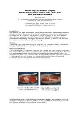
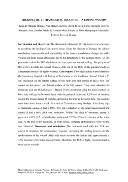
Santos, V., Santos, A. (2014) Providing Optimum Wound Healing Conditions during the
Proliferative Stage using Kerrafibre, Journal of Aging & Inovation, 3 (3): 50-55
ESTUDO DE CASO / CASE STUDY
DEZEMBRO, 2014
PROVIDING OPTIMUM WOUND HEALING CONDITIONS DURING THE PROLIFERATIVE STAGE USING
KERRAFIBRE
PAPEL DO KERRAFIBRE NA OBTENÇÃO DE ÓPTIMAS CONDIÇÕES PARA A CICATRIZARÃO EM FERIDAS
NA FASE PROLIFERATIVA
PROPORCIONAR OPTIMAS CONDICIONES DE CURA EN HERIDAS DURANTE LA FASE PROLIFERATIVA
USANDO KERRAFIBRE
Autores
Vítor Santos 1; Ana Sofia Teixeira Santos 2; César Fonseca3
1
Enfermeiro, CNS, MsC, Centro Hospitalar do Oeste, IberWounds Oeste, Caldas da Rainha, Portugal, 2
Enfermeiro, Centro Hospitalar de Lisboa Norte, Lisboa, Portugal,
Corresponding Author: [email protected]
Abstract : In order to analyze the role of a new structured fibrous dressing in the proliferative stage of chronic
wound healing in hard to heal wounds, a case study was performed on a stagnant venous ulcer, which remained
non-healed in the past 7 months. All chronicity factors that could affect wound healing were excluded, including
biofilm (with the use of polihexanide+betaine, Prontosan range), and compression therapy was provided. The
results were very interesting with healing achieved in 7 weeks of treatment. Most of times it is not easy to find/select
a dressing to promote granulation and epithelisation, once ideal cleaning/debridement and bioburden control are
achieved. Some dressings do not provide a good healing rate, lead to bioburden elevation during time and
recurrence in the use of antimicrobials. Other options to promote proliferation are very expensive and need a
secondary dressing. Treatment with kerrafibre showned to be very cost effective. Its is also implicit the important
role of advanced wound care centers versus conventional care.
This case study was originally presented as a poster at Wounds UK 2014 Conference, at Harrogate, England,
United Kingdom.
Keywords: Fibrous Dressings; Proliferative Stage, Hard to Heal Wounds.
Resumo: A fim de analisar o papel de um novo penso fibroso na fase proliferativa da cicatrização da ferida crônica
de difícil cicatrização, um estudo de caso foi realizado numa úlcera venosa estagnada, que permaneceu por
cicatrizar nos últimos 7 meses. Todos os factores de cronicidade que pudessem afectar a cicatrização foram
excluídas, incluindo o biofilme (com o uso de polihexanida + betaína, da gama Prontosan) sendo também utilizada
terapia compressiva. Os resultados foram muito interessantes com a obtenção da cicatrizarão ao fim de 7 semanas
de tratamento. Na maioria das vezes, não é fácil de encontrar / selecionar um curativo para promover a granulação
e epitelização, uma vez alcançada a limpeza ideal / desbridamento e controle de carga biológica. Alguns pensos
não fornecem uma boa taxa de cicatrização, e podem ocasionar elevação carga microbiana ao longo do tempo e a
reincidência no uso de antimicrobianos. Outras opções para promover a proliferação são muito dispendiosas e
precisam de um penso secundário. O tratamento com kerrafibre revelou ser muito rentável. Fica também implícita a
importância do papel dos centros avançados de tratamento de feridas versus o tratamento convencional. Este
estudo de caso foi originalmente apresentado como um Poster no Congresso Wounds UK 2014, em Harrogate,
Inglaterra, Reino Unido.
JOURNAL OF AGING AND INOVATION
(EM LINHA) ISSN: 2182-696X / (IMPRESSO) ISSN: 2182-6951
Volume 3. Edição 3
Página 51
Introduction
having mixed aetiology of both venous and
3
arterial disease.
Approximately 1 to 2% of the population will
suffer from leg ulceration, with lower limb chronic
venous insufficiency affecting up to 50% of the
1
adult population. There are many direct risk
factors for venous ulceration including: varicose
veins, deep vein thrombosis, chronic venous
insufficiency, poor calf muscle function, arteriovenous fistulae, obesity, and history of leg
fracture. Recurrent venous ulceration occurs in
up to 70% of those at risk. Many venous ulcers
are painful, so appropriate pain relief and advice
2
should be given. It is recognised that about
70% of ulcers are venous in origin, 10–15% are
arterial, with about 15% of leg ulcer patients
JOURNAL OF AGING AND INOVATION
(EM LINHA) ISSN: 2182-696X / (IMPRESSO) ISSN: 2182-6951
This case study describes the management of
an 86-year-old female who lived independently
and was socially active in her community. She
had hypertension, venous insufficiency, and had
suffered 2 episodes of venous ulcers in the
same location. She had an active venous leg
ulcer, which had been present for 7 months
(Figure 1). It was evaluated in line with clinical
guidelines, which suggest that all patients with
chronic venous leg ulcers should have an ankle
brachial pressure index (ABPI) performed prior
to treatment and the ulcer edge should be
measured as it often gives a good indication of
4
progress of wound healing.
Volume 3. Edição 3
Página 52
Method
The patient’s leg ulcer was located mainly in the
retromalleolar region, on the distal third of the
medial aspect of the left leg. The leg had been
eczematous, and became itchy and blistered
The patient wore compression stockings and
had previously been treated with a sodium
carboxymethylcellulose dressing, both with and
without silver.
2
before ulcerating. The ulcer measured 3.7cm
and the ABPI measurement was 0.91,
2
suggesting only mild arterial disease. There
was some slough present, with signs of infection
and critical colonisation. The wound was
malodorous, with moderate fibrinous exudate.
JOURNAL OF AGING AND INOVATION
(EM LINHA) ISSN: 2182-696X / (IMPRESSO) ISSN: 2182-6951
The treatment plan was to clean the wound and
so exclude biofilm, remove debris and slough.
Prontosan Solution (B. Braun) was used to clean
the wound during the first 3 weeks. Prontosan
Gel (B. Braun) was then used in conjunction with
a silver alginate foam dressing, twice weekly for
2 weeks, to prevent biofilm re-organisation,
reduce signs of infection, and manage exudate.
Volume 3. Edição 3
Página 53
JOURNAL OF AGING AND INOVATION
(EM LINHA) ISSN: 2182-696X / (IMPRESSO) ISSN: 2182-6951
Volume 3. Edição 3
Página 54
Subsequently, KerraFibre (Crawford Healthcare)
was used twice a week for 2 weeks and weekly
for 3 weeks. KerraFibre was chosen due to the
ability of its alginate fibres to help absorb and
wick away debris from the wound surface,
helping to keep the wound clean and so
reducing bioburden. It was also chosen due to
its ability to absorb and retain moderate to large
amounts of fibrinous exudate, and so maintain
the ideal moist wound healing environment to
allow granulation and epithelialisation. During
treatment the patient also wore 2-layer
compression bandages (Urgo K2). Compression
therapy may be safely used in leg ulcer patients
4
with ABPI≥0.8.
Results
Granulation and epithelialisation were achieved
after 7 weeks of treatment (Figure 6). By limiting
wound secretions and minimising bacterial
contamination KerraFibre helped to create the
ideal healing environment for the wound bed and
the wound healed before 2 months of treatment.
The dressing was accepted well by the patient,
who described it as comfortable.
JOURNAL OF AGING AND INOVATION
(EM LINHA) ISSN: 2182-696X / (IMPRESSO) ISSN: 2182-6951
Discussion
2
Venous leg ulcers often become infected so it is
important to control the bioburden and manage
the wound exudate. KerraFibre combines a highcalcium alginate fibre with a high-sodium
superabsorbent sulphonated co-polymer gel.
The combination of the alginate wicking layer
that forms a gel on contact with wound exudate,
and the superabsorbent co-polymer gel,
maintains the moist wound environment whilst
enhancing fluid management.
KerraFibre is suitable for moderate to highly
exuding wounds with no need for a secondary
dressing. Integrated into 1 structure, each of its
3 layers performs a different role – but when
combined with the innovative hexagonal
structure, the result is a dressing that absorbs
exudate and locks it away.
Volume 3. Edição 3
Página 55
Conclusion
Granulation and epithelialisation were assisted
by KerraFibre in the management of this
recurrent venous ulcer. After debridement,
cleaning of the wound and bioburden control are
achieved, dressings such as KerraFibre, which
reduce slough and exudate in a wound, help to
minimise bacterial contamination, promote
granulation and epithelisation. Their effective
wound healing is also very cost-effective in
managing chronic venous leg ulcers.
3.
Briggs M, Closs SJ (2003) The
prevalence of leg ulceration: a review of
the literature. EWMA Journal 3(2): 14-20 4.
Scottish Intercollegiate Guidelines
Network. (2010) SIGN Guideline 120:
Management of chronic venous leg
ulcers. August 2010 References
1. Venous Forum of the Royal Society of
Medicine, Berridge D, Bradbury AW, Davies AH
et al (2011) Recommendations for the referral
and treatment of patients with lower limb chronic
venous insufficiency (including varicose veins)
Phlebology 26(3): 91-3
2. Grey JE, Harding K, Enoch S (2006) ABC of
wound healing: venous and arterial leg ulcers.
British Medical Journal 332(7537): 347-50
JOURNAL OF AGING AND INOVATION
(EM LINHA) ISSN: 2182-696X / (IMPRESSO) ISSN: 2182-6951
Volume 3. Edição 3
Download