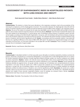
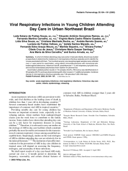
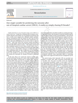
2/7/11
AUTONOMIC CONTROL OF THE VERTEBRATE HEART
E.W. (Ted) Taylor,
School of Biosciences,
University of Birmingham UK
“Hello folks/Bom dia gentes!”
The Golden Rule of
Vaudeville:
Always put a comedy act on
last and maybe finish with a
comic song.
So this is it!
Brasil (1999-now) with Tobias to work with Augusto
2 beers Wang
Denis Augusto Wilfried
Homer
Neto
“I distrust camels, and anyone else who can go a week
without a drink” Joe Lewis
1
2/7/11
Pharmacology of ANS
ACh receptors
Nicotinic receptors in the
autonomic nervous system
can be blocked by
hexamethonium
Nicotinic receptors on the
skeletal muscle can be
blocked by curare
Muscarinic receptors on the
heart can be blocked by
atropine
Adrenergic receptors
β receptors can be blocked by
propranolol, or stimulated by
isoproteronol
Ach acetylcholine
NE noradrenaline
stimulators are “agonists” blockers “antagonists”
α receptors can be blocked by
phentolamine or stimulated
by phenylephrine
Phylogeny of Cardiac Innervation
Taylor and Wang 2009
CONTROL OF THE HEART and of CARDIORESPIRATORY INTERACTIONS
The sympathetic nervous system is primarily responsible for increases in
heart rate in emergencies the “fight or flight response” or in
“stressed”animals. It does not exert beat-to-beat control of the heart.
The parasympathetic nervous system exerts a dominant role in the tonic
control of the heart and can exert beat-to-beat control that generates heart
rarte variability (HRV) including changes in heart rate in phase with
ventilation or cardiorespiratory interaction (CRI).
This control is exerted via the Xth cranial nerve, the vagus.
THIS IS OUR PRIMARY INTEREST!
2
2/7/11
CARDIORESPIRATORY INTERACTIONS
We are investigating:
•
• the evolution of the mechanisms for nervous control of the heart in
vertebrates using classic physiological and neuro-anatomical techniques .
•
• the roles of “feed-forward control” from the central nervous system (CNS)
and of reflex or “feedback control” from peripheral receptors in determining
heart rate variability (HRV) and in particular cardiorespiratory interactions
(CRI).
MAMMALS
In healthy mammals (e.g Marina!) heart rate increases
during inspiration, a phenomenon known as:
respiratory sinus arrythmia (RSA)
MAMÍFEROS
Em mamíferos saudáveis, a freqüência cardíaca aumenta
durante a inspiração, fenômeno conhecido como:
Arritmia sinusal respiratoria (RSA)
RSA developes in utero in human babies
It is present at full-term (40 weeks) but absent in premature babies (<
30 weeks)
It can be detected by Power Spectral Analysis (PSA)
RSA desenvolve-se dentro do útero em bebês humanos
Ele está presente durante 40 semanas, mas é ausente em bebês
prematuros (menos de 30 semanas)
Pode ser detectado por Power Spectral Analysis (PSA)
3
2/7/11
Power spectral analysis of heart rate variability (HRV) reveals respiratory
sinus arrythmia (RSA) in the human neonate
Thompson, Brown, Gee and Taylor (1993) Early Human Devel. 31, 217-228
Premature babies in intensive
care were monitored (from the
back of the hospital equipment)
with HRV and breathing
movements recorded to a pc for
power spectral analysis
Bebês prematuros foram monitorados
com HRV tendo os movimentos
respiratórios registrados num pc
The onset of
RSA:
Premature babies
were born without a
respiratory peak in
the HRV power
spectrum - A
This appeared at
33-36 weeks as
ventilation became
more regular - B, C
Babies compromised
at birth often did not
develop RSA (cot
death?)
30 wks
33 wks
36 wks
Taylor, Leite and Skovgaard (2010) Braz. J. Med. Biol. Res. 43, 600.
4
2/7/11
The mechanism:
Activity in cardiac vagal efferent (CVE) fibres is gated
during inspiration, including their response to
baroreceptor stimulation, leading to respiratory sinus
arrythmia (RSA)
A atividade das fibras cardíacas eferentes do vago (CVE) para
durante a inspiração, causando “respiratory sinus arrythmia” (RSA)
Differential modulation of reflex cardioinhibitory
responses by lung inflation in the cat
Lung inflation
Daly and Kirkman (1989) J. Physiol. 417, 323
cyanide
stimulation
pressure
veratridine
phenylbiguanide
J-receptors
5
2/7/11
In mammals over 30% of VPN
(vagal preganglionic neurones)
and over 70% of cardiac VPN
(CVPN) are located in the NA
(nucleus ambiguus) with the rest in
the dorsal motor nucleus (DVN)
DVN
NA
Ranson, Butler and Taylor (1993)
J. auton. Nerv. Syst. 43, 123- 138
Rat: compound cardiac action potentials
anodal
block
B – fibres from NA
C – fibres from DVN
Jones, Wang and Jordan (1995) J. Physiol. 489, 203
Rato
Coelho
Gato
Jones, Wang and Jordan (1995) J. Physiol. 489, 203
6
2/7/11
RSA is generated in the brainstem of mammals in an area called the nucleus
ambiguus (NA). Activity in cardiac vagal preganglionic (motor) neurones
(CVPN/CVM) that exercise a dominant role in control of the heart, is gated
by activity in neighbouring inspiratory neurones
RSA se desenvolve no tronco
encefálico dos mamíferos,
num local chamado nucleus
ambiguus (NA).
Mike Spyer and Dave Jordan (Spyer, 1989 TINS 12, 506-513)
Many physiologists and psychobiologists (because they
work on mammals!) consider RSA to be a mammalian
function (have the NA) not seen in “lower” vertebrates.
Muitos fisiologistas e psicobiólogos, por trabalharem com
mamíferos, consideram RSA como uma função exclusiva desse
grupo (tem NA) e ausente nos vertebrados “inferiores”.
As Comparative Physiologists we recognise various
forms of cardiorespiratory interactions (CRI) in a range
of species
Como estamos interessados em fisiologia comparativa, nós
reconhecemos formas diferentes de interação cardiorespiratória
(CRI) em muitas espécies
CHORDATE PHYLOGENY
7
2/7/11
BIRDS:
In the duck only 5% of VPN but 30%
of CVPN are in the ventro-lateral NA
Duck:
cardiovascular and
respiratory variables:
effect of water on
laryngeal receptors
(Event)
Butler and Taylor, 1983
Respir. Physiol. 53, 109
REPTILES
often breath discontinuously, heart rate varies with ventilation
8
2/7/11
REPTILES:
Rattlesnake - Crotalus durissus terrificus
Complete vagotomy slows ventilation,
increases heart rate and abolishes HRV
Wang, Warburton, Abe and Taylor (2001) Exp. Physiol. 86, 777
Estas mesmas respostas aparecem em mamíferos
Vagal control of the cardiac shunt Efferent electrical
stimulation
anaesthetised
Taylor et al. 2009
Pharmacological study of parasympathetic and sympathetic tone
on the heart of the rattlesnake
9
2/7/11
Rattlesnake = cascavel:
diurnal changes in body temperature and
heart rate (fH)
mudanças diárias na temperatura do corpo e na freqüência cardíaca
Rattlesnake: Power spectral analysis of heart rate variability shows
evidence for RSA with peaks in the spectrum appearing, then shifting as
heart rate (fH) slows during recovery from handling stress
Campbell, others and Taylor (2006) J. exp. Biol. 209, 2628
Rattlesnake: TS brainstem 0.5 mm caudal of obex
VPN labelled with true blue are in 2 locations: 96% in the DVN (núcleo
motor dorsal) and 4% scattered ventrolaterally
DVN
4V
Ventro-lateral cell
Edge of
brain
10
2/7/11
Caiman - TS brainstem to show VPN labelled with True blue
A
B
D2
V
V
D2
D1
D1
VL
OTHER REPTILES – Brasil!!!
Distribution of VPN in TS of brainstem Caiman latirostris 12%
(nº cells / section) (nº cells / section)
Boa constrictor
30
25
20
15
10
5
0
30
25
20
15
10
5
0
2.52 1.68 0.84
A
rattlesnake
B
Group 1 Group 2 Ventrolateral caiman
0
-0.84 -1.68 -2.52
Distance from obex (mm)
Taylor, Leite and Skovgaard (2010) Braz J Med Biol Sci.
11
2/7/11
Uromastyx aegyptius microlepis
From Saudi Arabia
VPN were located in the DVN or
DMNX and in the NA but only in
small numbers (5%)
Mohammed Al-Ghamdi, 1995
Boa constrictor – cardiovascular responses to a meal or a bad time
Effects of autonomic blockade on heart rate during forced activity and
digestion in Boa constrictor
Wang, Taylor, Andrade and Abe (2001) J Exp Biol. 204, 3553
12
2/7/11
Heart of doubled-blocked animals increases during digestion
1.  Release of an excitatory NANC factor
from cardiac nerve terminals ?
2.  Increased circulating levels of a
NANC factor(s) ?
AMPHIBIANS: Toad/Bullfrog
A
Respiratory muscles: inserted
around the buccal cavity(A),
innervated by cranial nerves V,
VII and X plus XII (B)
The vagus (X) provides:
. afferent nerves to chemo and
baroreceptors plus lung stretch
receptors (PSR) (C)
. efferent nerves to the lungs, glottis,
heart plus a sphincter on the
pulmonary artery (D)
B
C
D
Reflex control of respiratory
and cardiovascular systems is
similar to mammals
Wang, Hedrick, Ihmied and Taylor
(1999) CBP
13
2/7/11
Bufo: heart rate and pulmonary blood
flow increase during bouts of breathing
Bullfrog: effects of autonomic antagonists on
heart rate at 10, 20 and 30oC
Bullfrog: calculated autonomic tonus on
the heart
Filled columns, cholinergic/vagal tonus
Open columns, adrenergic/sympathetic
tonus
14
2/7/11
Bullfrog, black columns
Xenopus, grey columns
Bullfrog 3
TS brainstem
0,12 mm rostral to obex
Scale bar represents 100µm
V
V, 4th ventricle
Bullfrog 5
TS brainstem
0,34 mm rostral to obex
Scale bar represents 100µm
V, 4th ventricle
V
15
2/7/11
Bullfrog: Rostro-caudal distribution of cell bodies of
VPN, with respect to obex
no cells / sec9on 40
30
20
10
0
-2.7
-1.8
-0.9
0
0.9
1.8
2.7
3.6
Distance from obex (mm) Xenopus:
Vagal projections into the brainstem located
by axonal transport of HRP. There are 2
populations of VPN of differing in size and
location, inside or outside central grey area.
TS medulla 1 mm rostral of obex
Xenopus
Rostro-caudal distribution of VPN in the
medial DVN and in the lateral NA that
innervate different organs.
About 30% of VPN are located outside the
DVN
This is the same as mammals.
There is evidence of a residual sequential
distribution.
16
2/7/11
Axolotl, Ambystoma mexicanum
before and after induced metamorphosis
Axolotl: rostrocaudal distribution of VPN
Conclusions:
All tetrapod vertebrates (amphibians, reptiles, birds and
mammals) studied have neural mechanisms causing the heart to
beat in relation to ventilation (CRI). This relationship is
mediated via the vagus nerve.
2 locations for CVPN in the brainstem may be important for the
generation of CRI but is not present in all species
Todos os vertebrados estudados possuem mecanismos neurais que fazem
o coração bater de acordo com os ritmos ventilatórios (CRI). Essas
relações são mediadas pelo vago.
CVPN em dois locais do tronco encefálico talvez explique a produção de
CRI mas naõ tem de todos especies.
17
2/7/11
PEIXE
Take five
Levar cinco
FISH:
We have conducted a long-term study on the control of cardiorespiratory
interactions in fish. Sometimes they show cardiorespiratory synchony
(CRS), with the heart beating in phase with ventilation.
This may increase the effectiveness of respiratory gas exchange across the
counter-current between blood on water at the gills
Há vários anos, estamos estudando o controle da interação
cardiorespiratória em peixes. Algumas vezes, os resultados mostram
sincronia cardiorespiratória (CRS), com o coração pulsando em fase
com a ventilação.
Isso pode aumentar a eficiência da troca respiratória de gases, nas
brânquias, entre o sangue e a água
Counter-current exchange of O2 over fish gills
18
2/7/11
Counter-current exchange of O2 over fish gills
Cardiorespiratory synchrony:
Elasmobranch Dogfish: Ventilation (VR)
and heart (HR) rates recorded as pressures
A. From resting dogfish (normal) and
following injection of atropine and
B. during a spontaneous period of activity
Teleost Cod: Blood pressure and
flow in the dorsal aorta plus
water pressure in the buccal
cavity
Cod: polar diagrams of heart beat related to
respiratory cycle
normoxia
hypoxia
Jensen, Varne, Findorf, Dominici, McKenzie, Wang and Steffensen unpublished
19
2/7/11
When trout ram ventilate heart rate rises. This may indicate loss
of a respiration related vagal component
How can fish generate beat-to-beat changes in heart rate?
In mammals:
C – fibres conduct at around 2 metres / second (DVN)
B – fibres conduct at around 20 metres / second (NA)
Jones, Wang and Jordan (1995) J. Physiol. 489, 203
Dogfish cardiac vagus compound action potentialconduction velocity 7-35 metres/second
3 cm of nerve
5 volts
10 volts
25 volts
Barrett and Taylor (1985) J. exp. Biol. 117, 459
20
2/7/11
The Chondrichthyes (cartilaginous, elasmobranch fish)
are phylogenetically a “primitive” group (>300M years)
They are the first group to possess an autonomic nervous system
BUT – the sympathetic nervous system does not extend into the “head”
consequently the heart only has parasympathetic vagal innervation
Eles são os primeiros (Chondricthyes) com sistema nervoso autônomo,
MAS – o sistema nervoso simpático não atinge a “cabeça” e,
conseqüentemente, o coração somente possui inervação parassimpática
Thus, the dogfish brainstem provides a model for vagal control of
cardiorespiratory interactions
Dessa forma, o tronco encefálico do dogfish representa um modelo
para estudar o controle do vago na interação cardiorespiratória
Innervation of the
heart:
Elasmobranchs
Chondrichthyes (cartilaginous
fish)
brain
vagus
heart
solely parasympathetic supply
Teleosts
Osteichthyes (bony fish)
parasympathetic and
dual sympathetic supply
brain
Sympathetic
ganglia
heart
as “advanced”
as mammals!
Dogfish: Schematic
diagram of branches of
cranial nerves V, VII, IX
and X, innervating the
respiratory system
and heart
X br c branchial cardiac
X visc c visceral cardiac
A neural tracer (HRP)
was injected into
branches of cranial
nerves V, VII, IX and X
21
2/7/11
cobalt
chloride
True
blue
Horseradish
peroxidase
Dogfish: Sequential
rostrocaudal distribution of
neurones supplying the
respiratory and cardiac
branches of the cranial nerves
22
2/7/11
Dogfish: spontaneous
activity in central
cut branches of the
cranial nerves
supplying respiratory
muscles and the heart
Atividade espontânea dos nervos
cranianos controlam os músculos
respiratórios e o coração
Dogfish: decerebrate and paralysed
Efferent activity in cardiac and branchial vagi
(non-bursting activity seems responsible for reflex control of heart
rate and generation of vagal tone, which increases during hypoxia)
There are 2 types of efferent activity recorded from the
cardiac vagus of the dogfish.
Neuranatomical study revealed 2 locations for CVPN in the
brainstem: a medial group in the dorsal vagal motor nucleus
and a scattered ventro-lateral group outside the DVN
23
2/7/11
Dogfish:
Schematic dorsal view and TS of hindbrain, shaded
areas denote motor nuclei to V, VII, IX and Xth cranial nerves
Having located
neurone cell bodies in
the brainstem we
recorded from them
using microelectrodes
Dogfish: central recordings of
activity in CVPN
Cells located in the DVN
showed respiration-related
activity (A)
Those outside the DVN were
routinely active (B) or silent (C)
Activity in all CVPN could be
generated by touching gill septa
(stim)
Fish were paralysed and
decerebrated, removing any
phasic influences from
peripheral receptors or centres
higher in the CNS
RF reticular formation; Xs vagal sensory nucleus;
Xm, vagal motor nuclei; X4 4th branch of vagus nerve
occ. sn. occipital and spinal nuclei; hypobranchial
24
2/7/11
Respiration-related, phasic activity was recorded from
CVPN in the DVN of paralysed and decerebrated fish
implying that it was generated by feed-forward control
from the central respiratory pattern generator
Mechanical stimulation of a gill septum caused CVPN
to fire, implying that in the normally breathing fish
mechanoreceptor stimulation would induce reflex
activation of CVPN
The effects on heart rate of phasic activity in the
cardiac vagus was tested by peripheral stimulation
Dogfish:
Peripheral electrical stimulation of a cardiac nerve
A, with a continuous train of impulses
B, with continuous and phasic trains of pulses
Dogfish: Recruitment of heart rate to phasic peripheral
stimulation of the right cardiac branch of the vagus
Taylor et al (2006) PBZ
25
2/7/11
Putative conclusions:
Elasmobranch fishes have no excitatory sympathetic innervation of the
heart
They do have a parasympathetic inhibitory supply to the heart
It arises from CVPN in 2 locations in the brainstem:
A medial group in the dorsal vagal motor nucleus DVN show
respiration related activity and may generate CRI
A lateral group do not and may generate phasic changes in
heart rate (e.g. hypoxic bradycardia)
Both groups of CVPN respond to mechanical stimulation so will be
recruited by ventilation
Bursting efferent activity in the cardiac vagus can drive the heart, overriding the cardiac pacemaker
A teleost model:
Pacu – Piaractus mesopotamicus
2006 - Tadeu Rantin’s laboratory
UFSCar Sao Carlos, SP. Brasil
Pacu is unique?
A.  Does not respond to hypoxia or injection of nicotine with a significant increase
in circulating catecholamines (adrenaline/epinephrin) implying inoperative or
absent humoral adrenergic response
B.  Injection of propranolol (beta-adrenergic antagonist) after atropine injection
revealed that the excitatory adrenergic tone on the heart in pacu is below 10%.
C.  Thus, the predominant factor controlling heart rate in pacu is inhibitory
parasympathetic control, imposed via the vagus nerve, innervating
muscarinic cholinoceptors.
A. Não responde à hipoxia ou injeção de nicotina com um significante aumento na
circulação das catecolaminas (adrenalina/epinefrina) - resposta humoral adrenérgica
inoperante ou ausente
B. Injeção de propanolol depois de injeção de atropina – estímulo adrenérgico no
coração do pacu é menor que 10%.
C. Sendo assim, o principal fator que controla o ritmo cardíaco, em pacu, é o controle
parassimpático inibidor, determinado pelo nervo vago nos receptores muscarínicos
26
2/7/11
Pacu - Recordings from the cardiac vagus, ventilation and ECG
•  No routinely respiration-related bursts in cardiac vagus in normoxic fish
* increased ventilatory effort
** cough
A
**
B
*
*
**
Pacu - Effect of stopping the water flow irrigating the gills
•  Increased activity in cardiac vagus is associated with increased
ventilatory effort and generates a bradycardia
Pacu - Restricting water flow
•  Increased ventilation amplitude generated bursting activity in the
cardiac vagus that recruited the heart
27
2/7/11
SO:
The heart can be recruited by bursting activity in the cardiac
vagus, generated by reducing the supply of oxygen.
O ritmo cardíaco corresponde à atividade fásica do vago
cardíaco, fenômeno causado pela redução do nível de oxigênio.
Can this effect be simulated by peripheral electrical stimulation
of the cardiac vagus?
Pode esse efeito ser simulado por estimulação elétrica periférica
no vago cardíaco?
Pacu - Pulsed efferent stimulation of the cardiac vagus can
drive the heart both slower or faster than its intrinsic rate
If bursting activity recorded from the cardiac vagus is dependent on phasic
peripheral feedback from receptors in the branchial apparatus when oxygen
supply is limited (most likely to be mechanoreceptors on the gill arches,
opercula or jaws),
Then, can phasic central stimulation of cranial nerves innervating the
respiratory apparatus recruit the heart?
Se a atividade fásica do nervo vago cardíaco depende do feedback fásico
emitido pelos receptores do aparelho branquial quando o suprimento de
oxigênio é limitado,
Então, poderia a estimulação fásica e central dos nervos cranianos, que
inervam o aparelho respiratório, fazer o coração bater sincronicamente
com os estímulos?
28
2/7/11
Pacu – effects on
heart rate of central
stimulation of cranial
nerves IX, X, VII, and
V to respiratory
muscles
Phasic stimulation of
VII, IX and X
recruited the heart
but V was without
effect **
**
Pacu
Cardiac vagal preganglionic neurones (CVPN) and respiratory
vagal motor neurones (RVMN) located in the dorsal vagal motor
nucleus (DVN) in the brainstem by fluorescent tracers.
G1 dorsal DVN
(40%)
G2 ventral DVN
(60%)
LC lateral CVPN
(2%)
CVPN
V 4th ventricle
RVMN
There seems to be abundant evidence for the supremacy of feedback from
peripheral receptors on the gills or opercula (but not the jaws!) in the
generation of cardiorespiratory interactions (CRI) in pacu, a teleost fish.
VPN are predominantly located in DVN
But, is this the complete answer?
Existem muitas evidências sobre o papel do feedback dos receptores
periféricos, das brânquias ou opérculos (mas não da mandíbula), em
promover uma interação cardiorespiratória (CRI) em pacus.
Mas, essa pergunta está completa?
29
2/7/11
Pacu - Rostro-caudal distribution of RVMN and CVPN
in the brainstem. Respiratory neurones innervating the
Vth cranial nerve do not overlap with other neurones
**
Pacu
Recordings of ventilation
as opercular movements
and integrated activity in
cranial nerves V, IX, X 1st
and 2nd branchial and 4th
cardiac
Pacu – sequence of bursting activity
in cranial nerves.
Bursts in cardiac nerve are
synchronous with activity in Vth
cranial nerve, innervating the jaw.
A atividade fásica dos nervos
cardiacos é sincrônica com a
atividade do 5º nervo craniano,
que inerva a mandíbula (d)
Both anticipate activity in
other respiratory branches.
Ambos antecipam a atividade em
outros ramos respiratórios (e)
30
2/7/11
SO:
•  There is evidence of an important role for peripheral feedback in
generating CRI in pacu.
•  There remains a possibility that central, feed-forward drive from a
central respiratory pattern generator can drive both the respiratory
neurones innervating the jaw and CVPN innervating the heart, when
respiratory drive is high.
•  This may determine the phase relationships between heart beat and
ventilation, optimising oxygen delivery across the gills.
•  Existem evidências de que o feedback periférico promove CRI em pacus
•  Sobra a possibilidade do feed-forward do controle central gerar CRI,
por inervar tanto neurônios respiratórios como o CVPN.
•  Isso pode determinar a relação sincrônica entre o batimento cardíaco e a
ventilação, otimizando a captura do oxigênio nas brânquias
In elasmobranchs CRS seems to be primarily determined by
central, feed-forward control when vagal tone on the heart is
low (Taylor, 1992)
Nos elasmobrânquios, CRS parece ser determinada
por um controle central, feed-forward, quando o
tonus vagal no coração é baixo (Taylor, 1992)
In teleosts control seems to depend on feedback from
peripheral receptors generating high levels of cardiac vagal
tone (Randall and Smith, 1967)
Nos teleósteos, o controle parece depender do feedback
dos receptores periféricos gerando altos níveis de tonus
vagal cardíaco (Randall and Smith, 1967)
31
2/7/11
A schematic model of possible connections in the central and
peripheral nervous systems generating cardiorespiratory
interactions in fish
CPG, central pattern generator;
CVM, cardiac vagal motor neurone
CVS, cardiac sensory
RVM, respiratory motor;
RVS, respiratory sensory
We have some limited
insights into 1 - 8 but
cannot yet completely
delimit their roles
RF reticular formation; Xs vagal sensory nucleus;
Xm, vagal motor nuclei; X4 4th branch of vagus nerve
occ. sn. occipital and spinal nuclei; hypobranchial
32
2/7/11
Evolution of the NA: There is a progressive increase in the proportion
of VPN in the ventro-lateral NA, from fish through amphibians to
mammals but reptiles are complicated and birds out of line.
However, 2 populations of CVPN may be needed to generate CRI
Grossman and Taylor (2007) Biol. Psychol. 74, 26
33
Download