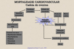
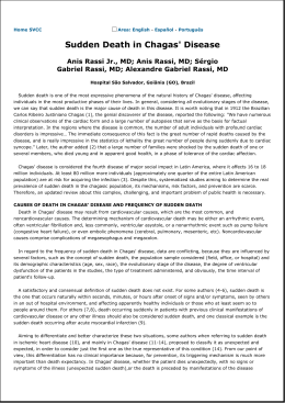
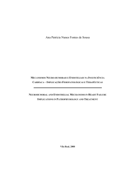
Pesq. Vet. Bras. 29(4):291-294, abril 2009
Evaluation of left ventricular diastolic echocardiographic
parameters in healthy dogs by pulsed-wave Doppler1
Guilherme G. Pereira2*, Liliam C. Petrus2, André L.F. Santos2, Fernanda L.
Yamaki2 and Maria Helena M.A. Larsson3
ABSTRACT.- Pereira G.G., Petrus L.C., Santos A.L.F., Yamaki F.L. & Larsson M.H.M.A.
2009. Evaluation of left ventricular diastolic echocardiographic parameters in
healthy dogs by pulsed-wave doppler. Pesquisa Veterinária Brasileira 29(4):291-294.
Departamento de Clínica Médica, Faculdade de Medicina Veterinária e Zootecnia,
Universidade de São Paulo, Av. Prof. Orlando Marques de Paiva 87, São Paulo, SP
05508-900, Brazil. E-mail: [email protected]
Left ventricular diastolic dysfunction plays an important role on heart failure progression.
In order to obtain additional reference values of left ventricular diastolic parameters and
investigate influence of common variables, peak E wave (peak E), peak A wave (peak
A), E/A ratio (E/A), E wave deceleration time (EDT) and isovolumic relaxation time (IRVT)
were studied in 40 clinically healthy dogs, by pulsed wave Doppler. The following values
were obtained: peak E = 0.747 ± 0.117 m/s, peak A = 0.487 ± 0.062 m/s, E/A = 1.533 ±
0.198, EDT = 88.7 ± 9.2 ms and IRVT = 0.080 ± 0.009 s. Some parameters were influenced
by heart rate (peak E, peak A and IRVT), by age (peak A and E/A) and by body weight
(TRIV). Gender influence was absent. Values obtained can be used as reference for
canine specimens but its interpretation should consider on the influence of related
variables.
INDEX TERMS: Diastole, pulsed-wave Doppler, Dogs, Left ventricle
RESUMO.- [Avaliação de parâmetros ecocardiográficos
diastólicos do ventrículo esquerdo de cães saudáveis
por meio de Doppler pulsátil.] A disfunção diastólica do
ventrículo esquerdo contribui de maneira importante para a
progressão da insuficiência cardíaca. No intuito de obter
valores de referência adicionais para os parâmetros
diastólicos do ventrículo esquerdo e para investigar a influência de variáveis comuns, estudou-se, por meio de Doppler
pulsátil, a velocidade máxima da onda E (Vmáx.E), a velocidade máxima da onda A (Vmáx.: A), a relação E/A (E/A), o
tempo de desaceleração da onda E (TDE) e o tempo de
relaxamento isovolumétrico (TRIV) em 40 cães clinicamente saudáveis. Os seguintes valores foram obtidos: Vmáx.E
= 0,747±0,117m/s, Vmáx.A = 0,487±0,062m/s, E/A =
1,533±0,198, TDE = 88,7±9,2ms e TRIV = 0,080±0,009s.
Alguns parâmetros foram influenciados pela freqüência cardíaca (Vmáx.E, Vmáx.A e TRIV), pela idade (Vmáx.A e E/A)
e pelo peso corpóreo (TRIV). Não houve influência pelo sexo.
Os valores obtidos podem ser utilizados como referência
para a espécie canina, porém devem ser interpretados considerando-se a influência das variáveis descritas.
TERMOS DE INDEXAÇÃO: Diástole, Doppler pulsátil, cães,
ventrículo esquerdo.
INTRODUCTION
Investigations concerning diastolic cardiac function have
experienced increasing relevance in the last years because
of diastolic dysfunction´s role on heart failure progression
(Dougherty et al. 1984, Rihal et al. 1994, Rossi et al. 2004).
Diastolic dysfunction is not only found on primary heart
disease but also as a consequence of systemic diseases,
such as hyperadrenocorticism, chronic renal failure,
obesity, and others (Atkins 1999). The study of ventricular
diastole allows identification of prognostic indicators and
is a valuable tool on therapy follow-up (Werner et al. 1993).
1
Received on April 8, 2008.
Accepted for publication on January 6, 2009.
2 Postgraduate student, Departamento de Clínica Médica, Faculdade
de Medicina Veterinária e Zootecnia (FMVZ), Universidade de São Paulo
(USP), Av. Prof. Orlando Marques de Paiva 87, São Paulo, SP 05508900, Brazil. *Corresponding author: [email protected]
3 Departamento de Clínica Médica, FMVZ-USP, São Paulo, SP.
291
292
Guilherme G. Pereira et al.
There are many useful pulsed-wave Doppler derived
parameters that allow diastolic function indirect evaluation.
Some of them are extremely relevant and easily obtained,
like maximum velocities of E (peak E) and A (peak A) waves,
E:A ratio (E:A), E wave deceleration time (EDT) and
isovolumic relaxation time (IVRT) (Nishimura & Tajik 1997).
Reference values for left ventricular diastolic parameters
are obviously relevant for determination of left ventricular
diastolic function. However, the evaluation of flows by pulsed
wave Doppler may be influenced by a sort of variables, like
heart rate, age (Schober & Fuentes 2001), body weight (Sido
et al. 2000) and loading conditions (Yamamoto et al. 1993).
Some reference values are described for specific breeds,
like Doberman Pinscher (Minors & O’Grady 1998), German
Shepherd and Beagle (Kirberger et al. 1992), but there are
also reports including dogs representing a wide range of
breeds (Schober & Fuentes 2001).
Once pulsed-wave Doppler of left ventricular flows is a
very useful tool on diastolic function investigation the study
reported here proposes to obtain some additional reference
values for transmitral diastolic flow parameters in dogs
and investigate the influence of heart rate, body weight,
age and gender on the values obtained.
represented the time interval (s) between the end of left ventricular
systolic flow and begging of diastolic mitral flow (E wave).
Five consecutive measurements were performed, for each
parameter, in order of reducing effects from sinus arrhythmia
variation.
Data analysis were performed by ANOVA for determination
of means and standard deviations. Pearson correlation´s test
(r) was employed in order to investigate influence of heart rate,
age and body weight on these parameters. The mean value
from five consecutive measurements was considered for
correlation studies with age and body weight, in total of 40 points
of analysis. For heart rate study, were considered each one of
the five measurements, in total of 200 points of analysis. The
coefficients of determination (r2) were obtained to quantify the
variation explained by the influence of the variable investigated.
Gender influence was investigated by t-Student test for the
parametric data and by Mann-Whitney test for the non-parametric
data. A significance level of p<0.05 was considered.
RESULTS
Figures 1 and 2 show recordings of mitral flow and IVRT,
respectively.
MATERIALS AND METHODS
Fourty adult dogs, 20 males and 20 females, representing
different breeds (23 cross-breed, three Poodles, two Labrador
Retrievers, Cocker Spaniels, Golden Retrievers and Canadian
Shepherds, and one Schnauzer, Brazilian Terrier, Dachshund,
Beagle, Lhasa Apso and Yorkshire), aged between 2 and 6
years, weighing from 4,2 to 37,4 kg (mean 18,3 kg), were included
in this study. Among them, 14 were maintained at Faculdade de
Medicina Veterinária e Zootecnia, Universidade de São Paulo,
according to the ethics committee of Universidade de São
Paulo, and 26 were pet-owned.
Before data acquisition, animals were evaluated by physical,
radiographic, electrocardiographic and echocardiographic
examination, as well as complete blood count, urinalysis, serum
biochemical profiles, ELISA test for Dirofilaria immitis3 antigen
and indirect Doppler ultrasonographic arterial blood pressure.
Echocardiographic examination was performed according
to the Echocardiography Committee of the Specialty of Cardiology,
American College of Veterinary Internal Medicine, and the
American Society of Echocardiography recommendations (Boon
1998). Pulsed-wave Doppler transmitral flow was recorded from
left caudal parasternal long axis four chamber view, with the
sample located in the left ventricle, proximal to the mitral leaflets
opening. Simultaneous unidimensional (M) mode recording was
performed, at mitral valve level, for correlation of flow with heart
cycle (Morcerf 1996). The first diastolic flow was considered as
early diastolic flow wave (E wave) and the second flow, as atrial
contraction flow wave (A wave). Peak E and A waves velocities,
considered as the peak of E and A waves, respectively (m/s), E/
A ratio and EDT, considered as the time from peak to end of E
wave (ms), were obtained.
For IVRT determination flows were obtained by a left caudal
parasternal long axis five chamber view, with sample positioned
between septal mitral leaflet and left ventricular outflow tract. IVRT
3
Snap, Idexx.
Pesq. Vet. Bras. 29(4):291-294, abril 2009
Fig.1. Simultaneous M-mode and pulsed-wave Doppler
recording of mitral E and A waves.
Fig.2. Pulsed-wave Doppler recording of IVRT.
Evaluation of left ventricular diastolic echocardiographic parameters in healthy dogs by pulsed-wave doppler
Table 1. Left ventricular diastolic echocardiographic
parameters, obtained by pulsed-wave Doppler, in 40
healthy dogs
Ea(m/s)
A(m/s)
E:A
EDT(ms)
IVRT(s)
0.75± 0.117
(0.53-1.06)
0.49± 0.062
(0.39-0.67)
1.53± 0.198
(1.18-1.89)
89± 9.2
(74-108)
0.080± 0.009
(0.066-0.100)
a
E = peak E velocity; A = peak A velocity; E:A = E:A ratio; EDT = E
wave deceleration time; IVRT = isovolumic relaxation time.
Values are expressed as means ± standard deviation. Range is shown
in parentheses.
Table 2. Pearson's correlation coefficients (r) for heart
rate, age and body weight influence on left ventricular
diastolic echocardiographic parameters
a
Variable
Ea
A
E:A
EDT
IVRT
Heart rate
Age
Body weight
0.271b
- 0.408d
0.017
0.384b
- 0.051
- 0.154
- 0.064
- 0.428d
0.126
- 0.134
- 0.196
0.150
- 0.179c
0.012
0.364c
E = peak E velocity; A = peak A velocity; E:A = E:A ratio; EDT = E
wave deceleration time; IVRT = isovolumic relaxation time; bp<0.001;
cp<0.05; dp<0.01.
Values obtained for mentioned parameters studied are
expressed as means, standard deviation and range in
Table 1.
Pearson´s correlation coefficients were calculated and
have shown influence of some variables on most of the
parameters studied, only excluding EDT (Table 2).
The coefficients of determination (r2) for heart rate
influence was 0.073, 0.147 and 0.032 for peak E, peak A
and IVRT, respectively. The r2 values of age was 0.166
and 0.183 for peak E and E:A ratio, respectively. Body
weight was found to have r2=0.132 for IVRT.
Values of peak E and EDT had parametric distribution
in both males and females groups, being investigated by
Student´s t-test, while values of peak A, E:A ratio and IVRT
had a non-parametric one, being investigated by MannWhitney´s test. No difference was found between both
groups at a value of p<0.05.
DISCUSSION
The study revealed referencial values for some left ventricular diastolic parameters in canine specimens, including
peak velocities of E and A waves, E:A ratio, E wave
deceleration time and isovolumic relaxation time. The
examination was performed in conditions that reproduced
what is commonly found in clinical practice. The parameters are easily obtained by an experienced examiner
and no sedation was required. So, these can be employed
in investigations concerning diastolic function in clinical
practice. Such parameters are helpful in identification of
diastolic abnormalities due to primay or secondary cardiac
disorders.
An influence of heart rate, age and body weight on some
parameters was found. However, the contribution of these
variables alone was low, being the amount of variation in
E:A ratio explained by age (18.3%) the biggest one.
293
Different sources of variation, like loading conditions,
should be involved.
Most of the parameters evaluated were influenced by
heart rate. This could be explained by variation in left
ventricular filling time, leading to different filling pressures.
Although some studies indicated absent (Schober &
Fuentes 2001) or negative (Minors & O´Grady 1998)
correlation between peak E and heart rate, the present
study demonstrated a positive correlation. Some authors
suggested that atrial pressures are the most important
reason for this variation in peak E, being heart rate only
an indirect component (Yamamoto et al. 1993). Also peak
A values were positively correlated with heart rate. This
could be explained by the short time to left ventricular filling
and increase in residual volume in left atrium after the early
filling phase, leading to a higher pressure gradient between
both chambers and, consequently, a higher flow velocity.
The IVRT demonstrated to shorten with higher heart rates,
probably because of early beginning of early left ventricular
filling, leading to a premature opening of mitral leaflets.
Age influenced peak E and E:A ratio and older animals
showed lower values for the mentioned parameters. E:A
reversion was not found in the animals of the study. As
were included animals aged under 7 years, the E:A reversion can be considered uncommon and probably abnormal
in animals with similar age. Schober and Fuentes (2001)
have found reversion in animals aged above 6 years and
with no apparent diastolic dysfunction. These findings
indicate caution in interpretation of peak E and E:A ratio in
elderly animals. Because EDT and IVRT were not influenced by age, it is advisable the concomitant analysis of
these parameters when investigating diastolic function in
elderly animals.
Body weight positive correlation with IVRT in part could
be explained by higher heart rates found in small dogs,
once IVRT is also influenced by this variable.
The values obtained can be used as reference for both
males and females, once no difference was found between
them.
Only EDT was not influenced by any of the variables
investigated, on the contrary of some reports (Yamamoto
1993, Schober & Fuentes 2001). So, it provides valuable
information concerning diastolic function in dogs, associated with the others studied parameters.
CONCLUSION
The present study revealed comparative reference values
for left ventricular diastolic parameters in healthy dogs aged
between 2 and 6 years and indicated that heart rate, age
and body weight, but not gender, should be considered on
their interpretation, except for EDT. Additional studies
should be performed in order to investigate different
sources of influence on these parameters.
REFERENCES
Atkins C.E. 1999. Cardiac manifestations of systemic and metabolic
disease, p.757-780. In: Fox P.R., Sisson D. & Moïse N.S. (Eds),
Pesq. Vet. Bras. 29(4):291-294, abril 2009
294
Guilherme G. Pereira et al.
Textbook of Canine and Feline Cardiology: Principles and clinical
practice. W.B. Saunders, Philadelphia.
ventricle in health and disease: Doppler echocardiography is the
clinician’s Rosetta Stone. J. Am. Coll. Cardiol. 30(1):8-18.
Boon J.A. 1998. Manual of Veterinary Echocardiography. Williams and
Wilkins, Baltimore. 478p.
Rihal C.S., Nishimura R.A., Hatle L.K., Bailey K.R. & Tajik A.J. 1994.
Systolic and diastolic dysfunction in patients with clinical diagnosis of
dilated cardiomyopathy. Relation to symptoms and prognosis.
Circulation 90(6):2772-2779.
Dougherty A.H., Naccarelli G.V., Gray E.L., Hicks C.H. & Goldstein R.A.
1984. Congestive heart failure with normal systolic function. Am. J.
Cardiol. 54:778-782.
Kirberger R.M., Berg P.B. & Grimbeek R.J. 1992. Doppler echocardiography in the normal dog. Vet Radiol Ultrasound 33(6):380-386
Minors S.L. & O’Grady M.R. 1998. Resting and dobutamine stress
echocardiographic factors associated with the development of occult
dilated cardiomyopathy in healthy Doberman Pinscher dogs. J. Vet.
Int. Med. 12:369-380.
Morcerf F.A.P. 1996. Avaliação do tamanho e função do ventrículo esquerdo, p.193-225. In: ___ (Ed.), Ecocardiografia Uni-Bidimensional,
Transesofágica e Doppler. 2a ed. Revinter, Rio de Janeiro.
Nishimura R.A., Abel M.D., Hatle L.K., Holmes D.R., Housmans P.R.,
Ritman E.L. & Tajik A.J. 1989. Significance of Doppler indices of diastolic
filling of the left ventricle: Comparison with invasive hemodynamics in a
canine model. Am. Heart J. 118(6):1248-1258.
Nishimura R.A. & Tajik A.J. 1997. Evaluation of diastolic filling of left
Pesq. Vet. Bras. 29(4):291-294, abril 2009
Schober K.E. & Fuentes V.L. 2001. Effects of age, body weight, and
heart hate on transmitral and pulmonary venous flow in clinically normal dogs. Am. J. Vet. Res. 62(9):1447-1454.
Sido Z., Jako P. & Pavlik G. 2000. The effect of moderate weight loss on
echocardiographic parameters in obese female patients. Acta Physiol.
Hung. 87(3):241-251.
Yamamoto K., Mauyama T., Tanouchi J., Uematsu M., Doi Y., Naito J.,
Hori M., Tada M. & Kamada T. 1993. Importance of left ventricular
minimal pressure as a determinant of transmitral flow velocity pattern
in the presence of left ventricular systolic dysfunction. J. Am. Coll.
Cardiol. 21(3):662-672.
Werner G.S., Scaefer C., Dirks R., Figulla H.R. & Kreuzer H. 1993.
Doppler echocardiographic assessment of left ventricular filling in
idiopathic dilated cardiomyopathy during a one-year follow-up: Relation
to the clinical course of disease. Am. Heart J. 126(6):1408-1416.
Download