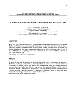
Close window to return to IVIS
Or
This manuscript is reproduced in the IVIS website with the permission of WSAVA
Or – Orthopaedics
ARTHROSCOPIC DIAGNOSIS OF ELBOW DYSPLASIA
Dominique J Griffon, DMV,
MS, PhD, DACVS,
DECVS Associate Professor,
Head, Small Animal Surgery
Director,Laboratory for
Orthopedic Research on
Biomaterials University of Illinois
Small Animal Clinic
1008 W Hazelwood drive
Urbana IL61802, USA
http://www.cvm.uiuc.edu/lorb/
[email protected]
What is elbow dysplasia?
“Elbow dysplasia” (ED) is a syndrome that
includes several conditions resulting in an
incongruency of the joint, eventually leading to
degenerative joint disease (DJD). Elbow dysplasia
is a common, inherited condition affecting 17%
of Labrador retrievers, and up to 70% of Bernese
Mountain dogs in the Netherlands. This high
prevalence and the devastating effects of elbow
DJD emphasize the need for early diagnosis,
individualized treatment and preventive measures.
In 1993, the International Elbow Working
Group (IEWG) agreed that “elbow arthrosis
caused by Fragmented Coronoid Process (FCP),
osteochondrosis (OCD), Ununited Anconeal
Process (UAP), articular cartilage anomaly and/or
joint incongruity is the manifestation of inherited
canine elbow dysplasia”.
2006 World Congress WSAVA/FECAVA/CSAVA
Conditions included in Elbow Dysplasia:
• Osteochondrosis (trochlea humeri)
• Ununited Anconeal Process (UAP)
• Fragmented Coronoid Process (medial, FCP)
• Incongruity:
1. Radio-ulnar incongruence
a. With or without angular limb deformity
b. Short ulna or short radius
2. Elliptical ulnar notch?
3. Local incongruity (deformed medial coronoid process)?
• Combination of the above
630
Why consider arthroscopy for the diagnosis of
elbow dysplasia?
The IEWG developed a radiographic protocol
and scoring system adopted as a screening system
to diagnose ED. The radiographic findings are
Elbow Dysplasia Grading
0
Normal elbow joint
I
Mild arthrosis
II
III
Moderate arthrosis
Severe arthrosis or 1° ED
scored according to severity of the arthrosis (DJD)
and/or presence of a primary lesion (derived from
Mark Flückiger, IEWG 2003 Estoril meeting):
Radiographic Findings
Normal elbow joint, no evidence of incongruency,
sclerosis or arthrosis
Sclerosis of ulnar trochlear notch or, step =/> 2 mm
between radius and ulna or, osteophyte formation less
than 2 mm high
Osteophyte formation 2 to 5 mm high
Osteophyte formation over 5 mm high and/or primary ED
such as LPA, FMCP or OCD
Close window to return to IVIS
Radiographs of both elbows should be obtained
in all cases diagnosed with elbow dysplasia,
because the condition is often bilateral. UAP and
OCD are typically diagnosed on radiographs, and
do not require advanced imaging.
Radio-ulnar incongruence has traditionally
been diagnosed based on standard radiography.
Degenerative joint disease secondary to elbow
dysplasia is mainly evaluated on hyperflexed
lateral, cranio-caudal and craniolateral /
caudomedial 15°oblique views. An UAP is best
visualized on a lateral hyperflexed radiograph.
The craniolateral / caudomedial 15°oblique view
improves the visualization of the medical coronoid
process compared to the cranio-caudal projection
(Wosar et al. 1999). However the sensitivity of
conventional radiography to image the medial
coronoid process has been estimated to only range
from 10 to 62% (Wosar et al. 1999). The effect
of radiographic positioning on interpretation of
cubital joint congruity has also been studied in
normal dogs. The authors of this study compared
three mediolateral (45°, 90° and 135° of flexion)
and three cranio-caudal views, and concluded that
elbow congruity was best assessed on a 90° flexed
lateral projection with the beam centered over
the joint (Murphy et al. 1998). The superiority
of mediolateral over craniocaudal projections
to evaluate radio-ulnar incongruence was later
confirmed with a surgical model of induced
radio-ulnar incongruence in cadaveric forelimbs
(Mason et al. 2002). However, radiologists were
only able to correctly identify congruent elbows
(specificity) in 86% and incongruent elbows
(sensitivity) in 78% of specimens. The radio-ulnar
step required for each of 4 American College of
Veterinary Radiology board-certified radiologists
to attain 90% sensitivity ranged from 1.5 to
greater than 4 mm. In comparison, the elevation
of the coronoid process in relationship with the
ulna in elbows with FCP has been reported to vary
between 1 and 2 mm (Wind 1986, Trostel et al.
2003) The results of this study did not support
the use of standard radiography for evaluation of
radio-ulnar incongruence.
Computed tomography has previously been used to
help diagnose elbow dysplasia when radiographs
did not provide a definitive diagnosis (Rovesti et
al. 2002). CT eliminated the false positive results
obtained with survey radiographs in the detection
of fragmented coronoid process. Compared to
standard radiographs, it also provided a better
definition of the disease n 46% of elbows examined.
CT scan has been reported to achieve the highest
accuracy (86.7%) and sensitivity (88.3%) when
compared to radiography, xeroradiography, linear
tomography and positive contrast arthrography
for diagnosing fragment medial coronoid process
(Carpenter et al. 1993). CT imaging protocols
have recently been described to measure elbow
incongruity (Holsworth 2004, Gemmill TJ et
al. 2004). Although the accuracy, sensitivity
and specificity of CT to diagnose radioulnar
incongruence has not been studied, this technique
is currently considered as a gold-standard to
diagnose radio-ulnar incongruence, and has been
applied in two clinical studies (Gielen 2004,
Holsworth 2004, Gemmill et al. 2004, Schulz
2004). Both studies compared radio-ulnar
incongruence in normal elbows and in dogs with
FCP (Gemmill et al. 2004, Kramer et al. 2004).
Unfortunately, the results and conclusions of
both studies differed drastically: Gemmill et al.
reported that radio-ulnar incongruity exists at
the apex of the coronoid process but not at its
base, whereas Kramer et al. described exactly the
opposite. Both groups used a similar protocol,
but the discrepancy between their results may
be due to the difference in positioning of the
elbow or in interpretation of images: one group
measured the humeroradial and humeroulnar
joint space (Gemmill et al. 2004), whereas the
other measured the actual step between the radius
and the ulna (Kramer et al. 2004). Although these
studies suggest that the form of incongruency
associated with FCP is more complex than a
simple proximal translation of the articular
surface of the ulna, they also illustrate the need
for further evaluation of CT as a diagnostic tool
for radio-ulnar incongruence. Ultimately, the
ability of computed tomography to diagnose
elbow incongruity will remain limited by the lack
of visualization of the cartilage, leading to an
evaluation of subchondral bone rather alignment
of joint surfaces. From a clinical standpoint,
this diagnostic tool currently requires general
anesthesia of the patient and may not be widely
accessible in private practices.
Arthoscopic evaluation of the elbow also requires
general anesthesia but it allows unparalleled
visualization of joint surfaces (Schulz 2004). In
addition, arthoscopy is minimally invasive and
can be combined with surgical treatment of the
lesion. One study found no difference in postoperative gait analysis of normal dogs undergoing
arthroscopic versus open exploration of the
elbow (Bubenik LJ et al, 2002). However, two
other studies documented improved function in
dogs immediately after and over the 21 months
following arthroscopic treatment of FCP,
compared to arthrotomy (Schwarz et al. 1993,
Meyer-Lindeberg A et al. 2003). Arthroscopic
management of FCP therefore appears to minimize
post-operative pain compared to open arthrotomy,
but further studies are needed to long term benefits
on degenerative joint disease.
2006 World Congress WSAVA/FECAVA/CSAVA
Or
631
Close window to return to IVIS
Or
What arthroscopic technique can be used to
diagnose FCP and radio-ulnar incongruence?
The dog is placed in dorsal recumbency with a
sandbag on the lateral aspect of the affected elbow.
An assistant maintains the limb in position over
the sand bag to provide adduction throughout the
study. A 2.7mm arthroscope may be used in large
breeds, although atraumatic insertion of the scope
between the humeral head and the ulna may be
difficult, especially in dogs with radio-ulnar
incongruity. A 1.9mm 30° fore obliqued scope
is therefore preferred. The entire joint is first
explored through a medial portal, including the
anconeal process, the medial and lateral coronoid
processes, the medial and lateral aspects of the
humeral condyle and the entire radial head. A
systematic evaluation of all compartments of
the elbow is crucial to complete the diagnosis of
elbow dysplasia. The egress portal consists of an
18-gauge needle directed in the joint pouch just
proximal to the anconeus. An instrument portal
located in the region of the medial collateral
ligament allows triangulation of a graduated
hook-probe. The probe can be used to mobilize
a fragmented coronoid process, cartilage flap in
OCD and appreciate radio-ulnar incongruence.
The arthroscopic appearance of OCD, UAP and
FCP are well established. In fact arthroscopy
has allowed the identification of variation in
appearance of FCP:
Arthroscopic appearance of fragmented coronoid process (FCP) in dogs:
• Fragment on the medial margin of the medial coronoid process (MCP)
• Erosion of the lateral rim of the MCP
• Incomplete fragmentation (fissure)
• Fragment in situ
• Minimally migrated FCP
• Fully migrated FCP (joint mouse)
• Chondromalacia – Eburnation of the MCP
2006 World Congress WSAVA/FECAVA/CSAVA
Elbows with FCP should be carefully evaluated
for degenerative joint disease (especially
cranial to the radial head), cartilage lesions on
the opposing articular surface of the humerus
(“kissing lesions”) and incongruity (Figure 1).
Unfortunately, objective assessment of elbow
incongruity via arthroscopy has not been fully
investigated.
632
Figure 1: Arthroscopic evaluation of radio-ulnar
incongruity in two dogs with medial FCP. Both
images show the humeral condyle (triangle), the
ulna commissure and lateral coronoid process
(arrow) and the radial head (block arrow). Left:
congruent radio-ulnar junction. Right: radioulnar incongruence.
We have recently studied the predictive value
of arthroscopy to diagnose experimentally
induced radio-ulnar incongruity (Wagner K
et al. 2006). A surgical model of radio-ulnar
incongruence was used to shorten the radius by
1, 2 and 3 mm increments in cadaveric forelimbs
obtained from adult Labrador Retrievers. Radio-
ulnar incongruence was blindly evaluated via
computed tomography (CT) and arthroscopy
before and after each modification. Radio-ulnar
incongruence was measured arthroscopically
with a graduated probe at 3 levels in each study:
the commissure of the medial and lateral portions
of the coronoid process, the apex and the midbody of the medial coronoid process. The
distance between the ulnar and radial surfaces in
unmodified elbows was equal to 1.18 ± 0.13 mm
on CT. The overall sensitivity of arthroscopy was
85% and 95%, when the radius was shortened by
1mm versus ≥2mm, respectively. The specificity
was equal to 50%. The ability to detect mild
incongruity (1 mm step) was greater at the
ulnar incisure than at other locations. Although
pronation subjectively appeared to modify the
appearance of the radio-ulnar junction, this factor
was not found to affect the overall predictive
value of arthroscopy. Intra-articular pressure did
not affect the examination. In this study, we found
arthroscopy to be very sensitive for detection of
radio-ulnar incongruence, especially at the ulnar
incisure. The greatest sensitivity obtained at
the ulnar incisure is due to the relative ease of
identifying landmarks compared to the mid body
and apex of the coronoid. The low specificity of
the technique (i.e. our ability to correctly identify
unmodified elbows as congruent) reflects the
physiological degree of radio-ulnar incongruence
previously reported in dogs, and confirmed by
the CT measurements in our study. The clinical
relevance of a mild radio-ulnar incongruence
Close window to return to IVIS
remains unclear in a normal elbow. However,
a diagnosis of mild incongruity in the presence
of other signs of elbow dysplasia would support
adjunctive osteotomies / ostectomies to modify the
distribution of loads in the diseased joint. Further
studies correlating radiographs, arthroscopy
and computed tomography in clinical cases of
elbow dysplasia are warranted to validate these
findings.
Selected references
Bubenik LJ, Johnson SA, Smith MM, et
al.: Evaluation of lameness associated with
arthroscopy and arthrotomy of the normal canine
cubital joint. Vet Surg 2002; 31: 23-31
Carpenter L, Schwarz P, Lowry J, et al.
Comparison of radiologic imaging techniques for
diagnosis of fragmented medial coronoid process
of the cubital joint in dogs. J Am Vet Med Assoc
1993; 203: 78-83.
Flückiger M: Radiographic diagnosis of elbow
dysplasia (ED) in the dog – Requirements for the
internationally standardized screening procedure
for ED. Proc International Elbow Working Group
May 22 2003, Estoril, Portugal.
Gielen I, Ven Ryssen B, van Bree Henri:
Arthrology – Diagnostic imaging: is CT the
answer. Proc 12th ESVOT Congress, Munich,
Germany, 10-12th September 2004b, pp 140.
Gemmill TJ, Clements DN, Clarke Sp et al.:
Investigation of elbow incongruency in dogs
suffering coronoid disease using reconstructed
computed tomography. Vet Surg, 2004; 33: E6.
Holsworth I: How I manage elbow incongruity,
Proc 12th ESVOT congress, Munich, Germany,
September 10-12, 2004, pp 60.
Kramer A, Filipowicz D, Hosworth IG et al.:
Computerized tomographic evaluation of canine radioulnar incongruence in vivo. Vet Surg 2004; 33: E13.
Mason DR, Schulz KS, Samii VF et al:
Sensitivity of radiographic evaluation of radioulnar incongruence in the dog in vitro. Vet Surg
2002; 31: 125-132.
Meyer-Lindenberg A, Langhann A, Fehr M et al.:
Arthrotomy versus arthroscopy in the treatment
of fragmented coronoid process of the ulna (FCP)
in 421 dogs. VCOT 2003; 16: 204-210.
Murphy ST, Lewis DD, Shiroma JT et al.: Effect
of radiographic positioning on interpretation of
cubital joint congruity in dogs. Am J Vet Res
1998; 59(11): 1351-1357.
Rovesti GL, Biasibetti M, Schumacher A et al:
The use of computed tomography in the diagnostic
protocol of the elbow in the dog: 24 joints. VCOT
2002; 15: 35-43.
Schulz KS: Diagnostic assessment of the elbow
(When in doubt, scope the elbow). Proc ACVS
symposium, Denver, Colorado, October 6-9
2004, pp 329-331.
Schwarz PD, Brevard SM, Baker CG: Arthroscopy
of the shoulder (OCD) and elbow (MFCP)- thirty
consecutive cases each: a comparative study of
the early postoperative period. Vet Surg, Proc 7th
Annual ACVS symposium; 1993: 21-22.
Trostel TC, McLaughlin RM, Pool RR: Canine
lameness caused by developmental orthopedic
diseases: Fragmented medial coronoid process
and united anconeal process. Comp Cont Ed
Pract Vet 2003; 25: 112-120.
Wind AP: Elbow incongruity and developmental
elbow disease in the dog. J Am Anim Hosp Assoc
1986; 22: 711-724.
Wosar M, Lewis D, Neuwirth L, Parker R, et
al. Radiographic evaluation of elbow joints
before and after surgery in dogs with possible
fragmented medial coronoid process. J Vet Med
Assoc 1999; 214: 52-58.
2006 World Congress WSAVA/FECAVA/CSAVA
Or
633
Download