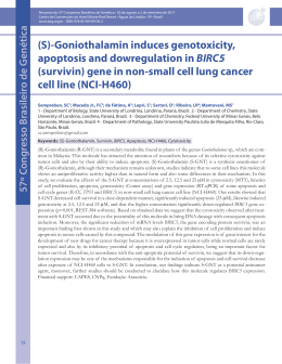
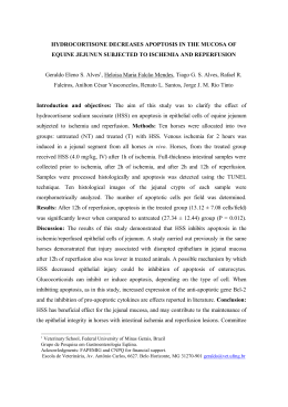
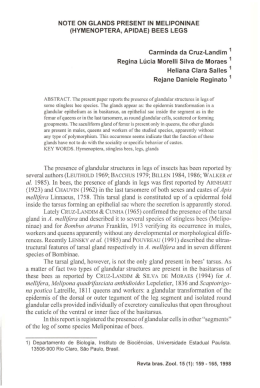
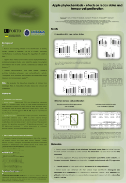
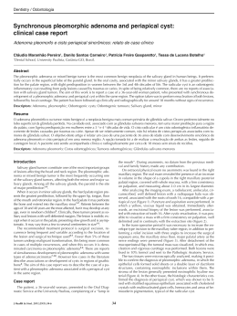
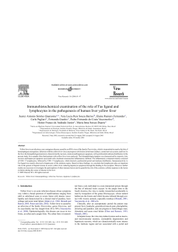
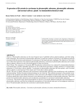
ARTIGO ORIGINAL
APOPTOSIS
THE
AND
PA R A D O X
IN
CELL
OF
P R O L I F E R AT I O N
S A L I VA R Y
SJÖGREN
’S
:
GLANDS
DISEASE
Rafael Herrera-Esparza, JuanJosé Bollain-y-Goytia, Claudia Ruvalcaba, MaGuadalupe Ruvalcaba,
Deyanira Pacheco-Tovar, Esperanza Avalos-Díaz
Sjögren primária.
Métodos: A apoptose foi estudada por imunohistoquímica utilizando anticorpos monoclonais anti-Fas, FasL e Caspase 3 e as características apoptóticas por TUNEL. Os estudos foram executados em
vinte e quatro glândulas salivares minor de doentes
com Síndroma de Sjögren primária e num igual número de controlos. A proliferação foi avaliada com
anticorpos monoclonais anti-PCNA e anti-Ki67.
Resultados: Todas as glândulas salivares dos doentes com Sjögren apresentavam moléculas apoptoticas no epitélio dos ductos salivares, e menos no
tecido acinar, consequentemente a presença do
caspase 3, Fas/FasL eram concordantes com a expressão da apoptose por TUNEL. Os marcadores de
proliferação foram encontrados nas células inflamatórias presentes, mas não no epitélio ductal nem
nos acinos. A expressão de marcadores de apoptose ou de proliferação nos tecidos das biopsias dos
controlos foi escassa.
Conclusão: Os dados actuais sugerem que as células do epitélio ductal e dos acinos das glândulas salivares dos doentes com doença de Sjögren têm aumento da apoptose. A proliferação foi observada
principalmente no infiltrado celular linfóide. Em
conjunto, estes eventos constituem um paradoxo
biológico relacionado com o processo inflamatório
das glândulas salivares na Síndrome de Sjögren.
Abstract
Aim: To assess apoptosis and proliferation in salivary glands of patients with primary Sjögren’s syndrome.
Methods: Studies were performed in twenty four
minor salivary glands from patients with primary
Sjögren’s syndrome and an equal number of controls. Apoptosis was studied by immunohistochemistry using monoclonal antibodies anti-Fas,
FasL and Caspase 3 and apoptotic features by
TUNEL. Proliferation was assessed with monoclonal anti-PCNA and anti-Ki67 antibodies.
Results: All salivary glands from Sjögren’s display
apoptotic molecules along the epithelia of salivary
ducts, and in a smaller amount in acinar tissue. The
presence of Caspase 3, Fas/FasL was concordant
with the expression of apoptosis by TUNEL. Proliferation markers were encountered in inflammatory emigrant cells, but not in ductal epithelia nor in
acini. Control biopsies poorly expressed apoptotic
or proliferation markers.
Conclusion: Present data suggests that the ductal
epithelial and acinar cells of salivary glands from
Sjögren’s disease patients exhibit increased apoptosis. Proliferation was mainly observed in infiltrating lymphoid cells. Both events constitute a biological paradox related to the inflammatory process of salivary glands in Sjögren’s disease.
Keywords: Apoptosis; cell proliferation ; Sjögren
disease; Salivary glands; PCNA; Caspase 3.
Introduction
The pathogenesis of primary Sjögren’s disease is
multifactorial but the antigen that drives the autoimmune response is still unknown. The disease
causes dry eyes and mouth, vasculitis, and neurological symptoms. Symptoms are the result of immune lymphocytic infiltration in lacrimal and salivary glands.1
B-cell hyperactivity, increased levels of B-cell-activating factor/B-lymphocyte stimulator and
Resumo
Este estudo avalia a apoptose e a proliferação nas
glândulas salivares dos doentes com Síndroma de
Centro de Biología Experimental (CBE) Universidad Autónoma
de Zacatecas. Guadalupe, Zacatecas. México.
Ó R G Ã O O F I C I A L D A S O C I E D A D E P O R T U G U E S A D E R E U M AT O L O G I A
299
-
A C TA R E U M AT O L P O R T . 2 0 0 8 ; 3 3 : 2 9 9 - 3 0 3
A P O P T O S I S A N D P R O L I F E R AT I O N I N S J Ö G R E N
tional groups as follows: those with less than two
foci/4 mm2 (1 ≤ FS < 2) and those with two or more
foci (FS ≥ 2).
Unstained tissue sections were used for immunohistochemistry.
Immunohistochemistry. Slides containing 4 µm
sections of minor salivary glands were dewaxed,
then permeabilized with 0.01% Triton X- 100/PBS
and washed three times with PBS. Endogenous peroxidase was blocked with 56°C heated horse serum. After washing, tissues were incubated for 1
hour with anti-FasL (sc-19681 Santa Cruz, Biotechnology, Inc. Santa Cruz.CA), anti-Fas (m3553,
Dako Carpinteria,CA), anti-Ki-67 antibody (cat 35-1600, Zymed, San Francisco, CA), and anti-PCNA
(cat 13-3900, Zymed). Antibodies were diluted
1:1000 in 10% BFS-PBS. After washings with PBS,
bound antibodies were tagged with goat anti-mouse IgG labelled with peroxidase. Colour reaction
was induced by 2, 2 diaminobenzydine-0.06% H2O2
(Sigma, San Luois, MO). Reaction was stopped with
2N sulphuric acid. The slides were then evaluated
by three observers; assays were done in triplicate.
TdT-mediated dUTP nick end labeling (TUNEL)
was done according to the manufacturer’s instructions (Roche Molecular Biochemicals. Penzberg,
Germany). Nuclear stripping was performed by
immersing the slides for 5 minutes in 10 mM TrisHCl, pH 8.0, followed by 15 minutes in 20 µg/ml
proteinase K dissolved in the same buffer, and finally the slides were washed with PBS. Elongation
of the DNA fragments was performed by incubation for 60 minutes at 37ºC with 75 µl of the reaction mixture [DDW, 10XTdT buffer (30 mM Tris base,
140 mM sodium cacodylate, pH 7.2, 1 mM cobalt
chloride, 1 mM DTT); 10% of the final volume],
fluorescein-11-dUTP (0.5 mg dissolved in 1 ml of
10 mM Tris-HCl, pH 7.0), and TdT enzyme (0.3 enzyme units/µl). The reaction was terminated by
adding the stop solution (300 mM NaCl, 30 mM
sodium citrate, pH 8.0).
anti-Ro or anti La autoantibodies’ production are
among the abnormalities of the immune system in
Sjögren’s disease. The glandular dysfunction results
from lymphocytic infiltration around the ductal
and acinar epithelium. Infiltrates of activated T cells
produce inflammatory cytokines, and these mediators cause tissue damage by cytotoxicity, or induce
glandular apoptosis; the apoptotic remains contain intracellular autoantigens that trigger autoantibody production. Another effect of cytokines is
the induction of functional quiescence, rather than
destructive damage; but in the majority of the patients different cytokines induce lymphocyte proliferation. Since pathophysiology of Sjögren’s disease is multifactorial, different environmental factors
have been implicated such as HTLV-1 and hepatitis
C viruses. However, the possible role of virus in the
pathogenesis of Sjögren’s is still under investigation.1-4
The present study addresses the issue of how
apoptosis and proliferation may coexist in salivary
glands of patients with primary Sjögren’s syndrome.
Material and Methods
Minor salivary gland biopsies from 24 patients with
primary Sjögren’s disease were studied; patients
fulfilled the revised version of the European criteria proposed by the American-European Consensus.5 There were 17 females and 7 males with a
mean age of 47 years (range 37 - 57). All the patients and controls were previously informed about
the biopsy and written consent was obtained. An
equal number of biopsies from controls without
Sögren’s disease were included; these samples were
obtained during oral surgery for other purpose. The
protocol was authorized and monitored by the
ethics committee of CBE and the study was conducted according to the World Medical Association’s Declaration of Helsinki. Specimens were fixed
in formaldehyde and included in paraffin, sectioned at 4 µm thin and stained with haematoxylin &
eosin. After microscopic evaluation by two pathologists, biopsies were classified according to the revised multilevel assessment of a cumulative focus
score (FS).6 Biopsies were considered negative in
case of absence of inflammatory infiltrates (FS=0).
The presence of one or more foci/4 mm2 was classified as positive in spite of the normal appearance of the adjacent glandular parenchyma (0 < FS
< 1). Positive FS biopsies were divided in two addi-
Results
Lymphocyte infiltrates: Biopsies from Sjögren’s
disease were classified as FS1=five biopsies,
FS<2=six, FS = 2=five and FS >2=eight biopsies.
Control biopsies from normal individuals were all
negative (FS 0).
Apoptotic features: All minor salivary glands show
apoptotic markers. As expected, the Fas receptor
Ó R G Ã O O F I C I A L D A S O C I E D A D E P O R T U G U E S A D E R E U M AT O L O G I A
300
-
A C TA R E U M AT O L P O R T . 2 0 0 8 ; 3 3 : 2 9 9 - 3 0 3
R A FA E L H E R R E R A - E S PA R Z A E C O L .
was widely distributed along ductal epithelium
and in a lesser amount along acini. No difference
between patients and controls was observed as Fas
is constitutive. However, normal salivary glands
displayed a faint signal of Fas receptor. Meanwhile,
Sjögren’s salivary glands exhibited a strong Fas signal. It seems that the glandular inflammation overexpresses the Fas receptor. In sharp contrast, the
FasL was expressed exclusively in Sjögren’s salivary
glands, but not in controls. Moreover, the FasL was
broadly distributed in inflammatory infiltrates,
suggesting that the main source of FasL is lymphocyte-dependent (Figure 1 and Table I).
Caspase 3 is a crucial molecule for apoptosis;
consequently it was important to define its presence in ductal epithelia and acini. Control biopsies
displayed a faint signal of caspase 3; Sjögren’s salivary glands expressed a strong signal of caspase 3
in ducts, acini and along inflammatory infiltrates.
Because the detection of Fas/FasL pair and Caspase 3 does not necessarily implicate functional ac-
tivity, a complementary TUNEL assay was performed with the following results: Sjögren salivary
glands showed positive TUNEL assays, apoptotic
tags were distributed along ductal epithelial and
acinar cells and were coincident with the Fas/FasL
and Caspase 3 presence (Figure 2).
Proliferation markers: PCNA and Ki-67 proteins
are considered reliable markers of proliferation.
No proliferation markers were detected in control
biopsies. PCNA was broadly expressed in Sjögren’s
tissues. Ki-67 was also detected in Sjögren’s salivary
glands, but the signal was weak. PCNA and Ki-67
were mainly detected along the inflammatory cell
infiltrates and ductal epithelia and were absent in
acinar cells (Figure 3).
Figure 1. Fas receptor distribution in salivary glands of
controls (A) and Sjögren disease (B). FasL distribution in
controls (C) and in Sjögren disease (D).
Figure 2. Caspase 3 distributions in salivary glands of
controls (A) and Sjögren disease (B).TUNEL in negative
controls (C) and positive in Sjögren salivary glands (D).
Discussion
The present study demonstrates that cells of the
salivary glands from Sjögren’s patients exhibit
Table I. Apototic and proliferation markers in salivary glands
Molecule
FasR
FasL
Caspase 3
TUNEL
PCNA
Ki-67
Controls
23/24
0/24
0/24
1/24
0/24
0/24
Distribution
Faint in epithelial ducts
Absent
Absent
Limited in ducts
Faint in ductal epithelium
Absent
Sjögren
24/24
24/24
17/24
24/24
24/24
10/24
Ó R G Ã O O F I C I A L D A S O C I E D A D E P O R T U G U E S A D E R E U M AT O L O G I A
301
-
Distribution
Broad, lymphoepithelial and acini
Broad, inflammatory cells
Broad, lymphoepithelial lesions
Broad, lymphoepithelial and acini
Broad, lymphoepithelial lesions
Faint, Lymphoepithelial lesions
A C TA R E U M AT O L P O R T . 2 0 0 8 ; 3 3 : 2 9 9 - 3 0 3
A P O P T O S I S A N D P R O L I F E R AT I O N I N S J Ö G R E N
Figure 3. Proliferation markers. PCNA expression in salivary glands of controls (A) and
in Sjögren disease (B). Ki-67 expression in Sjögren disease (C).
glands in Sjögren’s disease may develop benign
lymphoepithelial lesions and ductal lesions, which
are followed by basal cell hyperplasia and aberrant
differentiation into a multi-layered and reticulated
epithelium. All these changes are the result of cell
proliferation. Morphological changes are accompanied by alteration in the cytokeratin pattern and
enhanced Ki-67 protein expression.13 Interestingly,
a constant finding in target organs is a progressive
focal infiltration of mononuclear lymphoid cells
that replace the glandular epithelium. Different
cytokines and chemokines are involved in B cell
maturation and proliferation, the B cell activating
factor being involved in this process.14 In few cases
of Sjögren’s syndrome, the uncontrolled proliferation may result in malignant lymphoproliferative
diseases.15
In summary, there is growing evidence that
cytokines, chemokines and proliferation factors
create a favourable glandular environment that alters the cell cycle resulting in proliferation of few
epithelial areas and lymphoid foci.
Finally, the present investigation demonstrates
that inflammatory cells play an important role in
this paradox of the cell cycle, by which apoptosis
and proliferation can co-exist in the same tissue
target, leading to atrophy and loss of function.
apoptotic features; inflammatory infiltrates in Sjögren’s Syndrome display proliferation markers in
lymphoid cells and this biological paradox inflammation-apoptosis in Sjögren’s disease frequently
results in atrophy and secretor dysfunction.
Among the possible mediators of apoptosis in
Sjögren’s syndrome are the CD8 cells, who may release proteases such as perforin or granzyme B.7
Another route by which CD8 cell may induce apoptosis is by triggering the Fas pathway, because under stimulation FasL is expressed on its surface.
According to our findings, the apoptotic targets are
the epithelial and the acinar cells, rather than the
infiltrating lymphocytes, which seem to be unable
to cause autocrine or paracrine apoptosis. Simultaneous expression of Bcl-2 and FasL in CD8 lymphocytes may increase their resistance to apoptosis.8
The presence of CD4 cells in the inflammatory
infiltrates of Sjögren’s syndrome may be involved
in apoptosis. In the animal model of Sjögren’s
syndrome, glandular apoptosis is induced via CD4
cells. During this process, a cleavage of the 120 kDa
alpha-fodrin takes place. Interestingly, this autoantigen is important in triggering the disease in animal models and in humans. This effect is blocked
by inhibitors of caspases.9
Another mechanism of apoptosis has been proposed in Sjögren’s disease: anti-Ro and anti-La autoantibodies from Sjögren’s patients may induce
apoptosis of the A-253 cell line in a caspase-dependent manner, but this mechanism remains to
be clarified.10,11
Cell proliferation in Sjögren’s disease was recognized by Mickulicz in 1892, who reported a 42-year-old male farmer with lacrimal, parotid, and submandibular enlargement. The excised glands were
filled with small round cells (lymphocytes). The
patient died, possibly from a lymphoma, perhaps
even Waldenstrom’s macroglobulinemia.12 Salivary
Acknowledgements
Supported: by PROMEP-UAZ-CA5 Autoinmunidad.
Correspondence to
Rafael Herrera-Esparza, MD.
Chepinque 306, Col. Lomas de la Soledad.
Zacatecas. 98040. México.
E-mail: [email protected]
References
1. Fox RI, Stern M, Michelson P. Update in Sjögren syndrome. Curr Opin Rheumatol 2000;12:391-398
2. Hansen A, Lipsky PE, Dorner T. Immunopathogenesis of primary Sjögren's syndrome: implications for
Ó R G Ã O O F I C I A L D A S O C I E D A D E P O R T U G U E S A D E R E U M AT O L O G I A
302
-
A C TA R E U M AT O L P O R T . 2 0 0 8 ; 3 3 : 2 9 9 - 3 0 3
R A FA E L H E R R E R A - E S PA R Z A E C O L .
3.
4.
5.
6.
7.
8. Manganelli P, Fietta P. Apoptosis and Sjögren syndrome. Semin Arthritis Rheum 2003; 33:49-65
9. Hayashi Y, Arakaki R, Ishimaru N. The role of caspase
cascade on the development of primary Sjögren's
syndrome. J Med Invest 2003;50:32-38
10. Sisto M, Lisi S, Castellana D et al. Autoantibodies
from Sjögren's syndrome induce activation of both
the intrinsic and extrinsic apoptotic pathways in human salivary gland cell line A-253. J Autoimmun
2006;27:38-49
11. Mitolo V. Autoantibodies from Sjögren's syndrome
trigger apoptosis in salivary gland cell line. Ann N Y
Acad Sci 2007;1108:418-425
12. Mikulicz-Radeck Jv: Über eine eigenartige symmetrische Erkrankung der Thränen- und Mundspeicheldrüsen. Beiträge zur Chirurgie. Festschrift
gewidmet Theodor Billroth. Stuttgart 1892:610-630
13. Ihrler S, Zietz C, Sendelhofert A, Riederer A, Löhrs U.
Lymphoepithelial duct lesions in Sjögren-type
sialadenitis. Virchows Arch 1999;434:315-323
14. Jonsson R, Bolstad AI, Brokstad KA, Brun JG. Sjögren's syndrome a plethora of clinical and immunological phenotypes with a complex genetic background. Ann N Y Acad Sci 2007;1108:433-447
15. Grosbois B, Jego P, Leblay R. Gougerot-Sjögren syndrome and malignant lymphoproliferative syndromes. Rev Med Interne 1998;19:319-324
disease management and therapy. Curr Opin
Rheumatol 2005;17:558-565
Humphreys-Beher MG, Brayer J, Yamachika S, Peck
AB, Jonsson R. An alternative perspective to the immune response in autoimmune exocrinopathy: induction of functional quiescence rather than destructive autoagression. Scand J Immunol 1999; 49:7-10
Badillo Almaraz I, Avalos-Díaz E, Villalobos-Hurtado
R, Herrera-Esparza R. FasL and Bax genes are differentially expressed in acinar epithelium and inflammatory cells of primary Sjögren salivary glands. Minerva Med 2003;94:341-345
Vitali C, Bombardieri S, Jonsson R et al. European
Study Group on Classification Criteria for Sjögren's
Syndrome. Classification criteria for Sjögren's syndrome: a revised version of the European criteria
proposed by the American-European Consensus
Group. Ann Rheum Dis 2002;61:554-558
Morbini P, Manzo A, Caporali R et al. Multilevel examination of minor salivary gland biopsy for Sjogren's syndrome significantly improves diagnostic
performance of AECG classification criteria. Arthritis
Res Ther 2005;7:343-348
Fujihara T, Fujita H, Tsubota K et al. Preferential localization of CD8+ alpha E beta 7+ T cells around acinar epithelial cells with apoptosis in patients with
Sjögren's syndrome. J Immunol 1999;163:2226-2235
Jornadas de Outono da Sociedade
Portuguesa de Reumatologia 2008
Hotel Serra da Estrela, Covilhã
3-5 de Outubro de 2008
Data limite para envio de resumos:
18 de Setembro de 2008
Ó R G Ã O O F I C I A L D A S O C I E D A D E P O R T U G U E S A D E R E U M AT O L O G I A
303
-
A C TA R E U M AT O L P O R T . 2 0 0 8 ; 3 3 : 2 9 9 - 3 0 3
Download