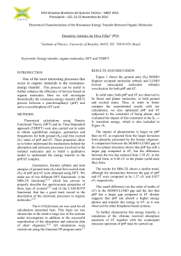
A
http://dx.doi.org/10.5935/0103-5053.20140149
J. Braz. Chem. Soc., Vol. 25, No. 10, 1848-1856, 2014.
Printed in Brazil - ©2014 Sociedade Brasileira de Química
0103 - 5053 $6.00+0.00
Article
Cobalt(III) Complexes with Thiosemicarbazones as Potential
anti‑Mycobacterium tuberculosis Agents
Carolina G. Oliveira,a Pedro Ivo da S. Maia,b Marcelo Miyata,c Fernando R. Pavan,c
Clarice Q. F. Leite,c Eduardo Tonon de Almeidad and Victor M. Deflon*,a
Instituto de Química de São Carlos, Universidade de São Paulo, 13566-590 São Carlos-SP, Brazil
a
Departamento de Química, Universidade Federal do Triângulo Mineiro, 38025-440 Uberaba-MG, Brazil
b
Faculdade de Ciências Farmacêuticas, Universidade Estadual Paulista, 14801-902 Araraquara-SP, Brazil
c
Instituto de Química, Universidade Federal de Alfenas, 37130-000 Alfenas-MG, Brazil
d
Complexos de CoIII derivados da 2-acetilpiridina N(4)-R tiossemicarbazona (Hatc-R, R = alquil
ou aril) foram caracterizados por análise elementar, espectroscopia na região do infravermelho,
UV-Visível e 1H RMN, voltametria cíclica (VC), medidas de condutividade e difração de raios X
em monocristal. Os resultados obtidos são consistentes com a oxidação do centro de CoII para CoIII
após a coordenação N,N,S-tridentada e monoaniônica dos ligantes tiossemicarbazonas, resultando
em complexos octaédricos iônicos do tipo [Co(atc-R)2]Cl. Os estudos de eletroquímica mostram
dois processos reversíveis, referentes aos pares redox CoIII/CoII e CoII/CoI, que são afetados pelo
efeito indutivo dos grupos substituintes na posição N4 dos ligantes. Dois complexos de CoIII se
mostraram satisfatoriamente ativos, com valores de concentração inibitória mínima abaixo de
10 μmol L–1 e um deles apresentou muito baixa citotoxicidade contra células VERO e J774A.1
(IC50), conferindo-lhe altos índices de seletividade (SI > 10).
CoIII complexes derived from 2-acetylpyridine N(4)-R thiosemicarbazone (Hatc-R, R = alkyl,
aryl) have been characterized by elemental analysis, FTIR, UV-Visible and 1H NMR spectroscopies,
cyclic voltammetry (CV), conductimetry measurements and single crystal X-ray diffractometry. The
results obtained are consistent with the oxidation of the CoII center to CoIII upon coordination of the
monoanionic N,N,S-tridentate thiosemicarbazone ligands, resulting in octahedral ionic complexes
of the type [Co(atc-R)2]Cl. Electrochemistry studies show two reversible processes referring to
the redox couples CoIII/CoII and CoII/CoI which can be modified by the inductive effects of the
substituents groups at the N4 position of the ligands. Two CoIII complexes showed satisfactory
activity with minimal inhibitory concentration value under 10 μmol L–1 and one presented quite low
cytotoxicity against VERO and J774A.1 cells (IC50), resulting in high selectivity index (SI > 10).
Keywords: cobalt(III), thiosemicarbazones, anti-mycobacterium tuberculosis activity
Introduction
Tuberculosis (TB) still causes the death of millions
of people every year, more than any other disease around
the world, as described by the World Health Organization
(WHO).1 The pathogenicity of Mycobacterium tuberculosis
(MTB), the human pathogen responsible for TB, is
based on the development of methods to survive inside
host cells, comprising the capacity to take possession
of macrophages.2 Despite a 95% efficacious 6-month
*e-mail: [email protected]
treatment, the TB problem is still expanding world-wide.3
It’s estimated that one-third of the world’s population
is infected with dormant forms of MTB, 10% of which
will develop the disease among their lives.4 The actual
research on TB has been focused on the increased number
of multidrug and extensively drug-resistant TB (MDR- and
XDR-TB),5 especially in HIV-positive patients, giving
rise to very high mortality.6 The number of promising
anti‑TB drugs following pre-clinical tests has increased
and although they involve diverse possible mechanisms, no
one targets dormant bacteria, which means that the latent
infections cannot be eliminated by the moment.7 Therefore,
Vol. 25, No. 10, 2014
Oliveira et al.
it is mandatory to develop new anti-MTB agents that can
solve the current therapy problems and inhibit the growth
of pathogenic microorganisms in their latent forms.
Biologically active transition metal complexes
constitute an increasing research area.8-11 Metals like Mn,
Fe, Co, Ni, Cu and Zn, even in a very low concentration
in human body, are responsible for a series essential of
biological functions10 being studied in the bioinorganic
chemistry field.12-14 Cobalt is an essential element for life
being the main component of Vitamin B12, which is an
essential micronutrient that is required for human health
and, more importantly, is required in large quantities by
cells that are replicating DNA prior to cell division.15
Moreover, a lot of new cobalt compounds have been
demonstrated to possess particular biological activities,
such as antitumor and antibacterial.16-20
The interest on the thiosemicarbazones (TSCs) and their
metal complexes is due to their versatile chemistry21 and
pharmacological activities,22-26 which also include anti-TB
properties.11 Frequently, the biological activity presented
by the free thiosemicarbazone ligands is enhanced upon
complexation.27 In an effort to select lead candidates for
treatment of TB, in the last few years we have studied
and reported complexes derived from thiosemicarbazones
which presented a high anti-MTB activity.11 Precisely,
VIV and VV,8 NiII 14 and MnII complexes28 derived from
2-acetylpyridine thiosemicarbazones have shown excellent
activity against MTB. In this context, we focused our
interest in a continuation of the previous studies with some
of the first transition metal row by developing now new
CoIII compounds, cationic instead of neutral complexes
as the NiII and MnII compounds studied before.14,28 Hence,
believing in the high potential of such compounds as
anti‑MTB agents and the biochemical features of cobalt,
here we describe the preparation of CoIII complexes derived
from 2-acetylpyridine-thiosemicarbazones, their full
characterization as well as the study of their anti-MTB
activity and cytotoxicity.
Experimental
Materials
2-Acetylpyridine, thiosemicarbazide, 4-methyl3-thiosemicarbazide, 4-ethyl-3-thiosemicarbazide,
4-phenyl-3-thiosemicarbazide and analytical reagents
grade chemicals and solvents were obtained commercially
and used without further purification. 4-Cyclohexyl-3thiosemicarbazide was prepared as previously described.27
The ligands Hatc, Hatc-Me, Hatc-Et, Hatc-Ch and Hatc-Ph
were prepared by refluxing equimolar ethanolic solutions
1849
containing the desired thiosemicarbazide (10 mmol)
and 2-acetylpyridine (10 mmol) for 1 h, as reported
elsewhere.29,30
Instruments
FTIR spectra were measured as KBr pellets on a
Shimadzu IR Prestige-21 spectrophotometer between
400 and 4000 cm−1. Elemental analyses were determined
using a Leco Instrument, model Truspec CHNS-O.
The conductivities of the complexes were measured in
1 × 10⁻³ mol L–1 MeOH or H2O solutions using an Orion Star
Series conductometer. UV-visible (UV-Vis) spectra were
measured with a Shimadzu UV-1800 spectrophotometer
in MeOH solutions. The electrochemical experiments
were carried out at room temperature in acetonitrile
containing 0.1 mol L–1 tetrabutylammonium perchlorate
(PTBA) (Fluka Purum) as supporting electrolyte, using an
electrochemical analyzer µAutolab III. Cyclic voltammetry
experiments were performed with a glassy carbon (CG)
working stationary electrode, a platinum auxiliary electrode
and an aqueous Ag/AgCl reference, carried out with a rate
sweep of 100 mV s–1 or 200 mV s–1. The 1H NMR spectra
were acquired using equipment Agilent 400/54 Premium
Shielded 9.4 T, working at 399.8 MHz for 1H. The NMR
spectra were internally referenced to TMS.
Crystal structure determination
Brown crystals of [Co(atc)2]Cl∙H2O (1·H2O) were
grown by slow evaporation of the ethanol mother solution
of 1. Crystals of [Co(atc-Ph)2]·MeOH (5·MeOH) were
obtained by recrystallization of 5 in MeOH/CH2Cl2 1:2 at
room temperature. The data collections were performed
using Mo-Kα radiation (λ = 71.073 pm) on a BRUKER
APEX II Duo diffractometer. Standard procedures were
applied for data reduction and absorption correction.
The structures were solved with SHELXS97 using direct
methods,31 and all non-hydrogen atoms were refined with
anisotropic displacement parameters with SHELXL97.32
The hydrogen atoms were calculated at idealized positions
using the riding model option of SHELXL97.32 Table 1
presents more detailed information about the structures
determination.
Determination of MICs
The anti-MTB activity of the compounds was
determined as MIC (minimal inhibitory concentration)
values by the REMA (Resazurin Microtiter Assay) method
according to Palomino et al.33
1850
Cobalt(III) Complexes with Thiosemicarbazones as Potential anti-Mycobacterium tuberculosis Agents
J. Braz. Chem. Soc.
Table 1. Crystal data and structure refinement for [Co(atc)2]Cl∙H2O (1∙H2O) and [Co(atc-Ph)2]Cl∙MeOH (5∙MeOH)
1∙H2O
5∙MeOH
Empirical formula
C16H20ClCoN8OS2
C29H30ClCoN8OS2
Formula weight
498.90
665.11
Temperature / K
296(2)
293(2)
Wavelength / Å
0.71073
0.71073
Crystal system
Monoclinic
Monoclinic
Space group
P21/n
P21/c
Unit cell dimensions
a = 10.0774(4) / Å α = 90°
b = 17.4487(7) / Å β = 91.012(2)°
c = 11.6658(4) / Å γ = 90°
a = 9.9167(2) / Å α = 90°
b = 23.4163(5) / Å β = 121.17(10)°
c = 15.2784(3) / Å γ = 90°
Volume / Å3
2050.97(14)
3035.55(11)
Z
4
4
Density (calculated) / (mg m )
1.616
1.455
Absorption coefficient / mm–1
1.197
0.829
F(000)
1024
1376
Crystal size / mm
0.19 × 0.17 × 0.06
0.971 × 0.386 × 0.37
Theta range for data collection
2.10 to 25.35°
1.74 to 25.05°
Index ranges
−12 ≤ h ≤ 11,
−20 ≤ k ≤ 20,
−14 ≤ l ≤ 14
−11 ≤ h ≤ 11,
−27 ≤ k ≤ 24,
−16 ≤ l ≤ 18
Reflections collected
12598
18887
Independent reflections
[R(int) = 0.0364]
[R(int) = 0.0181]
Completeness to theta = 25.14° / %
99.3
99.2
Absorption correction
Semi-empirical from equivalents
Semi-empirical from equivalents
Max. and min. transmission
0.7452 and 0.6388
0.7452 and 0.6775
Refinement method
Full-matrix least-squares on F
Full-matrix least-squares on F2
Data / restraints / parameters
Goodness-of-fit on F2
3725 / 3 / 270
5327 / 0 / 382
1.037
1.068
Final R indices [I>2sigma(I)]
R1 = 0.0402, wR2 = 0.0981
R1 = 0.0316, wR2 = 0.0864
R indices (all data)
R1 = 0.0642, wR2 = 0.1112
R1 = 0.0367, wR2 = 0.0895
Largest diff. peak and hole / (e Å–3)
1.043 and −0.275
0.290 and −0.220
–3
3
2
Cytotoxicity assay
Preparations
In vitro cytotoxicity assays (IC 50, half maximal
inhibitory concentration) were performed first on VERO
epithelial cells (ATCC CCL81). Following this approach,
the most selective compound (higher SI) was additionally
tested on the J774A.1 (ATCC TIB-67) murine macrophage
cell line. Both studies were recorded as reported before in
a previously work.28
The Co III complexes were synthesized by adding
0.25 mmol CoCl2·6H2O to solutions of 0.5 mmol of the
desired ligands in EtOH (15 mL). The resulting solutions
were stirred for 2 h under reflux. The solutions were kept
under slow evaporation at room temperature until brown
precipitates were formed. After 3 days the solids were
filtered off, washed with hexane and dried under vacuum.
Selectivity index
[Co(atc)2]Cl·H2O (1·H2O)
The selectivity index (SI) was calculated by dividing
IC50 for VERO cells by the MIC for the pathogen; if
SI ≥ 10, the compound is considered suitable for further
investigations.28
Yi e l d 0 . 0 8 5 g ( 6 8 % ) . A n a l y s i s : C a l c . f o r
C16H20ClCoN8OS2: C, 38.52; H, 4.04; N, 22.46%. Found:
C, 39.58; H, 4.19; N, 22.87%. IR (KBr) ν/cm−1 3259, 3101,
1620, 1598, 1035, 773; UV-Vis in 3.2 × 10⁻⁵ mol L–1 MeOH
solution [λ / nm (ε / L mol–1 cm–1)]: 424 (4801), 365 (7289),
Vol. 25, No. 10, 2014
Oliveira et al.
309 (14267), 233 (31028); Molar conductivity (Λm in H2O):
106.80 µS cm–1; 1H NMR (DMSO, 399.8 MHz): d 2.80
(s, 6H, 2CH3C=N), 7.45 (t, 2H, J 8.0 Hz, Py-H), 7.86‑7.90
(m, 6H, 2Py-H and 2NH2), 8.02 (d, 2H, J 8.0 Hz, Py-H),
8.07 (t, 2H, J 8.0 Hz, Py-H).
[Co(atc-Me)2]Cl (2)
Yield 0.085 g (67%). Analysis: Calc. for C18H22ClCoN8S2:
C, 42.48; H, 4.36; N, 22.02%. Found: C, 42.68; H, 4.55;
N, 21.61%. IR (KBr) ν/cm−1 3190, 1598, 1560, 1076,
773; UV-Vis in 1.96 × 10 –5 mol L –1 MeOH solution
[λ / nm (ε / L mol–1 cm–1)]: 415 (10178), 367 (18214), 314
(24897), 260 (40102); Molar conductivity (Λm in H2O):
160.00 µS cm–1; 1H NMR (DMSO, 399.8 MHz): d 2.85
(s, 9H, 2CH3C=N and 1CH3NH), 2.97 (s, 3H, CH3NH),
7.45-8.09 (m, 8H, Py-H), 8.16 (s, 1H, NHCH3), 8.80 (s,
1H, NHCH3).
1851
(DMSO, 399.8 MHz): d 1.06-1.95 (m, 20H, Ch-H), 2.83
(s, 6H, CH3C=N), 3.84 (s, 2H, CH-Ch), 7.44-8.05 (m, 8H,
Py-H), 8.86 (s, 2H, NH-Ch).
[Co(atc-Ph)2]Cl (5)
Yield 0.126 g (80%). Analysis: Calc. for C26H29ClCoN8S2:
C, 53.12; H, 4.14; N, 17.70%. Found: C, 52.80; H, 4.73; N,
17.25%. IR (KBr) ν/cm−1 3174, 1598, 1552 , 1074 , 752;
UV-Vis in 2.21 × 10⁻⁵ mol L–1 MeOH solution [λ / nm
(ε / L mol–1 cm–1)]: 385 (20588), 315 (18190), 253 (37239);
Molar conductivity (Λm in MeOH): 88 µS cm–1; 1H NMR
(DMSO, 399.8 MHz): d 2.99 (s, 6H, CH3C=N), 7.08 (t,
2H, J 6.0 Hz, Ph-H), 7.38 (t, 4H, J 8.0 Hz, Ph-H), 7.51 (t,
2H, J 6.0 Hz, Py-H), 7.69 (d, 4H, J 8.0 Hz, Ph-H), 8.02
(d, 2H, J 4.0 Hz, Py-H), 8.12-8.18 (m, 4H, Py-H), 10.37
(s, 2H, NH-Ph).
Results and Discussion
[Co(atc-Et)2]Cl·H2O(3·H2O)
Yi e l d 0 . 0 8 6 g ( 6 1 % ) . A n a l y s i s : C a l c . f o r
C20H28ClCoN8OS2: C, 43.28; H, 5.09; N, 20.19%. Found:
C, 43.83; H, 5.17; N, 20.22%. IR (KBr) ν/cm−1 3197, 1598,
1556, 1078, 773; UV-Vis in 2.98 × 10⁻⁵ mol L–1 MeOH
solution [λ / nm (ε / L mol–1 cm–1)]: 413 (10302), 369
(18993), 315 (25241); Molar conductivity (Λm in H2O):
100 µS cm–1; 1H NMR (DMSO, 399.8 MHz): d 1.00-1.20
(m, 6H, -CH2CH3), 2.84 (s, 6H, CH3C=N), 3.20-3.43 (m,
4H, -CH2CH3), 7.45-8.08 (m, 8H, Py-H), 8.16 (s, 1H,
NHCH2CH3), 8.86 (s, 1H, NHCH2CH3).
[Co(atc-Ch)2]Cl (4)
Yield 0.086 g (53%). Analysis: Calc. for C28H38ClCoN8S2:
C, 52.13; H, 5.94; N, 17.37%. Found: C, 51.71; H, 6.70;
N, 16.81%. IR (KBr) ν/cm−1 3201, 1598, 1556, 1072, 775;
UV-Vis in 2.48 × 10⁻⁵ mol L–1 MeOH solution [λ / nm
(ε / L mol–1 cm–1)]: 428 (10967), 372 (21129), 316 (25564);
Molar conductivity (Λm in MeOH): 100 µS cm–1; 1H NMR
Scheme 1. Synthesis of the CoIII complexes.
Synthesis of the complexes
Reactions of CoCl 2 ·6H 2 O with two equivalents
of Hatc-R in EtOH under reflux for 2 h results in
microcrystalline precipitates of the cobalt complexes 1‑5 in
good yields (Scheme 1). Elemental analyses are consistent
with the formation of cationic complexes [Co(atc-R)2]+, in
accordance with the observed molar conductivity values.
All the compounds except [Co(atc-Ph) 2]Cl are water
soluble. They are very soluble in methanol and dimethyl
sulfoxide and sparingly soluble in dichloromethane and
chloroform, demonstrating a high hydrophilic character.
Infrared, UV-Vis and 1H NMR spectroscopies
The IR spectra of the TSC ligands are characterized
by strong broad ν(NH) absorptions in the range
3365‑3153 cm−1. One of them, around 3300 cm–1, is absent
1852
Cobalt(III) Complexes with Thiosemicarbazones as Potential anti-Mycobacterium tuberculosis Agents
in the spectra of the CoIII cationic complexes, according to
the monodeprotonation of these ligands.
The ν(C=N) stretching band found around 1580 cm−1
for the free Hatc-R is observed in the 1552-1620 cm–1 range
for the complexes. The ν(N-N) band at higher frequencies
in the IR spectra of the complexes, between 1035 and
1078 cm–1, comparing to those observed for the ligands,
in the 989-995 cm–1 range, confirms coordination through
the azomethine nitrogen atom.13,34
T h e ν ( C = S ) b a n d s a p p e a r i n t w o r eg i o n s
(1118‑1074 cm –1 and 800-846 cm –1 ) for the free
thiosemicarbazones,35 while for the complexes the C=S
only one band is observed (752‑775 cm–1), indicating
coordination through the sulfur atom and being consistent
with the deprotonation and consequent formation of a
C–S single bond in the thiosemicarbazone ligands.11 The
IR absorption bands assigned for the free ligands and
their cobalt complexes are consistent with the tridentate
coordination of the thiosemicarbazone derivatives in
a N,N,S-tridentate mode, through the sulfur atom, the
azomethine nitrogen and the pyridine nitrogen atoms,
forming octahedral complexes.
The electronic spectra of the ligands show a band in
the 312-402 nm range, assignable to a combination of
internal n→π* and π→π*electronic transitions related to
the pyridine ring.30,36-39 The spectra of the CoIII complexes
show the pyridine ring transitions, with the n→π* occurring
at higher energies, below 300 nm, confirming the complexes
formation.40 Additional bands in the 360-400 nm range
are assignable as combinations of d→d transitions with
S→Co III and Py→Co III charge transfer transitions.40
Therefore, it was not possible to see the CoIII 1T1g ← 1A1g
and 1T2g ← 1A1g allowed transitions usually observed in
the visible region.41
J. Braz. Chem. Soc.
The 1H chemical shift values of the free ligands were
previously reported.8,30,35 The CoIII complexes show similar
1
H NMR behavior, with the hydrogen signals being found
as expected. The NH2 hydrogens of complex 1 are found
at 7.90 ppm as a broad singlet, while the spectra of the
complexes 4 and 5 showed the NH signals at 8.86 and
10.37 ppm, respectively. For the complexes 2 and 3, however,
two different signals relative to NH hydrogen atoms were
observed, at 8.81 and 8.16 ppm for 2 and at 8.86 and
8.17 ppm for 3. This is in accord with the non‑equivalence
also observed for the methyl and ethyl groups, suggesting
that rotation around the C−NHR (R = ‑CH3, −CH2CH3)
bond is not totally restricted.42 In fact, the methyl groups
attached to the NH fragments are also observed at different
chemical shifts for 2, one at 2.97 ppm and another one
around 2.85 ppm, overlapped with the signal for the methyl
group of the CH3−C=N moiety. The integration for this
broad peak (9 H) is consistent with this overlapping. In the
case of the complex 3, however, this observation is difficult
to confirm with certainty due to the overlapping of CH2
peak by residual water, while the methyl group appears as
a broad multiplet. For the complexes 1-5, the methyl group
(CH3−C=N) is observed in the 2.80‑2.99 ppm region. The
aromatic protons are observed between 7.08 and 8.18 ppm
for all the complexes.
Crystal structures
ORTEP drawings of complexes 1·H2O and 5·MeOH with
numbering scheme are represented in Figure 1. Crystal data
and structure refinement for both compounds are depicted in
Table 1. The cobalt complexes are monocationic, presenting
a chloride as counter ion. The thiosemicarbazone ligands
are coordinated to the CoIII center in N,N,S-tridentate mode
Figure 1. An ORTEP view of [Co(atc)2]Cl·H2O (1·H2O) (left) and [Co(atc-Ph)2]Cl·MeOH (5·MeOH) (right). Chloride and solvate molecules were omitted
for clarity.
Vol. 25, No. 10, 2014
1853
Oliveira et al.
and monoanionic form through the pyridine nitrogen atoms
N(1A) and N(1B), azomethine atoms N(2A) and N(2B) and
sulfur atoms S(1A) and S(1B).
The CoIII complexes are clearly characterized by the
smaller bond lengths compared to MnII [Mn(atc-Et)2]
compound previously reported.28 This fact is assigned to
the change of the oxidation state +II to +III, resulting in a
larger attraction of the electrons from donor atoms of the
ligand. This fact is easily observed comparing the distances
of Co(1)–N(1) and Mn–N(1) 1.952(3) e 2.2806(15) Å,
respectively. The distance Co(1)–S(1A) = 2.2035(6) Å is
also shorter compared to Mn–S(1A) = 2.5216(5) Å distance.
Furthermore, the bond distances C(8A)–S(1A), 1.745(3) and
1.745(2) Å for complexes 1·H2O and 5·MeOH, respectively,
are consistent with a single bond character. On the other
hand, the bond distance N(3A)–C(8A), 1.321(4) Å in 1·H2O
and 1.314(3) Å in 5·MeOH, shows a double bond character,
in accordance with the deprotonation of the TSCs ligands.
The coordination geometry around the metal
center is a distorted octahedron with the tridentate
thiosemicarbazone ligands perpendicular to each other
with N(1A)‑Co(1)‑N(2B) being close to 90º in both
complexes. A quite smaller distortion of the octahedral
angles is observed for the CoIII complexes when compared
with similar MnII compounds.28 This fact can be observed
through the bond angle N(2B)–M(1)–N(2A) that is around
159º in [Mn(atc-Et)2] and 178º in the CoIII complexes
studied here. The bond lengths are similar to those found for
other similar CoIII complexes.43 Selected data of interatomic
distances and main angles can be found in Table 2.
The crystal structure of 5·MeOH is stabilized by
intermolecular hydrogen bonds, as shown in Figure 2. The
nitrogen atom N(4A) is H-bonded through H(4a) to the
oxygen atom O(1c) from a methanol molecule, while the
nitrogen atom N(4b) is H bonded with a chloride ion Cl(1),
which also interacts with a solvate molecule. The interactions
build a zigzag alignment of the species parallel to the c axis.
The crystal of 1·H2O is built up by intermolecular
hydrogen bonds in diverse directions (see Supplementary
Information), which involve the NH2 groups, water solvate
molecules and chloride ions.
Electrochemical studies
All complexes presented a similar CV behavior,
showed exemplarily for 3 in Figure 3. One irreversible
process and two well-defined quasi-reversible (ipa/ipc ≈1)
waves are detected. The irreversible peak around 1.2 V is
assigned as an oxidation process involving the TSC ligand,
as previously reported for a similar compound.44 The two
cathode processes correspond to the CoIII/CoII and CoII/CoI
Table 2. Selected bond lengths (Å) and angles (º) refined from X-ray
for 1·H2O and 5·MeOH
1∙H2O
5∙MeOH
Bond Lengths
Co(1)–N(1A)
1.952(3)
1.9579(18)
Co(1)–N(2A)
1.893(2)
1.8861(18)
Co(1)–N(1B)
1.959(3)
1.9659(18)
Co(1)–N(2B)
1.893(2)
1.8826(18)
Co(1)–S(1A)
2.2185(10)
2.2035(6)
Co(1)–S(1B)
2.2151(10)
2.2125(6)
S(1A)–C(8A)
1.745(3)
1.745(2)
S(1B)–C(8B)
1.737(3)
1.741(2)
1.321(4)
1.314(3)
N(3A)–C(8A)
Bond Angles
N(2A)–Co(1)–N(1A)
82.82(11)
82.73(7)
N(2B)–Co(1)–N(1B)
82.44(11)
82.55(7)
N(2B)–Co(1)–N(1A)
95.85(11)
99.09(8)
N(2B)–Co(1)–N(2A)
178.54(12)
177.90(7)
N(2A)–Co(1)–N(1B)
98.15(11)
98.54(7)
N(1A)–Co(1)–N(1B)
90.23(11)
90.41(7)
N(2B)–Co(1)–S(1A)
95.43(9)
91.96(6)
C(8A)–S(1A)–Co(1)
94.58(12)
94.47(7)
N(1A)–Co(1)–S(1A)
168.72(8)
168.94(6)
Figure 2. Crystalline and molecular structure of [Co(atc-Ph) 2]
Cl·MeOH [N(4a)–H(4a)···O(1c) = 158.3º, N(4b)–H(4)···Cl(1) = 177.9º,
O(1)‑H(1)···Cl(1) = 159.2º]. Symmetry operation used to generate O(1c),
H(1c) and Cl(1c): x, –y – 1/2, z – 1/2.
couples, while the two anodic processes correspond to the
CoI/CoII and CoII/CoIII couples. The complexes presented
here have an electrochemical behavior similar to that
observed for other CoIII complexes already reported.44
Through the results depicted in Table 3, it is possible
to observe the inductive effects of the R group bonded
to the N(4) atom of the thiosemicarbazone ligand on
the redox potential values. The electron donating group
(R = Ch) tends to provide the more negative potential
(E1/2 = −1.10 V) while the electron withdrawing group
1854
Cobalt(III) Complexes with Thiosemicarbazones as Potential anti-Mycobacterium tuberculosis Agents
J. Braz. Chem. Soc.
Mycobacterium tuberculosis H37Rv ATCC 27294. Synthetic
compounds with MIC ≤ 12.5 μg mL–1 are considered of
interest to be further evaluated in cytotoxicity tests, which
were primarily evaluated using normal epithelial cells
(VERO). Complex 5, with SI ≥ 10 (SI = IC50/MIC) for
VERO cells, was further analyzed against macrophages
cells J774A.1 (immunologic system cells).
The biological results (anti-MTB activity and cytotoxicity
against VERO cells) are shown in Table 4. Two cobalt
complexes present MIC ≤ 12.50 µg mL–1, 4 and 5, with
values of 2.41 µmol L–1 and 9.87 µmol L–1, respectively.
Complex 4 presented a similar activity as the free Hatc-Ch
ligand (MIC = 2.82 µmol L–1)27 while complex 5 was more
active than the free Hatc-Ph ligand (MIC = 57.75 µmol L–1),27
in this case improving the activity by complexation. In
the other cases the complexation to the CoIII didn’t lead
to improvement on the activities in relation to the free
ligands. The cobalt salt CoCl2·6H2O was not effectively
active (MIC > 105 µmol L–1) showing that the activity of the
complexes cannot be merely associated to the presence of
the metal ion. Complex 5 presented quite low cytotoxicity
against VERO cells and therefore was also investigated on
macrophages cell line J774A.1 (IC50 = 988.79 µmol L–1)
resulting in high selectivity (SI = 100).
NiII and MnII structurally related compounds studied
before14,28 showed to be more active in vitro than the
CoIII analogs studied here. This fact can be explained
by the increased polarity of the ionic CoIII compounds
compared with the neutral NiII and MnII complexes, which
can influence the permeability through the lipid layer of
bacterial membrane resulting in a lower cellular inflow
of the active species.45 Otherwise, the cationic cobalt
complexes are very selective and also more water soluble
than the neutral nickel or manganese species, which could
enhance their absorption in vivo, compensating the eventual
lower cellular permeation.
The high SI found for complex 5 shows its potential
for clinical use, with a wide difference between the
concentrations regarding the activity on the pathogen
and the cytotoxicity on normal epithelial VERO cells,
respectively. Furthermore, at the concentration the complex
is active on the pathogen it remains innocuous front the
Figure 3. Cyclic voltammogram of [Co(atc-Et)2]+ (scan rate 100 mV s–l)
full amplitude and narrow amplitudes.
(R = Ph) shifted the process to a less negative potential
(E1/2 = −0.70 V), according to the order: −cyclohexyl <
−ethyl < −hydrogen < −methyl < −phenyl relative to
CoIII/CoII couple. In this context, the process relative to the
CoII/CoI couple presents the same trend, demonstrating
the same behavior to the first couple. In relation to the
second redox pair CoII/CoI the lower half-wave potential
is equal to −1.57 V (complex 4) and the higher potential
is E1/2 = −1.46 V (complex 5). Finally it is evidenced that
phenyl stabilizes better the oxidation state +II, while the
cyclohexyl group, with electron donating effect, reaches the
oxidation state +III easier than the other groups.
We previously reported a similar trend with MnII
complexes relative to oxidation of Mn II /Mn III and
MnIII/MnIV 28 with four of those ligands. However the
influence of the groups bonded to N(4) atom observed
for MnII complexes is different for each redox pair. By
comparing the values found for manganese complexes,
it is possible to conclude that cobalt compounds oxidize
much more easily than the manganese ones, since cobalt
complexes are stabilized in oxidation state +III.
Biological activity
The biological activity of the compounds was verified
by determining the values of MIC against strains of
Table 3. Cyclic voltammetry for the redox couples CoIII/CoII and CoII/CoI for all four complexes, measured in acetonitrile with 0.1 M PTBA as the electrolyte
Complex
ECoIII/CoII
ECoII/CoIII
E1/2 / V
ECoII/CoI
ECoI/CoII
E1/2 /V
1
−0.86
−0.76
−0.80
−1.54
−1.50
−1.52
2
−0.80
−0.73
−0.77
−1.54
−1.47
−1.50
3
−0.86
−0.75
−0.80
−1.60
−1.48
−1.54
4
−1.15
−1.02
−1.10
−1.60
−1.54
−1.57
5
−0.73
−0.65
−0.70
−1.52
−1.41
−1.46
Vol. 25, No. 10, 2014
1855
Oliveira et al.
Table 4. Anti-MTB activity (MIC), cytotoxicity (IC50), and selectivity index (SI) of the complexes
Compound
MIC
IC50 (VERO cells)
SIa
/ μg mL
/ μmol L
/ μg mL–1
/ μmol L–1
[Co(atc)2]Cl∙H2O (1∙H2O)
> 25
> 50
434.30
872.05
—
[Co(atc-Me)2]Cl (2)
> 25
> 49
483.50
950.69
—
[Co(atc-Et)2]Cl (3)
> 25
> 46
95.90
178.89
—
[Co(atc-Ch)2]Cl (4)
1.56
2.41
11.80
18.31
7.5
[Co(atc-Ph)2]Cl (5)
6.25
9.87
105.30
166.59
17
CoCl2·6H2O
> 25
> 105
368.40
3409.2
—
Isoniazid
0.03
0.21
—
—
—
–1
–1
The selectivity index (SI) was calculated by the ratio IC50VERO/MIC. MIC values for the ligands: Hatc = 31.3 μg mL–1 (161.1 μmol L–1); Hatc‑Me = 7.8 μg mL–1
(37.4 μmol L–1); Hatc-Et = 3.13 μg mL–1 (14.08 μmol L–1); Hatc-Ch = 0.78 μg mL–1 (2.82 μmol L–1); Hatc-Ph = 15.6 μg mL–1 (57.75 μmol L–1).27
a
macrophage cells (J774A.1), which represent the first
immune response to the infection.
2. Li, R.; Sirawaraporn, R.; Chitnumsub, P.; Sirawaraporn, W.;
Wooden, J.; Athappilly, F.; Turley, S.; Hol, W. G. J.; J. Mol.
Biol. 2000, 295, 307.
Conclusions
3. Ananthan, S.; Faaleolea, E. R.; Goldman, R. C.; Hobrath,
J. V.; Kwong, C. D.; Laughon, B. E.; Maddry, J. A.; Mehta, A.;
A series of CoIII compounds with thiosemicarbazones
ligands, changing the N(4) substituent group by H,
methyl, ethyl, cyclohexyl and phenyl could be synthesized
in satisfactory yields and fully characterized. The
coordination of Hatc-Ph to the CoIII was able to enhance
the anti‑M. tuberculosis activity when compared with the
free ligand. Moreover the results confirm that structural
changes in peripheral group of the ligands can affect
significantly the activity against M. tuberculosis as well
as the cytotoxicity. Complex 5 showed potential biological
results, not being the most active on the pathogen, but acting
more selectively and thus showing higher potentiality as
anti-M. tuberculosis agent.
Rasmussen, L.; Reynolds, R. C.; Secrist III, J. A.; Shindo, N.;
Showe, D. N.; Sosa, M. I.; Suling, W. J.; White, E. L.;
Tuberculosis 2009, 89, 334.
4. Lin, P. L.; Dartois, V.; Johnston, P. J.; Janssen, C.; Via, L.;
Goodwin, M. B.; Klein, E.; Barry, C. E.; Flynn, J. L.; PNAS
2012, 109, 14188.
5. Lun, S.; Guo, H.; Onajole, O. K.; Pieroni, M.; Gunosewoyo, H.;
Chen, G.; Tipparaju, S. K.; Ammerman, N. C.; Kozikowski, A. P.;
Bishai, W. R.; Nat. Commun. 2013, 4, 2907.
6. Hong, X.; Hopfinger, A. J.; Biomacromolecules 2004, 5,
1066.
7. Lång, H.; Quaglio, G.; Olesen, O. F.; Tuberculosis 2010,
90, 1.
8. Maia, P. I. S.; Pavan, F. R.; Leite, C. Q. F.; Lemos, S. S.; de Sousa,
Supplementary Information
G. F.; Batista, A. A.; Nascimento, O. R.; Ellena, J.; Castellano,
E. E.; Niquet, E.; Deflon, V. M.; Polyhedron 2009, 28, 398.
Supplementary data are available free of charge at
http://jbcs.sbq.org.br as PDF file.
9. Tarallo, M. B.; Urquiola, C.; Monge, A.; Costa, B. P.; Ribeiro,
R. R.; Costa-Filho, A. J.; Mercader, R. C.; Pavan, F. R.; Leite,
C. Q. F.; Torre, M. H.; Gambino, D.; J. Inorg. Biochem. 2010,
Acknowledgments
104, 1164.
10. Alessio, E.; Bioinorganic Medicinal Chemistry, 1 st ed.;
The authors thank FAPESP (Grants 2009/54011-8,
2011/16380-1 and 2013/14957-5), CNPq and CAPES for
supporting this work. This work is also a collaboration
research project of a member of the Rede Mineira de
Química (RQ-MG) supported by FAPEMIG (Project:
REDE-113/10).
WILEY‑VCH: Trieste, Italy, 2011.
11. Maia, P. I. S.; Graminha, A.; Pavan, F. R.; Leite, C. Q. F.;
Batista, A. A.; Back, D. F.; Lang, E. S.; Ellena, J.; Lemos, S. S.;
Salistre-de-Araujo, H. S.; Deflon, V. M.; J. Braz. Chem. Soc.
2010, 21, 1177.
12. Chen, C.; Zhu, X.; Li, M.; Guo, H.; Niu, J.; Russ. J. Coord.
Chem. 2011, 37, 435.
References
13. Batista, D. da G. J.; Silva, P. B.; Lachter, D. R.; Silva, R. S.;
Aucelio, R. Q.; Louro, S. R. W.; Beraldo, H.; Soeiro, M. N. C.;
1. http://www.who.int/tb, accessed in June 2014.
Teixeira, L. R.; Polyhedron 2010, 29, 2232.
1856
Cobalt(III) Complexes with Thiosemicarbazones as Potential anti-Mycobacterium tuberculosis Agents
J. Braz. Chem. Soc.
14. Maia, P. I. S.; Pavan, F. R.; Leite, C. Q. F.; Abram, U.; Lang,
29. Richardson, D. R.; Kalinowski, D. S.; Richardson, V.; Sharpe,
E. S.; Batista, A. A.; Deflon, V. M. In Metal Ions in Biology and
P. C.; Lovejoy, D. B.; Islam, M.; Bernhardt, P. V.; J. Med. Chem.
Medicine and Aqueous Chemistry and Biochemistry of Silicon.
John Libbey Eurotext: Paris, 2011, p. 164–171.
15. Bertini, I.; Gray, H. B.; Lippard, S. J.; Valentine, J. S.;
Bioinorganic Chemistry, 1st ed.; University Science Books Mill
Valley: California, USA, 1994.
16. García-Tojal, J.; García-Orad, A.; Díaz, A. A.; Serra, J. L.;
Urtiaga, M. K.; Arriortua, M. I.; Rojo, T.; J. Inorg. Biochem.
2001, 84, 271.
17. Fan, X.; Dong, J.; Min, R.; Chen, Y.; Yi, X.; Zhou, J.; Zhang, S.;
J. Coord. Chem. 2013, 66, 4268.
2009, 52, 1459.
30. West, D. X.; Billeh, I. S.; Jasinski, J. P.; Jasinski, J. M.; Butcher,
R. J.; Transit. Metal Chem. 1998, 23, 209.
31. Sheldrick, G. M.; SHELXS97; Program for the Solution of
Crystal Structures; University of Göttingen, Germany, 1997.
32. Sheldrick, G. M.; SHELXL97; Program for the Refinement of
Crystal Structures; University of Göttingen, Germany, 1997.
33. Palomino, J. C.; Martin, A.; Camacho, M.; Guerra, H.;
Swings, J.; Portaels, F.; Antimicrob. Agents Chemother. 2002,
46, 2720.
18. Ramachandran, E.; Thomas, S. P.; Poornima, P.; Kalaivani, P.;
34. Maia, P. I. S.; Nguyen, H. H.; Ponader, D.; Hagenbach, A.;
Prabhakaran, R.; Padma, V. V.; Natarajan, K.; Eur. J. Med.
Bergemann, S.; Gust, R.; Deflon, V. M.; Abram, U.; Inorg.
Chem. 2012, 50, 405.
19. Mondelli, M.; Pavan, F. R.; Souza, P. C.; Leite, C. Q. L.;
Ellena, J.; Nascimento, O. R.; Facchin, G.; Torre, M. H.; J. Mol.
Struct. 2013, 1036, 180.
20. Hoffman, A. E.; DeStefano, M.; Shoen, C.; Gopinath, K.;
Warner, D. F.; Cynamon, M.; Doyle, R. P.; Eur. J. Med. Chem.
2013, 70, 589.
21. Maia, P. I. S.; Nguyen, H. H.; Hagenbach, A.; Bergemann, S.;
Gust, R.; Deflon, V. M.; Abram, U.; Dalton Trans. 2013, 42,
5111.
22. García-Gallego, S.; Serramía, M. J.; Arnaiz, E.; Díaz, L.;
Muñoz-Fernández, M. A.; Gómez-Sal, P.; Ottaviani, M. F.;
Gómez, R.; Javier de la Mata, F.; Eur. J. Inorg. Chem. 2011,
1657.
23. Santos, D.; Parajón-Costa, B.; Rossi, M.; Caruso, F.; Benítez, D.;
Varela, J.; Cerecetto, H.; González, M.; Gómez, N.; Caputto,
M. E.; Moglioni, A. G.; Moltrasio, G. Y.; Finkielsztein, L. M.;
Gambino, D.; J. Inorg. Biochem. 2012, 117, 270.
24. Klayman, D. L.; Bartosevich, J. F.; Griffin, T. S.; Mason, C. J.;
Scovill, J. P.; J. Med. Chem. 1979, 22, 855.
25. Stefani, C.; Punnia-Moorthy, G.; Lovejoy, D. B.; Jansson, P. J.;
Kalinowski, D. S.; Sharpe, P. C.; Bernhardt, P. V.; Richardson,
D. R.; J. Med. Chem. 2011, 54, 6936.
26. Soares, M. A.; Lessa, J. A.; Mendes, I. C.; Silva, J. G.; Santos,
R. G.; Salum; L. B.; Daghestani, H.; Andricopulo, A. D.; Day,
Chem. 2012, 51, 1604.
35. Wiles, D. M.; Gingras, B. A.; Suprunchuk, T.; Can. J. Chem.
1967, 45, 469.
36. Chan, J.; Thompson, A. L.; Jones, M. W.; Peach, J. M.; Inorg.
Chim. Acta 2010, 363, 1140.
37. Enyedy, E. A.; Primik, M. F.; Kowol, C. R.; Arion, V. B.;
Kiss, T.; Keppler, B.; Dalton Trans. 2011, 40, 5895.
38. West, D. X.; Beraldo, H.; Nassar, A. A.; El-Saied, F. A.; Ayad,
M. I.; Transition Met. Chem. 1999, 24, 595.
39. Pavia, D. L.; Lampman, G. M.; Kriz, G. S.; Introduction to
Spectroscopy. A Guide for Students of Organic Chemistry; 3rd
ed.; Bellingham, USA, 2001.
40. West, D. X.; Lockwood, M. A.; Transition Met. Chem. 1997,
22, 447.
41. Housecroft, C. E.; Sharpe, A. G. Inorganic Chemistry; 2nd ed.;
Prentice Hall: Harlow, England, 2005.
42. Nguyen, H. H.; Jegathesh, J. J.; Maia, P. I. S.; Deflon, V. M.;
Gust, R.; Bergemann, S.; Abram, U.; Inorg. Chem. 2009, 48,
9356.
43. Zhu, X. F.; Fan, Y. H.; Wang, Q.; Chen, C. L.; Li, M. X.; Zhao,
J. W.; Zhou, J.; Russ. J. Coord. Chem. 2012, 38, 478.
44. Rodić, M. V.; Leovac, V. M.; Jovanović, L. S.; Vojinović-Ješić,
L. S.; Divjaković, V.; Češljević, V. I.; Polyhedron 2012, 46, 124.
45. Maurya, M. R.; Kumar, A.; Abid, M.; Azam, A.; Inorg. Chim.
Acta 2006, 359, 2439.
B. W.; Vogt, A.; Pesquero, J. L.; Rocha, W. R.; Beraldo, H.;
Bioorg. Med. Chem. 2012, 20, 3396.
27. Pavan, F. R.; Maia, P. I. S.; Leite, S. R. A.; Deflon, V. M.;
Submitted: March 31, 2014
Published online: June 25, 2014
Batista, A. A.; Sato, D. N.; Franzblau, S. G.; Leite, C. Q. F.;
Eur. J. Inorg. Chem. 2010, 45, 1898.
28. Oliveira, C. G.; Maia, P. I. S.; Souza, P. C.; Pavan, F. R.; Leite,
C. Q. F.; Viana, R. B.; Batista, A. A.; Nascimento, O. R.; Deflon,
V. M.; J. Inorg. Biochem. 2014, 132, 21.
FAPESP has sponsored the publication of this article.
Baixar