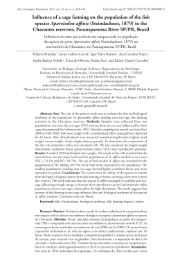
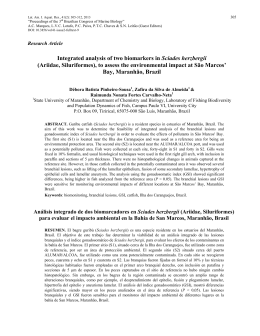
Water quality, toxicity and gill lesions caused by intraperitoneally administered water-bloom crude extract in Brycon cephalus (Günter, 1896; Characidae) from fee-fishing ponds in São Paulo state, Brazil Qualidade de água, toxicidade e lesões branquiais causadas por meio ensaio com extrato seco de floração algal em Brycon cephalus (Günter, 1896; Characidae), coletadas em um pesque-pague, estado de São Paulo, Brasil Eler, MN.1, Campagna, AF.2, Minillo, A.3, Ribeiro, MAP.4 and Espíndola, ELG.1 1 Programa de Pós Graduação em Ecologia e Recursos Naturais, Centro de Ciências Biológicas e da Saúde – CCBS, Departamento de Hidrobiologia – DH, Universidade Federal de São Carlos – UFSCar, Rodovia Washington Luís SP-310, km 235, CP 676 São Carlos, SP, Brazil e-mail: [email protected] 3 Departamento de Engenharia Civil, Universidade Estadual Paulista Júlio de Mesquita Filho – UNESP, Campus de Ilha Solteira, Alameda Bahia, 550 CEP 15385-000, Ilha Solteira, SP, Brazil e-mail: [email protected] 4 Programa de Pós Graduação em Zootecnia, Faculdade de Zootecnia e Engenharia de Alimentos – FZEA, Universidade de São Paulo – USP, Av. Duque de Caxias Norte, 225, Campus da USP, CEP 13635-900, Pirassununga, SP, Brazil e-mail: [email protected] Abstract: Aim: The aim of this study was to evaluate the water quality, cyanobacteria toxins in a water-bloom and acute toxicity of intraperitoneally administered crude extract of water-bloom to Brycon cephalus (Günter, 1896; Characidae), as well as the histological changes of the gill tissues of fish exposed for 26 hours. Methods: B. cephalus is a native Brazilian fish, popular with anglers. Samples were collected from two ponds (fishing and hatchery) from fee-fishing facilities located in the Mogi-Guaçu basin, SP, Brazil. The physical, chemical and biological parameters were measured. The Elisa competitive immunoassay was used to ascertain the microcystin content in intracellular cyanobacteria water-bloom samples from Hatchery Pond (HP), and tissue sections were stained with haematoxylin-eosin and examined using light microscopy. Results: According to the Modified Carlson Index, both ponds were classified as being hyper-eutrophic (>80), with chlorophyll-a of 327.4- 420.6 µg.L–1, TP of 248-340 µg.L–1, and Secchi disc depth of 50-30 cm). The Elisa technique showed evidence of microcystin-LR in the water bloom (242 µg.g–1p.s). There were two species of Anabaena; two of Aphanocapsa; one each of Aphanotece sp.; and Cylindorospermopsis rarciborkii, and four species of Microcystis. In a 26 hours laboratory acute test, doses of 1000, 500, 250 and 125 ug.kg–1 of crud extract caused 100, 80, 60 and 20% mortality in B. cephalus. The LC50 was 229 mg.kg–1 of fish body weight. No histopathological changes were found in the control fish. The results showed that the gills exposed to the different concentrations of crude extract suffered modified morphology, lesions, total fusion of lamellae, dilated sanguineous capillaries of secondary lamellae and displacement of the epithelium. Conclusions: We suggest that fish farms and fee- fishing facilities should be monitored for algae blooms, to minimize the exposure of fish to potent cyanotoxins and the probability of fish kills as a consequence of biologics intoxication. Keywords: cyanobacteria bloom, fee-fishing ponds, toxicity, mycrocistin, gill lesion, Brycon cephalus. Resumo: Objetivo: O objetivo deste trabalho foi avaliar a qualidade de água, a possível toxicidade de um florescimento de Cynobateria via administração intraperitoneal do extrato seco em peixes da espécie Brycon cephalus (Günter, 1896; Characidae), bem como as alterações histológicas nas brânquias dos peixes em teste agudo de 24 horas. Métodos: O B. cephalus é uma espécie da fauna nativa brasileira e com grande aceitação no mercado. As amostras foram coletadas em pesque-pague localizado na cidade de Espírito Santo do Pinhal, bacia hidrográfica do rio Mogi-Guaçu, SP, Brazil. Foram tomadas de um lago de pesca (FP) e de um viveiro de criação de peixes (HP) com intensa floração de Cyanobacteria. Foram medidas as variáveis físicas, químicas e biológicas da água. Aplicou-se o teste Elisa de imunuensaio competitivo para determinação da presença de Microcistina no conteúdo celular das espécies presentes no florescimento. Os materiais para estudos histológicos foram corados com hematoxilina-eosina e analisados em microscopia Acta Limnol. Bras., 2009, vol. 21, no. 1, p. 89-100. Biological Limnology 2 Centro de Recursos Hídricos e Ecologia Aplicada – CRHEA, Departamento de Hidráulica e Saneamento – SHS, Escola de Engenharia de São Carlos – EESC, Universidade de São Paulo – USP, Av. Trabalhador Sãocarlense, 400, CP 292, CEP 13560-970, São Carlos, SP, Brazil e-mail: [email protected], [email protected] 90 Eler, MN. et al. óptica. Resultados: De acordo com o índice de estado trófico(modificado de Carlson) os dois corpos estudados foram considerados super-eutróficos (>80), as concentrações de clorofila foram 327.4 (FP) –420.6 µg.L–1 (HP); fósforo total 248 (FP) –340 µg.L–1(HP); profundidade do disco de Secchi 50 (FP) –30 (HP) cm. Os resultados do teste Elisa mostraram evidências da presença de microcystina_LR nos extrato do florescimento (242 µg.g–1p.s). Identificou-se duas espécies do gênero Anabaena, dois de Aphanocapsa, Aphanotece SP, Cylindorospermopsis rarciborki e duas espécies do gênero Microcystis. Os resultados do ensaio agudo, nas doses de 1000, 500, 250, 125 ug.kg–1 do extrato bruto do florescimento apresentaram as seguintes mortalidades 100, 80, 60, 20% de mortalidade dos peixes testados, não ocorreu mortalidade no controle. A CL50 foi de 229 mg.kg–1. Não ocorreram alterações histológicas nas brânquias dos peixes do tratamento controle. As brânquias dos peixes, expostos nas diferentes concentrações do extrato bruto sofreram modificações morfológicas. Dentre estas modificações estiveram: lesões, fusão total da lamela, dilatação dos capilares sangüíneos da lamela secundária e deslocamento do epitélio. Conclusões: Sugerem-se monitoramentos periódicos das pisciculturas e dos pesque-pague com foco na presença de cyanobacteria. O objetivo do monitoramento em viveiros de piscicultura seria o de evitar perdas econômicas por mortandades e comercialização de peixes comprometidos a fim de evitar biointoxicação. Palavras-chave: floração de cyanobacteria, pesque-pague, toxicicidade, lesões branquiais, Brycon cephalus. 1. Introduction Fee fishing, where an angler pay for the right to fish or for the fish they catch, is becoming increasingly popular in Brazil. Fee-fishing started in marginal areas unsuitable for agriculture or other activities, but it has recently expanded rapidly to occupy former farmland, as landowners realize that ponds can be turned into alternative sources of revenue. (Graziano-Silva et al., 1999; Lopes, 2005) The highest potential demand for fee-fishing in Brazil is in the Southeastern and Southern regions, especially close to large urban centers (Kitamura et al., 1999, 2002). This demand is related to several factors, such as the search for leisure and tourist activities in natural environments, and location and esthetics are often the most important selling points. Many people fish to relax and to escape the hustle and bustle of everyday life (Schuett and Pierskalla, 2007). They want to be assured of catching fish, which is becoming less likely as natural stocks are over-fished in more accessible locations traditionally visited by anglers. Fee-fishing ponds have been expanding in number in the state of São Paulo, along with the growing development of fish farming. According to Venturieri (2002) there are 1000 fee-fishing ponds on a database, out of which 250 locations were visited. The most crucial factor affecting fish health in ponds is the oxygen depletion resulting from blooms of phytoplankton. It is a common cause of mortality to fish and crustaceans in aquaculture ponds (Boyd, 1990), and the maintenance of water quality in fish culture crawls is a basic requirement for the economic success of productive systems. This quality can be affected by several factors, such as the water source and feeding management (Eler et al., 2001). Improving water sanitary conditions in fee-fishing and fish culture not only enhances production efficiency, but protects the health of those eating the fish caught and of the environment in general. Acta Limnol. Bras., 2009, vol. 21, no. 1, p. 89-100. Cyanobacteria are naturally occurring components of the freshwater phytoplankton community, but are frequently associated with eutrophication. Most cyanobacteria blooms have significant adverse effects on water quality. The high eutrophication in the pond promotes the occurrence of increasingly frequent cyanobacterial blooms, which in turn produce toxic substances (cyanotoxins) that are either excreted or released into the water when algal cells decompose. It is well known that some cyanobacteria, such as Microcystis aeruginosa, produce hepatotoxins such as microcystin-LR (MC-LR) (Sivonen and Jones, 1999) and have been linked with poisoning and the death of livestock, wildlife and pets (Chorus, 2001; Codd, 2000). Although suspected as a possible cause or contributing factor of fish kills, there are few direct links between such events and microcystins (Andersen et al., 1993; Rodger et al., 1994, Jewel et al., 2003). High mortality in channel catfish, Ictalurus punctatus (Rafinesque), and production ponds in South Carolina, USA because of a toxic bloom of Anabaena flos-aquae was reported by English, Schwedler and Dyck (1994). Channel catfish kills occurring in ponds in Arkansas have been attributed to dense blooms of Microcystis aeruginosa that produce lethal levels of the algal toxin microcystin (Zimba et al., 2001). Honda et al. (2006), found the presence of Aphanocapsa sp., Microcystis aeruginosa, M. panniformis, Microcystis sp., Phormidium sp. and Radiocistis fernandoi in 14 fee-fishing ponds around the São Paulo metropolitan region, and the authors concluded that the species obtained were toxic (hepatoxins), mailing Microcystis. Moreover, under experimentally essay, Magalhães et al. (2001) and Soares et al. (2004) verified accumulation of microcystins in muscle tissue of Tilapia rendalli, a fish species frequently found in Brazilian fish ponds and reservoirs that experience heavy Microcystis blooms. These authors also detected microcystins in muscle tissue of fish collected from a brackish lagoon. Water quality, toxicity and gill lesions caused by intraperitoneally administered water-bloom crude extract in Brycon cephalus... In São Paulo State, two cases of cyanobacteria blooms with suspected toxicity occurred in 1979. In the first, death occurred among pigeons at the edges of an ornamental lake, and in the second, ducks and teals in a zoo died. Microcystis aeruginosa and Microcystis sp. prevailed in the bloom, and toxicity tests were carried out with mice, which were inoculated with the algae by intraperitoneal injection. The presence of microcystin was verified. These two cases were the first published in Brazil in which the death of animals was proved to be caused by algal toxins (Beyruth, 1992). Eler et al. (2001) first reported fish kills in a fee-fishing pond (Descalvado, SP, Brazil) associated with a cyanobacteria bloom in 1998. The authors highlighted the urgent need to monitor fish farming and fee-fishing operations, and warned that fish from such systems may constitute a new route of human intoxication due to the direct and cumulative action of toxins, such as the microcystins produced by cyanobacteria. Building on these works, the purpose of this paper is to measure cyanobacteria toxins in a water-bloom at a feefishing facility and the acute gill toxicity of intraperitoneally administered water-bloom material to Brycon cephalus (Günter, 1896; Characidae). We chose B. cephalus because it is a native Brazilian fish, popular among anglers, and one the most common fish species found in fee-fishing facilities (Eler et al., 2006; Lopes et al., 2005; Esteves and Ishikawa, 2006). 2. Material and Methods 2.1. Environmental variables The water samples were collected from a fee-fishing pond and hatchery located in the Mogi-Guaçu watershed, in the municipality of Espírito Santo do Pinhal, São Paulo state, Brasil. The pond was stocked with Piaractus mesopotamicus, Oreochromis niloticus, and the hatchery with Piaractus mesopotamicus (pacu). Both ponds were stocked at high fish density (4 kg.m–2). Samples from the two ponds were taken during the intermediary season (November/02) at 2:00 PM. to measure the following variables: pH, conductivity, dissolved oxygen and temperature, measured directly in the ponds with a Horiba U-10 multiline instrument; NO2--N and NO3-N, according to the technique described by Mackereth et al. (1978); NH3--N according to Koroleff (1976); total organic-N according to Golterman et al. (1978); phosphorus according to the American Public Health Association (1980); orthophosphates, total inorganic-P and SiO-2 according to Golterman et al., (1978); total suspended solids according to Wetzel and Likens (1991), chlorophyll-a according to Nusch (1980); and water transparency using a Secchi disk. The Modified Carlson Index (MSI), described in Toledo-Jr. et al. (1983), was applied to the results obtained for phosphorus, chlorophyll-a and water transparency, in order to assess the trophy levels of the 91 environments studied. The cyanobacteria were determined according to the current taxonomy literature descript by Komárek and Anagnostidis (1985, 1986, 1999, 2005), Komárek and Komárková (2002), Sant’Anna and Azevedo (2000), Sant’Anna et al. (2004, 2006). The evaluation of water quality in the ponds was based on the classifications recommended by Brazil’s National Environmental Council (CONAMA 357, 2005) for special class and class 2, which ranks the water in natural environments or aquaculture operations from which aquatic organisms for human consumption are taken. For the qualitative analysis of phytoplankton, 50 liter water samples were concentrated through a plankton net (20 µm mesh) and fixed with formaldehyde. 2.2. Microcystin LR analysis The dense (with scum) water bloom cyanobacteria were collected with a bucket, and concentrated through a plankton net (20 µm mesh) to obtain 5 L of material. The samples were frozen and lyophilized at –30 °C, and the dried material was weighed (10-4 g) and stored at –20 °C. An Elisa competitive immunoassay kit (Envirologix, Inc.®) was used to analyze the microcystin content in intracellular cyanobacteria water bloom samples. Typically, 30 g of a well-mixed freeze-dried algae sample was homogenized and resuspended in 1 mL distilled water and broken (with intermittent freezing and defrosting), and submitted to ultrasonification (50 watt high intensity ultrasonic processor) for 3 minutes at 20 x 100 kHz under cooling on ice. The supernatant was filtered (millipore, ester and cellulose), two analyses were performed, and triplicates were run. The results were expressed as microcystin-LR equivalents (µg.g-1 dry wt freeze-dried cyanobacteria). 2.3. Fish bioassay Brycon cephalus fingerlings (19 ± 5 g) were obtained from the Águas Claras fish farm, Mococa, São Paulo State, Brazil. The fish were placed in a flow-through tank containing 22-25 °C of dechlorinated farm reservoir water and allowed to acclimate for seven days prior to the experiment. The fish were fed with commercial food once daily at a rate of 1% body weight. Feeding was terminated 48 hours prior to toxin exposure and throughout the experiment. The experiment was conducted at the School of Animal Science, University of São Paulo. The water bloom (scum) crude extracted, collected from Hatchery Pond was dissolved in 0.9% NaCl immediately before use. The fish were maintained according to standard husbandry procedures. Using 1 cm, 22 gauge needles, the following doses were injected intraperitoneally along the ventral midline: two fish (controls) received 0 µg.kg–1 extracted (0.5 mL, 0.9% NaCI), and six fish were injected with 125, 250, 500, 1000 mg.kg–1 (dried extracted/kg of body weight). The fish were continuously monitored for Acta Limnol. Bras., 2009, vol. 21, no. 1, p. 89-100. 92 Eler, MN. et al. changes in swimming and other behavior. At the end of the experiment (26 hours), the surviving fish (whether moribund or still active) were killed by a sharp blow to the head. They were then weighed and a complete necropsy was performed (Kotak et al., 1996). 2.4. Histological analysis The histological studies were carried out on the gills of both the fish that survived and died in the experiment. After necropsy, the specimens were fixed in Bouin’s liquid, dehydrated and embedded in paraffin, sliced into 4.0 to 6.0 µm seriated cuts using a Micron HM 340E microtome and steel blade, stained with hematoxylin-eosin (HE), and then studied under an optical microscope. The assessment of the type and degree of alterations was based on Poleksic and Mitrovic-Tutundisic (1994). 3. Results 3.1. Environmental conditions In the fish farming pond, the measurements and collections were carried out under clear skies and intense sunlight, with a light breeze, between 2:00 and 3:00 PM. We observed the presence of algal blooms covering half the pond, concentrated in the direction of the monk. The water coloration was blue-green, with the presence of surface scum, and some points were dark at the surface, which could have been related to the collapse of the algae cells. According to the owner, this had lasted for a month. The algae bloom gave off a strong odor of BHC (CHC). The bloom sample concentrated in the plankton net (20 µm mesh) had the same smell, which is characteristic of cyanobacteria blooms. The Secchi disk depth was 30 cm, which is in line with the presence of these algae blooms. Although we found no fish morality in this pond, according to the owner, previously the fish had been swimming erratically and in circles at the surface with their mouths open. Because of this, these fish had been transferred to two other ponds on the property. In the fee-fishing pond, we did not observe the presence of a similar bloom. Although the water was greenish, we did not note the characteristic smell of cyanobacteria blooms. Table I presents the water quality (physical, chemical and biological characteristics) and Carlson index obtained at each pond. The water temperature was 27.5 °C in the fee-fishing pond (FP) and to 29.2 °C in the hatchery pond (HP). According to the Modified Carlson Index (MCI), both ponds were classified as hyper-eutrophic (>80). Although the concentrations of NT, PT, total, orthophosphate and chlorophyll-a were higher in the HP than the FP. According to the standard Brazilian guidelines (CONAMA Resolution 357/05) for class 2, the concentrations of total phosphorus were up to 10 and 14 times higher in the FP and HP, respectively. The nitrogen/phosphorus ratio (N/P) ranged from 29:1 to 17:1 in the FP and HP, respectively. Acta Limnol. Bras., 2009, vol. 21, no. 1, p. 89-100. The result of NO3--N concentration was up to 17 times higher in the FP. The TSS concentrations ranged from 320 mg.L–1 (FP) to 350 mg.L (HP), and the form of organic matter in suspension was two times higher than in inorganic form in both ponds. The result of the Elisa test showed evidence of microcystin-LR in the water bloom sample from the HP, with a concentration of 242 µg.g–1 (d.w.). The phytoplankton biomass varied from 327.4 (HP) to 254 µg.L–1 (FP) of chlorophyll-a. The cyanobacteria species identified were Anabaena spiroides, Anabaena sp., Aphanocapsa delicatissima, Aphanocapsa sp., Aphanotece sp., Cylindorospermopsis rarciborkii, Microcystis aeruginosa, Microcystis novacekii, Microcystis poniformiis, Microcystis sp., Pseudoanabaena muscicola and Pseudoanabaena sp. 3.2. Acute exposure tests The control fish became slightly lethargic following injection but resumed normal swimming and feeding approximately 60 minutes afterwards. In contrast, the fish injected with algae extract remained lethargic for extended periods, interrupted by quick bursts of frenetic swimming. The depression and lethargy became more pronounced with time and they began to avoid food. The treated fish also lost swimming coordination and buoyancy control, which increased with time and was most severe in the 1000, 500 and 250 mg.kg–1 dose groups. Fish in these groups frequently floated near the surface of the water, either on their side or upright with their heads facing down. Terminally, opercular ventilatory movements gradually decreased in frequency and magnitude, although the fish receiving a dose of 250 mg.kg–1 maintained opercular ventilatory movement throughout the bioassay period. Fish in the 125 mg.kg–1 dose group also exhibited sluggish swimming and loss of buoyancy control, but to a lesser degree. 100% fishes in the 1000 mg.kg–1 dose group died (2, 5, 18 and 24 hours after treatment). 80% fishes in the 500 mg.kg–1 group died (6, 18 and 24 hours after treatment). 60% fishes in the 250 mg.kg–1 group died (all 18 hours after treatment). 10% fishes in the 125 mg.kg–1 group died 24 hours after treatment. Thus, there were four survivors after 26 hours in the treatment groups. There was no mortality in the controls over the same 26 hours period. The necropsy revealed that the fish killed by the toxin had mucous in the operculum and on the skin. There was hemorrhaging near the eyes and between the ventral fins. The gills and livers showed the effects of anemia, with rose coloring, and the livers also showed hyperplasia. In two individuals from the 1000 mg.kg–1 and one from the 500 mg.kg–1 treatment there were dark biliary crystals spread throughout the abdominal cavity. The kidneys were rose colored and the biliary vesicle was dark. Besides this, we observed hemorrhaging in the muscles, along with an Water quality, toxicity and gill lesions caused by intraperitoneally administered water-bloom crude extract in Brycon cephalus... olive green tinge to the flesh of the fish killed by high doses of the algal extracts. 3.3. Gill histopathology The gills of the fish in the control group had normal morphology. We could see the primary filament, well-developed secondary lamellae with pronounced interlamellar spaces, the arrangement of the pillar cells and erithrocytes in the secondary lamellae, as well as the presence of the cartilaginous support (Figure 1a). The gills of the fish exposed to different concentrations of algal extract had altered gill morphology, whose lesions in general consisted of intense proliferation of epithelial cells between the secondary lamellae, resulting in reduced interlamellar spaces and total fusion of some lamellae; dilation of sanguineous capillaries and displacement of the epithelium covering the secondary lamellae (Figure 1b-l). In some cases there were more severe alterations, such as total fusion of all the secondary lamellae, aneurisms, rupture of secondary lamellae and hemorrhaging, character- E a PC SL x D izing second-stage alterations (Figure 1c, d, e, h, j, l). Even at the lowest concentration (125 mg.kg–1) we observed the presence of chloride cells in the secondary lamellae (Figure 1b). The histological analysis showed second-stage alterations in the exposed specimens at all concentrations, which indicates the potential toxicity of MCYT-LR. The proliferation of cells between the secondary lamellae became more accentuated with increasing concentrations, leading to the occurrence of total fusion of the lamellae and reduction of the respiratory area at the intermediate concentrations (250 and 500 mg.kg–1) and highest one (1000 mg.kg–1) (Figure 1e, h, j). However, the severity of the lesions (regarding the degree of change) did not increase with the rising concentrations, since even at the lowest concentration (125 mg.kg–1) there was rupture of the secondary lamellae and hemorrhaging, which are considered to be irreversible alterations (Figure 1d). We also found this at the highest concentration (Figure 1l). These results indicate that cyanotoxins can cause severe effects in fish even at low concentrations. c b R 20 Mm 20 Mm 10 Mm e f g c x SL c h F D 20 Mm 5 Mm TF F SL d R D x F C c 93 D i 20 Mm c TF D 20 Mm j c k SL TF 20 Mm R l TF TF 20 Mm 10 Mm 10 Mm 20 Mm Figure 1. Cross-section of the gill filament of Brycon cephalus. a) Control. Normal development of the primary filament (F) and secondary lamellae (SL), well-defined interlemellar spaces (X); stratified epithelium of the filament (→); pillar cells (PC); erythrocytes (E) and cartilaginous support (C). b-d) 125 mg.kg–1. Dilation of the sanguineous capillaries (D); lifting of the epithelium (→); chloride cells (); aneurism (); rupture of secondary lamellae and hemorrhaging (R). e-f ) 250 mg.kg–1. Proliferation of epithelial cells and total fusion of the all the lamellae (→); reduced interlamellar spaces (X); dilation of sanguineous capillaries (D). g-h) 500 mg.kg–1. Proliferation of epithelial cells and total fusion of all the lamellae (→) and total fusion of all the lamellae (TF); lifting of the epithelium that covers the secondary lamellae (); dilation of capillaries (D). i-l) 1000 mg.kg–1. Proliferation of epithelial cells (→) and total fusion of all the lamellae (TF), making it impossible to see the secondary lamellae; lifting of the epithelium that covers the secondary lamellae (); rupture of lamellae (R). HE coloration. Acta Limnol. Bras., 2009, vol. 21, no. 1, p. 89-100. 94 Eler, MN. et al. 4. Discussion Blooming of Cyanobacteria in fee-fishing ponds and fish farms has become a more frequent phenomenon in the last years in Brazil. In this research, a monitored fee-fishing ponds, pond indicated blooms composed by different species in the hatchery pond. The diversity of Cyanobateria in the blooming could be associated to the combination of physicals and chemicals variables described in this research. For instance, high temperatures (above of 20 °C), lightly alkaline pH (7.9), total phosphorus (248-340 μg.L–1) and N/P ratio (29:1 and 17:1) might have promoted favorable conditions for the growing and maintenance of the bloom in the studied place (Table 1). Such conditions were similar to those described by Kotak et al. (2000) and Zurawell et al. (2005) in their reviews on the main environmental factors associated with the development and maintenance of Cyanobacteria blooms in fresh waters environments. According to many published reports, the high phosphorous load derived from fish food increases the phytoplankton biomass in fee-fishing ponds, indicating an intense eutrophication process. The Modified Carlson Index classifies these ponds as hyper-eutrophic environments. This can render them unfeasible as a consequence of poor water Table 1. Morphometric parameters, major chemical, physical and biological data on the fee-fishing pond and hatchery in November/02, 2:00 PM. Parameters Area (m2) Average depth (cm) Temperature (°C) Dissolved O2 (mg.L–1) Conductivity (µS.cm–1) Secchi disc depth (cm) pH TN (mg.L–1) Total N organics (mg.L–1) NO3-N (µg.L–1) NO2-N(µg.L–1) NH4-N(µg.L–1) Soluble reactive silica (mg.L–1) TP (µg.L–1) Total inorganic-P (µg.L–1) Ortho phosphate (µg.L–1) TN/TP Total suspended solids (mg.L–1) Inorganic. suspended solids (mg.L–1) Organic suspended solids (mg.L–1) Mycrocystin-LR (µg.g–1 – d.w.) Chlorophyll-a (µg.L–1) Modified Carlson Index (MSI) Fishing pond (FP) 500 200 27.5 15.8 11 50 6.9 4.1 4 116 2.2 23.4 7 248 30 57 17 32 10 22 327.4 80.6 Acta Limnol. Bras., 2009, vol. 21, no. 1, p. 89-100. Hatchery (HP) 2000 180 29.2 15.1 12 30 7.4 9.8 9 7 4.3 28 6.9 340 85 139 29 35 11 24 242 420.6 86.8 quality. Many studies have found that the quality of the water used in recreational fisheries is poor and that many problems result from inadequate management. Anoxia is one of the main effects of eutrophication, which causes the appearance of algae blooms and the release of toxic substances by certain species of cyanobacteria (Kitamura et al., 1999, Eler et al., 2003, Mercante et al., 2004). Studies have found that the toxins released by cyanobacteria plays (cause) intoxication and death of fish (Phillips et al., 1985; Rabergh et al., 1991; Rodger et al., 1994; Zimba et al., 2001). Although some species of toxic cyanobacteria can cause harmful effects on fish when ingested, they can be components of the diet of certain species of the Cyclidae and Cyprinidae families (Rabergh et al., 1991; Paerl and Tucker, 1995). A study by Carmichael and Safferman (1992) found that some species of carp and tilapia are tolerant to microcystins, which can indicate a possible route for human exposure to the harmful effects of cyanotoxins from eating contaminated fish. Because of this possible threat, the World Health Organization (WHO) has established an acceptable limit for the “tolerable daily intake” (TDI) of microcystins, which is 0.04 µg.kg–1 of body weight (Chorus and Bartram, 1999). In the present study the cyanobacteria bloom had a mixed composition, with predominance of the genus Microcystis. Besides microcystin-LR, other cyanotoxins may also be present in the lyophilized algal extract. The presence of some species that are potential producers of these substances was confirmed by the identification of the phytoplankton in the samples. For example, we identified Cylindorospermopsis rarciborkii, which are producers of saxitoxins and cylindrospermopsin, and species of the genus Anabena, which produce anatoxin-a. Microcystins are the most commonly studied toxins in vivo and in vitro conditions. These studies have been more focused on acute toxicity. Extensive studies of the LC50 have shown that this is approximately >500 µg.kg for fish and around 50 µg.kg–1 (one-tenth as much) for mammals, for example mice (Zurawell et al., 2005) In the present study, the LC 50 was established at 229.0 mg.kg of body weight over a period of 26 hours. This value can be considered extremely high when compared with others obtained for Cyprinus carpio L. by intraperitoneally administered, between 300 and 550 µg.kg–1 of MC-LR in a seven-day experiment (Andersen et al., 1993) and for Oncorhynchus mykiss, by intraperitoneally administered, between 400 and 1000 µg.kg–1 of MC-LR in 26 hours (Kotak et al., 1996). Doses around 6600 and 1700 µg.kg–1 of MC-LR cause mortality to the same species mentioned before within 10 and 24-48 hours, respectively (Tencalla et al., 1994). However, it appears that this difference in sensitivity can be a consequence of the susceptibility to MC-LR of various fish species (Kotak et al., 1996; Fischer Water quality, toxicity and gill lesions caused by intraperitoneally administered water-bloom crude extract in Brycon cephalus... and Dietrich, 2000), or even can vary within a single species (carp) (Rabergh et al., 1991). Besides the intrinsic differences of each species, the nutritional state and age of the test fish, exposure time and form of administering the toxins must be considered as factors influencing the toxicity. Thus, the tolerance of the test fish in this study in relation to the species studied in other reports can be the result of a lower concentration of MC-LR and the detoxification capacity of the individual fish. According to Kotak et al. (1996), the high tolerance of fish to MC-LR and the low mortality found in their study can be explained by the slow rate of perfusion of the blood in the liver (less than 25% of the capacity of mammals), which can reduce the effectiveness of the initial dose administered. Studies conducted with trout suggest that cyanobacteria blooms can cause cellular degeneration, necroses, edemas, changes in the consistency of the mucous and gill irritation caused by the toxic or physical effects, or a combination of these factors (Rodger et al., 1994). In the present study, the lesions observed in the test organisms were severe even at the lowest concentrations. We found that the intensity of the damage was not related to an increase in the dose injected into the fish. This may have occurred because of the way the cyanotoxin was intraperitoneally administered, the capacity of the fish to metabolize the compounds and the nature of the substance itself (hepatotoxin). Some authors have discussed the effects of cyanotoxins on fish gills in function of the factors mentioned above. Kotak et al. (1996) found that the severity of hepatocellular changes was not due to an increase in the tested concentration of microcystin LR (MC-LR). They also found that even though this is considered a hepatoxin, the gills were also affected, but to a lesser degree than the other organs analyzed (liver and kidneys). The authors attribute these results to the type of exposure in their study (oral). According to Cazenave et al. (2006), C. paleatus exposed to MC-RR presented two responses to the activity of CAT in the liver, first an increase in response to low concentrations and depletion at high levels of exposure. Gupta and Guha (2006), investigating the histological and biochemical effects in Heteropneustes fossilis exposed to lethal and sublethal contaminations by means of intraperitoneally administered, found that the most-affected organ was the liver, followed by the kidneys and gills. The most frequent changes in the gills were proliferation of epithelial cells, edemas, lamellar fusion, hyperplasia, necroses, aneurisms, hypertrophy, epithelial displacement, proliferation of chloride and mucous cells, dilation of the sanguineous capillaries and apoptoses. They pointed out that the gills were less affected than the other organs because of the method of administering the toxin (intraperitoneally administered), which reaches the gills by blood circulation, mediated by filtering in the kidneys. They 95 also considered that the toxic concentrations can bioaccumulate in the liver and only later reach the gills. This observation was also made by Molina et al. (2005) in investigating the biochemical and histological effects on Oreochromis sp. exposed sub-chronically to the microcystin LR. The authors evaluated the toxic effects comparing two types of toxin administration: 1) oral dose mixed with food (14 days); and 2) oral dose ground up with food (21 days). The biochemical and histological changes were more pronounced in the liver and kidneys, but the gills and stomach also showed negative effects. The exposure time was the determining factor of the effects found, since the tissue lesions and levels of acid and alkaline phosphatases increased with the exposure period. The histological changes in the gills only appeared after 21 days of exposure (hyperemia in the central venous sinus). Possibly the gills were less affected because of the method of exposure (oral), allowing the MC-LR to be metabolized first in the liver and gut and only later carried to the gills. In trying to metabolize and eliminate stress agents, fish use physiological and biochemical mechanisms, which can vary from species to species. In the present study we used high concentrations of cyanotoxins, since our objective was to evaluate the mortality from acute exposure. Nevertheless, the specimens showed 75% survival at 125 mg.kg–1 for 26 hours of testing. In contrast, Carbis et al. (1996) noted histopathological changes in the pancreas and gills of carp exposed to 250 µg.kg–1 of MC-LR for seven days, but did not observe any mortality in that period. Molina et al. (2005) found that tilapia exposed to 60 µg MC-LR/fish/ day changed their antioxidant defense system according to the time of exposure. Simultaneously, the level of lipid peroxidation increased significantly in the liver, kidneys and gills of fish exposed for 21 days, particularly when they were fed with cyanobacteria cells ground up in their food. Studies of the morphology of fish gills exposed to various toxic agents have shown two types of response: defensive (inflammatory) and compensatory (cell proliferation, mucous secretion) (Takashima and Hibiya, 1995). Both responses serve to block the entrance of toxic substances and prevent their distribution through the blood, and thus prevent harm caused by direct effects of the agent, with these being considered the first signals of intoxication. However, because the gills are responsible for maintaining the gas exchanges and other important functions, such as the ionic and osmotic balance and the acid-base balance, histopathological changes in the gill structure can trigger respiratory and electrolytic disturbances. The proliferation of cells can lead to a notable reduction in the respiratory surface, caused by the disappearance of the secondary lamellae, which consequently hampers the exchange of gases and other gill functions (Poleksic and Mitrovic-Tutundzic, 1994). Total fusion of the secondary lamellae is induced by Acta Limnol. Bras., 2009, vol. 21, no. 1, p. 89-100. 96 Eler, MN. et al. high doses of toxic agents, and can also be caused by infections by parasites and bacteria (Temmink et al., 1989). Hence, it is possible to consider that in our study the gill changes preceded the death of the fish in the toxicity tests, since the lesions noted probably triggered an imbalance in the physiological and metabolic processes of the fish. The increase in the number of chloride cells in the secondary lamellae at a concentration of 125 mg.kg–1 represents a compensatory response to the decrease in ions. The chloride cells are involved in ionic regulation, taking in Cl-1 and excreting HCO3–. These cells are mainly distributed in the primary filament. The increase in the number of chloride cells suggests a dysfunction in the osmoregulatory equilibrium, which was reflected in changes in the plasma ion concentrations. The increase of chloride cells has been described by other authors (Rajbanshi, 1988; Mazon et al., 2002) as a response to pollution. We only found this cellular change at the lowest concentration, representing the start of intoxication. The changes in the blood vessels (capillary dilation, aneurisms and hemorrhaging) found in the present study demonstrate the toxic potential of cyanotoxins. Aneurisms can hinder the gill function by interfering in the blood composition. If the unfavorable condition persists for extended periods, the aneurisms can rupture, causing hemorrhaging, which may well provoke anemia, besides providing an entryway for pathogens such as bacteria and fungi. These effects eventually kill the fish, diminishing the population density of the animals exposed to the toxic agent (Rand and Petrocelli, 1985; Poleksic and MitrovicTutundzic, 1994). According to Verbost et al. (1989), the entrance of Ca+2 in the gills is facilitated by a calcium channel in the apical membrane of the chloride cells. The extrusion of Ca+2 from the cells is mediated by the high affinity of Ca+2 – ATPase and possibly by the Na+/ Ca+2 exchange (Flik et al., 1993; Verbost et al., 1994). The concentration of intracellular Ca+2 can regulate the extrusion process by means of the Ca+2 – protein bond, through the Ca+2 in the endoplasmic reticulum (Somlyo, 1985) or sequestration by mitochondria (Gunter et al., 1994). Some xenobiotics can inhibit Ca+2 in the gills and cause hypocalcemia. Bury et al. (1996) found that concentrations of 86.4 µg.L–1 (27 mg/dry mass/L) of MC-LR 7820 or MC CYA 43 (oral exposure) produced compounds that inhibit the intake of Ca+2 by the gills and the K+ - PnppASE activity, as well as the transport of Ca+2 in tilapia (Oreocrhomis mossambicus). Studies such as this enable suggesting that the morphological changes in gills exposed to cyanotoxins are preceded by physiological and biochemical changes. Cazenave et al. (2005), in analyzing the detoxification capacity of different organs of Corydoras paleatus orally exposed to MC – RR, at concentrations of 0.5, 2, 5 and 10 µg.L–1 for 24 hours, found that at lower concentrations Acta Limnol. Bras., 2009, vol. 21, no. 1, p. 89-100. there was a reduction in the production of GST in the liver and brain, while this reduction appeared in the gills only at the intermediate concentrations. The authors attributed the different responses observed in the organs analyzed to the entry route, along with each organ’s capacity for biotransformation and bioaccumulation. Both detoxification and the activity of antioxidants were inhibited in the gills, suggesting that these organs are not able to compensate for the presence of MC-RR. They concluded that this toxin is not metabolized in the gills and considered this behavior a natural defense against the toxin, explainable by the fact the gills are constantly in direct contact with the outside environment and thus there is a constant flow of toxic solutions, while the other organs are exposed to a determined quantity of the toxin. According to Egaas et al. (1999), high concentrations of toxins produce a secondary effect due to the inhibition of the substrate. The first step is the biotransformation by enzymes of the cytochrome P450, which can produce a cocktail of different metabolites, competing with the GST substrates for binding sites on the GST enzyme. On the other hand, the inhibition can be characterized by a covalent modification of the enzyme, leading to an irreversible loss of activity. Alternatively, low activity might be caused by a decrease in the synthesis of the GST protein at the molecular level. Considering the importance of GST in detoxifying microcystins, this inhibition implies a risk to the fish’s health. Further, if the fish are not able to eliminate the toxins, they will be susceptible to bioaccumulation, which can trigger cell damage, physiological alterations and behavioral changes, among other effects. Jos et al. (2005) and Prieto et al. (2006) evaluated the effects of MC-LR on the oxidative stress in the liver, kidneys and gills of Nile tilapia (Orecochromis sp.) exposed to 60 µg/fish/day under oral administration (mixed and ground with food), over periods of 14 and 21 days. They concluded that the toxicity depends on the exposure time and type of administration of the toxin, since the stress levels were higher when given with ground-up food and during the 21 day exposure period. Some authors have evaluated intraperitoneally administered. exposure to microcystins in fish with emphasis on histological aspects. All these studies show necrosis and degeneration of the liver, kidneys and also indicate damage to the gill epithelium (Carbis et al., 1997; Kotak et al., 1996, Snyder et al., 2002; Molina et al., 2005; Gupta and Guha, 2006). The changes noted in the gills examined by these authors corroborate the present study’s findings: hyperplasia, fusion of secondary lamellae, epithelial dislocation, and production of chloride cells, aneurisms and dilation of the sanguineous capillaries. Similar alterations have been observed when fish are exposed to oral doses of microcystins (Fischer and Dietrich, 2000). Water quality, toxicity and gill lesions caused by intraperitoneally administered water-bloom crude extract in Brycon cephalus... Some studies have shown that the gills and epithelium function as a barrier to the transport of MCs, and that probably the main route of absorption is by ingestion. The production of toxins by cyanobacteria can have great ecological importance, due to the potential impacts ranging from the level of the nanoplankton peak to the upper trophic levels (Chorus, 2001). Associated with this problem is the risk of bioaccumulation of cyanotoxins in organisms higher up the aquatic food chain (Vasconcelos, 1999; Buynder et al., 2001; Sipiä et al., 2001; Magalhães et al., 2003). 5. Conclusions Nutrient concentrations in the hatchery pond were relatively high, which contributed to proliferation of a toxic cyanobacterial bloom leading to the degradation of the water quality. Crude extract of cyanobacteria from fishing and hatchery ponds was toxic, and promoted expressive alterations in the gill’s tissue. These findings indicate another way that toxins could be manifested. It is important to point out the changes in gill’s tissue during the fish bioassay, and mainly when massive fish kill took place at the farm following the Cyanobacterial bloom event. We found a pattern of dominance among the cyanobacteria, with high densities of representatives Microcystis genus. We also identified the presence of microcystin-LR (hepatotoxins) in the sample. This finding indicates the potential risk to human health of consuming fish contaminated with cyanotoxins, as well as the urgent need to control the eutrofication of fishing pond and hatchery ponds to minimize the occurrence and proliferation of toxic cyanobacteria blooms. Acknowledgements We are grateful to Fundação de Amparo a Pesquisa do Estado de São Paulo (FAPESP) for the financial support and fellowship during the course of this research. We thank the Águas Claras fish farm for fish donation for the bioassay, and the Animal Science Institute (FZEA/USP) and Center of Hydrological Resource and Applied Ecology (CRHEA/ EESC/USP) for the support in the laboratory assays. To Doctor Liany Printes Biehl for the manuscript review. References American Public Health Association – APHA. American water works association, water pollution control federation: standard methods for the examination of water and wastewater. 19 ed. Washington: Byrd Prepress Springfield, 1985. 1134p. ANDERSEN, RJ., LUU, HA., CHEN, DZX., HOLMES, CFB., KENT, ML., BLANC, MLE., TAYLOR, FJR. and WILLIAMS, DE. Chemical and biological evidence links microcystins to salmon netpen liver disease. Toxicon, 1993, vol. 31, no. 10, p. 1315–1323. 97 BEYRUTH, Z., SANT´ANNA, CL., AZEVEDO, MTP., CARVALHO, MC. and PEREIRA, HASL. Toxic Algae in freshwaters of São Paulo State. In CORDEIRO-MARINO, M., SANT’ANNA, CL., PAIVA, MTA., TOMITA, N.; PLASTINO, S. (Org.). Algae and environment: a general approach. São Paulo: Sociedade Brasileira de Ficologia, 1992. p. 53-63. BOYD, CE. Water quality in ponds for aquaculture. Auburn: Auburn University Press, 1990. 482p. BURY, NR., FLIK, G., EDDY, FB. and CODD, GA. The effects of cyanobacteria and the cyanobacterial toxin microcystin-lr on ca2+ transport and na+/k+-atpase in tilapia gills. J. Exp. Biol. 1996, vol. 199, no. 6, p. 1319-1326. CARBIS, CR., RAWLIN, GT., GRANT, P., MITCHELL, GF., ANDERSON, JW. and McCAULEY, I. A study of feral carp, Cyprinus carpio L., exposed to Microcystis aeruginosa at Lake Mokoan, Australia, and possible implications for fish health. J Fish Dis. 1997, vol. 20, no. 1, p. 81-91 CARBIS, CR., MITCHELL, GF., ANDERSON, JW. and McCAULEY, I. The effects of microcystins on the serum biochemistry of carp, Cyprinus carpio L., when the toxins are administered by gavage, immersion and intraperitoneal routes. J. Fish Dis. 1996, vol. 19, p. 151-159. CARMICHAEL, WW. Status Report on Planktonic Cyanobacteria (Blue-Green Algae) and their Toxins. EPA / 600 / R-92 / 079, Jun/92. CAZENAVE, J., WUNDERLIN, DA., BISTONI, MA., AME, MV, KRAUSE, E., PFLUGMACHER, S. and WIEGAND, C. Uptake, tissue distribution and accumulation of microcystin-RR in Corydoras paleatus, Jenynsia multidentata and Odontesthes bonariensis field and laboratory study. Aquat. Toxicol. 2005, vol. 75, no. 2, p. 178-190. CAZENAVE, J., BISTONI, MA., PESCE, SF. and WUNDERLIN, DA. Differential detoxification and antioxidant response in diverse organs of Corydoras paleatus experimentally exposed to microcystin-RR. Aquat. Toxicol. 2006, vol. 76, p. 1-12. CODD, GA. Cyanobacterial toxins, the perception of water quality, and the prioristisation of eutrophication control. Ecolog. Engineer. 2000, vol. 16, p. 51-60. Conselho Nacional do Meio Ambiente – CONAMA. Resolução CONAMA 357 de 17 de Março de 2005. Diário Oficial da República Federativa do Brasil, Brasília, 2005. p. 58-63. CHORUS, I. and BARTRAM, J. (Eds.). Toxic Cyanobacteria in water: a guide to their public health consequences, monitoring and management. New York: E & FN Spon, Inc., 1999. 416p. CHORUS, I. Introduction: Cyanotoxins - research for environment safety and human health. In ______. Cyanotoxins. Germany: Springer, 2001. p. 1-4. EGAAS, E., SANDVIK, M., FJELD, E., KALLQVIST, T., GOKSOYR, A. and SVENSEN, A. Some effects of the fungicide propiconazole on cytochrome P450 and glutathione S-transferase in brown trout (Salmo trutta). Comp. Biochem. Physiol. 1999, vol. 122, no. 3, p. 337-344 ELER, MN., CECCARELLI, PS., BUFON, AGM. and ESPINDOLA, ELG. Mortandade de peixes (matrinxã, Brycon Acta Limnol. Bras., 2009, vol. 21, no. 1, p. 89-100. 98 Eler, MN. et al. cephalus, e pacu, Piaractus mesopotamicus) associada a uma floração de cianobactérias em pesque-pague, município de Descalvado, Estado de São Paulo, Brasil. Bol. Téc. CEPTA, 2001, vol. 14, p. 35-45. ELER, MN., PARESCHI, DC., ESPÍNDOLA, ELG. and BARBOSA, DS. Ocorrência de Rotífera e sua relação com o estado trófico da água em pesque-pague na bacia do rio MogiGuaçu - SP. Bol. Téc. do CEPTA, 2003, vol. 16, p. 41-56. ELER, MN., MINILLO, A., RIBEIRO, MAR., DEBEM, THC., SANTOS, NP., BLAZQUEZ, FJH., MILANI, TJ., ESPINDOLA, ELG. and NOGUEIRA, AAM. Cianobactérias, toxinas e bioensaios ecotoxicológicos nos pesque-pague na Bacia Hidrográfica do Rio Mogi-Guaçu. In ELER, MN. and ESPINDOLA, ELG. (Org.). Avaliação dos impactos de pesque-pague: uma análise da atividade na bacia hidrogáfica do rio Mogi-guaçu. São Carlos: Rima Editora, 2006. p. 145-162. ENGLISH, WR., SCHWEDLER, TE. and DYCK, LA. Aphanizomenon flos-aquae, a toxic blue green alga in commercial channel catfish, Ictalurus punctatus, ponds: a case history. J. Appl. Aquacult. 1993, vol. 3, no. 5, p. 195-209. ESTEVES, KE. and ISHIKAWA, CM. Características gerais e práticas de manejo em pesqueiros da Região Metropolitana de São Paulo. In ESTEVES, KE. and SANT’ANNA, CL. (Org.). Pesqueiros sob uma visão integrada de meio ambiente, saúde pública e manejo: um estudo na Região Metropolitana de São Paulo. São Carlos: RiMA, 2006. p. 1-18. FISCHER, WJ. and DIETRICH, DR. Pathological and biochemical characterization of microcystin-induced hepatopancreas and kidney damage in carp (Cyprinus carpio). Toxicol. Appl. Pharmacol. 2000, vol. 164, no. 1, p. 73‑81. FLIK, G. and VERBOST, PM. Calcium transport in fish gills and intestines. J. Exp. Biol. 1993, vol. 184, p. 17–29. GRAZIANO-SILVA, JDA., DEL GROSSI, ME. and LAURENTI, AC. The evolution of rural nonagricultural activities and intersectorial linkages in Brazil. Campinas: IE/UNICAMP, 1999. p. 75. Text for Discussion. GOLTERMAN, HL., CLYMO, RS. and OHNSTAD, MAM. Methods for physical and chemical analysis of fresh water. Oxford: Blackwell Scientific Publications, 1978. 213p. GUPTA, US. and GUHA, S. Microcystin toxicity in a freshwater fish, Heteropneustes fossilis (Bloch). Current Science, 2006, vol. 91, no. 9, p. 1261-1271. HONDA, RY., MERCANTE, CTJ., VIEIRA, JMS., ESTEVES, KE., CABIANCA, MAA. and AZEVEDO, MTP. Cianotoxinas em pesqueiros da Região Metropolitana de São Paulo. In ESTEVES, KE. and SANT’ANNA, CL. (Org.). Pesqueiros sob uma visão integrada de meio ambiente, saúde pública e manejo: um estudo na Região Metropolitana de São Paulo. São Carlos: RiMa, 2006, p. 105-120. JEWEL, MAS., AFFAN, MA. and KHAN, S. Fish mortality due to cyanobacterial bloom in an aquaculture pond in Bangladesh. Pakistan J. Biol. Sc. 2003, vol. 6, no. 12, p. 1046‑1050. JOS, AMG., PICHARDO, SS., PRIETO, AIO., KUHN, GR., VAZQUEZ, CMC., MORENO IMN. and CAMEÁN, AMF. Acta Limnol. Bras., 2009, vol. 21, no. 1, p. 89-100. Toxic cyanobacterial cells containing microcystins induce oxidative stress in exposed tilapia fish (Oreochromis Sp.) under laboratory conditions. Aquatic Toxicol. 2005, vol. 72, no. 3, p. 261-271. KITAMURA, PC., LOPES, RB., CASTRO Jr., FG. and QUEIROZ, JF. Avaliação ambiental e econômica dos lagos de pesca esportiva na Bacia Do Rio Piracicaba. Bol. Indústria Animal, 1999, vol. 56, n. 1, p. 95-107. KITAMURA, PC., QUEIROZ, JF., LOPES, RB., CASTRO Jr., FG. and BOYD, CE. Environmental and economic assessment of fee-fishing in São Paulo, Brazil. J. Appl. Aquaculture, 2002, vol. 12, n. 4, p. 23-41. KOMÁREK, J. and AGNOSTIDES, K. Modern approach to the classification systems of cyanophites. II Chroococcales. Arch. Hydrobiol. Suppl. 1985, vol. 73, no. 2, p. 157-226. KOMÁREK, J. and AGNOSTIDES, K. Modern aproach to the classification systems of cyanophites. IV Nostocales. Arch. Hydrobiol. Suppl. 1989, vol. 82, no. 3, p. 247-345. KOMÁREK, J. and ANAGNOSTIDIS, K. Cyanoprocaryota. 1.Teil Chroococcales. In ETTL, H., GÄRTNER, G., HEYNIG, H. and MÖLLENHAUER, D. (Eds.). Süsswasserflora von Mitteleuropa. Sttutgart: Fisher Verlag, 1999, vol. 19, n. 1, p. 1-548. KOMÁREK, J. and KOMÁRKOVA, J. Review of the European Microcystis-Morphospecies (Cyanoprokaryotes) from nature. Czech Phycology, Olomouc. 2002, vol. 2, p. 1-24. KOMÁREK, J. and ANAGNOSTIDIS, K. Süßwasserflora von Mitteleuropa: Band 19 (2): Cyanoprokaryota: Part 2: Oscillatoriales. [S.L.]: Elsevier, 2005. 759p. KOROLEFF, F. Determination of nutrients. In GRASSHOFF, K. (Ed.). Methods of seawater analysis. Weinhein: Verlag Chemie, 1976. p. 117-181. KOTAK, BG., SEMALULU, S., FRITZ, DL., PREPAS, EE., HRUDEY, SE. and COPPOCK, RW. Hepatic and renal pathology of intraperitoneally administered Microcystin-Lr in rainbow trout (Oncorhynchus Mykiss). Toxicon, 1996, vol. 34, no. 5, p. 517-525. KOTAK, BG., LAM, AKY., PREPAS, EE. and HRUDEY, SE. Role of chemical and physical variables in regulating microcystin-LR concentration in phytoplankton of eutrophic lakes. Can. J. Fish. Aquat. Sci. 2000, vol. 57, no. 8, p. 1584‑1593. LOPES, RB., LANDELL-FILHO, LC. and DIAS, CTS. Feefishing operation evaluation at northwest São Paulo State, Brazil. Sci. Agric. 2005, vol. 62, no. 6, p. 590-596. MacKERETH, FGH., HERON, J. and TALLING, JF. Water analysis some revised methods for Limnologists. Freshwater Biological Association Scientific Publication, 36; Kendall: Titus Wilson & Son Ltd, 1978. 117p. MAGALHÃES, VF., SOARES, RM. and AZEVEDO, SMFO. Microcystins contamination in fish from the Jacarepaguá Lagoon (Rio de Janeiro, Brazil): ecological implication and human health risk. Toxicon. 2001, vol. 39, no. 7, p. 1077‑1085. Water quality, toxicity and gill lesions caused by intraperitoneally administered water-bloom crude extract in Brycon cephalus... MAGALHÃES, VF., MARINHO, MM., DOMINGOS, P., OLIVEIRA, AC., COSTA, SO., AZEVEDO, LO. and AZEVEDO, SMFO. Microcystins (Cyanobacteria Hepatotoxins) bioaccumulation in fish and crustaceans from Sepetiba Bay (Brazil - RJ). Toxicon. 2003, vol. 42, no. 3, p. 289-295. MERCANTE, CTJ., CABIANCA, MA., SILVA, D., COSTA, SV. and ESTEVES, KE. Water quality in fee-fishing ponds located in the metropolitan region of São Paulo city, Brazil: an analysis of the eutrophication process. Acta Limnol. Bras. 2004, vol. 16, no. 1, p. 95-102. MOLINA, R., MORENO, I., PICHARDO, S., JOS, A., MOYANO, R., MONTERDE, JG. and CAMEÁN, A. Acid and alkaline phosphatases activities and pathological changes induced in Tilapia fish (Oreochromis sp.) exposed subchronically to microcystins from toxic cyanobacterial blooms under laboratory conditions. Toxicon. 2005, vol. 46, no. 7, p. 725-735. NUSCH, EA. Comparison of different methods for Chlorophyll and phaeopigment determination. Arch. Hydrobiol. Beih. Ergebn. Limnol. 1980, vol. 14, p. 14-36. PAERL, HW. and TUCKER, CS. Ecology of Blue-Green Algae in Aquaculture Ponds. J.World Aqu. Soc. 1995, vol. 26, no. 1, p. 109-131. PHILLIPS, MJ., ROBERTS, RJ., STEWART, JA. and CODD, G. The toxicity of the Cyanobacterium Microcystis aeruginosa to rainbow trout Salmo gairdneri Richardson. J. Fis. Dis. 1995, vol. 8, p. 339-344. POLEKSIC, V. and MITROVIC-TUTUNDIZIC, V. Fish gills as monitor of sublethal and chronic effects of pollution. In MULLER, R. and LLOYD, R. (Eds.). Sublethal and chronic effects of pollutants on freshwater fish. United Nations: Fishing News Books, 1994. p. 339‑352. PRIETO, AI., JOS, A., PICHARDO, S., MORENO, I. and CAMEAN, AM. Differential oxidative stress responses to microcystins LR and RR in intraperitoneally exposed tilapia fish (Oreochromis sp.). Aquatic Toxicol. 2006, vol. 77, no. 3, p. 314‑321. RABERG, CMI., BYLUND, G. and ERIKSSON, JE. Histological effects of MC-LR, a cyclic Peptide Toxin from the Cyanobacterium (Blue-Green Alga) Microcystis aeruginosa, on common carp (Cyprinus carpio L.). Aquat. Toxicol. 1991, vol. 20, p. 131-146. RAJBANSHI, VK. and GUPTA, AK. Alterations in the architecture of gill surface produced by water-borne copper in Hepteroneusters fossilis (Bloch). Acta Hydrochem. Hydrobiol. 1988, vol. 16, p. 325–332. RAND, GM. and PETROCELLI, SR. Fundamentals of aquatic toxicology: methods and application. London: Hemisphere Publishing Corporation, 1985. RODGER, HD., TURNBULL, T., EDWARDS, C. and CODD, G. Cyanobacterial (Blue-Green Algal) Bloom Associated Patology in Brow Trout, Salmo trutta L. in Loch Leven, Scothand. J. Fis. Dis. 1994, vol. 17, p. 177-181. SANT`ANNA, CL. and AZEVEDO, MTP. Contribution to the knowledge of potentially toxic Cyanobacteria from Brazil. Nova Hedwigia. 2000, vol. 71, no. 3-4, p. 359-385. 99 SANT’ANNA, CL., AZEVEDO, MT., SENNA, PAC., KOMÁREK, J. and KOMÁRKOVÁ, J. Planktic Cyanobacteria from São Paulo State, Brazil: Chroococcales. Rev. Brasil. Bot. 2004, vol. 27, no. 2, p. 213-227. SANT`ANNA, CL., AZEVEDO, MTP., AGUJARO, LF., CARVALHO, MC., SOUZA, RC. and CARVALHO, LR. Manual ilustrado para identificação e contagem de cianobactérias planctônicas de águas continentais brasileiras. 1 ed. Rio de Janeiro: Interciência, 2006. 58p. SIPIÄ, V., KANKAANPÄÄ, H., LAHTI, K., CARMICHAEL, WW. and MERILUOTO, J. Detection of Nodularin in flounders and cod from the Baltic Sea. Environ Toxicol. 2001, vol. 16, p. 121-126. SCHUETT, MA., PIERSKALLA, CD. Managing for desired experiences and site preferences: the case of fee-fishing anglers. Environ Manage. 2007, vol. 39, no. 2, p. 164-177. SIVONEN, K. and JONES, G. Cyanobacterial toxins. In CHORUS, I. and BARTRAM, J. (Ed.). Toxic cyanobacteria in water: a guide to their public health consequences, monitoring and management. London: E & FN Spon, 1999. p. 41-111. SIVONEN, K. Cyanobacterial toxins and toxin production. Phycol. 1996, vol. 35, no. 6, p. 12-24. SOARES, RM., MAGALHÃES, VF. and AZEVEDO, SMFO. Accumulation and depuration of microcystins (cianobactéria hepatotoxins) in Tilapia rendalli (Cichlidae) under laboratory conditions. Aquat. Toxicol. 2004, vol. 70, no. 1, p. 1‑10. TAKASHIMA, F. and HIBIYA, T. (Ed.). An atlas of fish histology: normal and pathological features. 2 ed. Tokio: Kodanska, 1995. 195p. TEMMINK, JHM., FIELD, JA., VAN HAASTRECHT, JC. and MERKELBACH, RCM. Acute and subacute toxicity of bark tannins in carp (Cyprinus carpio). Water Res. 1989, vol. 23, no. 3, p. 341-344. TENCALLA, FG., DIETRICH, DR. and SCHLATTER, C. Toxicity of Microcystis aeruginosa peptide toxin to yearling rainbow trout (Oncorhynchus mykiss). Aquatic Toxicol. 1994, vol. 30, no. 3, p. 215-224. TOLEDO, AP., TALARICO, M., CHINEZ, SJ. and AGUDO, EG. A aplicação de modelos simplificados para a avaliação do processo da eutrofização em lagos e reservatórios tropicais. In Anais do Congresso Brasileiro de Engenharia Sanitaria e Ambiental, 12., Novembro 20-25, 1983. Camburiu: CETESB, 1983. p. 1-34. VASCONCELOS, VM. Cyanobacterial toxins in Portugal: effects on aquatic animals and risk for human health. Braz. J.Med. Biol. Res. 1999, vol. 32, no. 3, p. 249-254. VENTURIERI, R. Pesque-pague no Estado de São Paulo: vetor desenvolvimento da psicultura e opção de turismo e lazer. São Paulo: Eco Associação para Estudos do Ambiente, 2002. 168p. VERBOST, PM., VAN ROOD, J., FLIK, G., LOCK, RAC., BONGA, SEW. The movement of cadmium through freshwater Trout branchial epithelium and its interference Acta Limnol. Bras., 2009, vol. 21, no. 1, p. 89-100. 100 Eler, MN. et al. with calcium transport. J. exp. Biol. 1989, vol. 145, no. 1, p. 185-197. WETZEL, RG. and LIKENS, GE. Limnological analyses. 2 ed. New York: Springer-Verlag, 1991. 391p. XIE, L., XIE, P., OZAWAB, K., HONMA, T., YOKOYAMA, A. and PARK, H. Dynamics of Microcystins-LR and RR in the Phytoplanktivorous Silver Carp in a Sub-chronic Toxicity Experiment. Environ. Pollution, 2004, vol. 127, no. 3, p. 431-439. ZIMBA, PV., KHOO, L., GAUNT, PS., BRITTAIN, S. and CARMICHAEL, WW. Confirmation of catfish, Ictalurus Acta Limnol. Bras., 2009, vol. 21, no. 1, p. 89-100. punctatus (Rafinesque), Mortality from Microcystis aeruginosa toxins. J. Fish. Dis. 2001, vol. 24, no. 1, p. 41-47. ZURAWELL, RW., CHEN, H., BURKE, JM. and PREPAS, EE. Hepatotoxic cyanobacteria: a review of the biological importance of microcystins in freshwater environments. J. Toxicol. Environ. Health B Crit Rev. 2005, vol. 8, no. 1, p. 1-37 Received: 14 October 2008 Accepted: 27 February 2009
Download