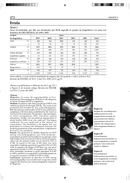
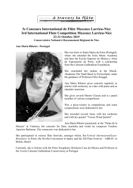
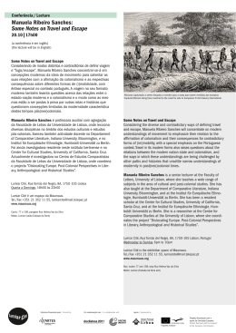
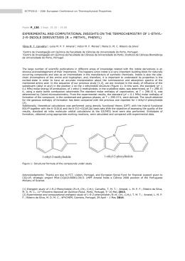
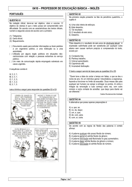
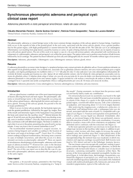
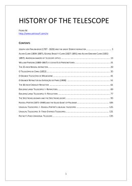
VISUALIZATION
FOR
CYTOCHEMICAL
INTRAMANDIBULAR
GLAND OF OLD MAJOR
(HYM.: FORMICIDADE).
COMPLEX
IN
GOLGI
OF
WORKERS OF Zacyptocerccs pusilh
E.M. RIBEIRO* e F.H. CAETANO*.
* Instituto de Biociencias - Department0 de Biologia, UNESP - Campus de Rio Claro (S.P.). Av. 24 A no
1515, CEP: 13.506-900 - Brasil. E-mail: [email protected]
The study on the intramandibular gland in Formicidaes is rare: few works was realized
about this subject. The intramandibular gland locates inside of the mandible, for that reason it is
denominated of intramandibular, and was described in some species of bees and ants. It possesses
unknown function until the moment. By hystological analysis it can be divided in 3 parts: one that it
is constituted by cells of oval format, that form the epithelium of the gland; another that it is
constituted by the secretory cells, properly said and the last formed by the cells fulfilling, the cells of
the fat body. Its secretion product passes for the outside, through intracuticulars ducts existent in the
mandible surface (1, 2 and 3). Already in the ultra-structure images, of the intramandibular gland of
Zacryptocerus pusihs, the cells form a cord that crosses the lumen of the mandible. It cytoplasm
contains several vesicles of lipids, rough and smooth endoplasmic reticulum, this last one in larger
amount, mitochondrias and other organelles. The membrane presents several invaginations in the face
gone back to the lumen (4). For the visualization of the Golgi complex we used the technique of the
iodeto of zinc and tetroxide osmium (ZIO) according to Reineck & Walther, 1978. The material was
inclueded in mixture of Epon-Araldite (l- 1) and cut with diamond razors, contrasted with uralina
acetate and lead citrate and examined in MET Philips CM 100. Through the use of that technique
synaptic vesicles, lysosomes, mitochondrias,
endoplasmic reticulum and Golgi complex can be
marked (5). The results was a demarcation in the Golgi complex which it showed very defined with a
very strong eletron-density, so much in the secretory cells as in the epithelial cell (fig. 1,3 and 4). All
the cisterns of Golgi had a reaction of the same intensity. The Golgi didn’t present any specific
location inside of the cells (fig. l), there was also demarcation in mitochondrias (fig.1) and in some
vesicles spread by the cytoplasm (fig. 2). It didn’t have any positive reaction inside of the nucleus (fig
1). It had smaller demarcations in the cytoplasm of some cells, so much in the fat body cells as in the
epithelial cells, even so, the largest amount of that demarcation type happened in the cytoplasm of the
fat body cells (rig 2), that demarcation type can indicates lysosomes.
References:
1. NEDEL, J.O. Morphologie und Physiologie der Mandibeldrtise eneiger Bienen-Arten (Apidae). Z. Morph. 6kel.
Tiere49 1960 p. 139 - 183.
2. TOLEDO, L.F.A. Histo-anatomia de glandulas de Atta sexdens rubropilosu Fore1 (Hymenoptera) - Arq. Inst Biol., 34,
1967 p. 321- 329.
3. RIBEIRO, E.M. e CAETANO, F.H. The mandibles glands in ants Zmyptocerus pusillus workes (Myrmicinae) - XVII
Congreso National de Entomologia. Santiago. 1995 p. Il.
4. RIBEIRO, E.M. e CAETANO, F.H. Ultra-estrutura da glandula intramandibular de soldados velhos de Zacryptocerus
pusillus workes (Formicidae: Myrmicinae) - Anais do VI International Pest Ant Symposium & XIII Encontro
de Mirmecologia - Mirmecologia Tropical. Ilheus. 1997 p. 40.
5. COSTA-LEONARDO, A.M. Glandulas intramandibulares em abelhas sociais. Ci&nc. Cult. 30, 1978, p 835 - 838.
6. BECHIMOL, M. e SOUZA, W. de Tritrichomonas fotus: Cytochemical Visualization of the Endoplasmic Reticulum Golgi Complex and lipids. Experimental Parasitology 59 1985 p. 51 - 5X.
I
/
Legendes:
Figura 1 - Secretory cells (CS) with marcation in the Golgi complex (G) and in some mitochondrias
(M). Observe the Golgi complex of the epithelial cell (arrow). C = cuticle; V = vacuole; N = nucleus.
Figura 2 - Observe the many smoller marcation (arrows) in the cytoplasm of the fat body cells (FB)
and epithelial (E). L = lipids.
Figura 3 - Figure showing many Golgi complex (G).
Figura 4 - Detail of the Golgi complex (G). Observe that all the cisterns of the Golgi possess the
same intensity of reaction.
Download