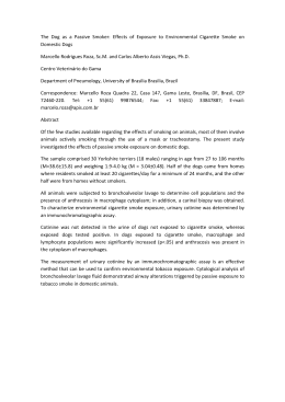
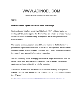
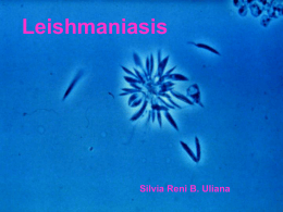
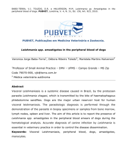
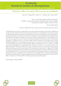
Veterinary Parasitology 204 (2014) 169–176 Contents lists available at ScienceDirect Veterinary Parasitology journal homepage: www.elsevier.com/locate/vetpar Antibody responses induced by Leish-Tec® , an A2-based vaccine for visceral leishmaniasis, in a heterogeneous canine population Miriam C. de Souza Testasicca a , Mariana Silva dos Santos a , Leopoldo Marques Machado a , Angela Vieira Serufo a , Daniel Doro a , Daniel Avelar b , Ana Maria Leonardi Tibúrcio c , Christiane de Freitas Abrantes d , George Luiz Lins Machado-Coelho e , Gabriel Grimaldi Jr. f , Ricardo Tostes Gazzinelli g,h,i , Ana Paula Fernandes a,∗ a Faculdade de Farmácia, Universidade Federal de Minas Gerais, Belo Horizonte, MG, Brazil Fundação Ezequiel Dias, Belo Horizonte, MG, Brazil Hertape Calier Saude Animal, Juatuba, MG, Brazil d Labtest Diagnostica, Belo Horizonte, MG, Brazil e Departamento de Ciências Médicas, Universidade Federal de Ouro Preto, Ouro Preto, MG, Brazil f Centro de Pesquisa Gonçalo Moniz/Fiocruz, Salvador, BA, Brazil g Departamento de Bioquímica e Imunologia, Instituto de Ciências Biológicas, Universidade Federal de Minas Gerais, Belo Horizonte, MG, Brazil h Centro de Pesquisas René Rachou/Fiocruz, Belo Horizonte, MG, Brazil i University of Massachusetts Medical School, Worcester, MA, USA b c a r t i c l e i n f o Article history: Received 6 March 2014 Received in revised form 15 April 2014 Accepted 22 April 2014 Keywords: Visceral leishmaniasis Vaccine Leish-Tec® Dogs A2 antigen Antibodies a b s t r a c t Zoonotic visceral leishmaniasis (VL) is a widespread disease, and dogs are the main reservoirs for human parasite transmission. Hence, development of an effective vaccine that prevents disease and reduces the transmission of VL is required. As euthanasia of seropositive dogs is recommended in Brazil for VL epidemiological control, to include anti-VL canine vaccines as a mass control measure it is necessary to characterize the humoral responses induced by vaccination and if they interfere with the reactivity of vaccinated dogs in serological diagnostic tests. Leish-Tec® is an amastigote-specific A2 recombinant protein vaccine against canine visceral leishmaniasis (CVL) that is commercially available in Brazil. Here, we tested the immunogenicity of Leish-Tec® in a heterogeneous dog population by measuring A2-specific antibody responses. Healthy dogs (n = 140) of various breeds were allocated to two groups: one group received Leish-Tec® (n = 70), and the other group received a placebo (n = 70). Anti-A2 or anti-Leishmania promastigote antigen (LPA) antibody levels were measured by ELISA in serum samples collected before and after vaccination. An immunochromatographic test (DPP) based on the recombinant K28 antigen was also used for serodiagnosis of CVL. Vaccinated animals, except one, remained seronegative for anti-LPA total IgG and anti-K28 antibodies. Conversely, seropositivity for anti-A2 total IgG antibodies was found in 98% of animals after vaccination. This value decreased to 81.13% at 6 months before rising again (98%), after the vaccination boost. Anti-A2 IgG2 and IgG1 titers ∗ Corresponding author at: Department of Clinical and Toxicological Analyses, Faculty of Pharmacy, Federal University of Minas Gerais, Av. Antônio Carlos, 6627, Belo Horizonte, MG CEP 31270 901, Brazil. Tel.: +55 31 34096884; fax: +55 31 34096985. E-mail addresses: [email protected], [email protected] (A.P. Fernandes). http://dx.doi.org/10.1016/j.vetpar.2014.04.025 0304-4017/© 2014 Elsevier B.V. All rights reserved. 170 M.C. de Souza Testasicca et al. / Veterinary Parasitology 204 (2014) 169–176 were also increased in vaccinated animals relative to control animals. These data indicate that Leish-Tec® is immunogenic for dogs of different genetic backgrounds and that humoral responses induced by vaccination can be detected by A2-ELISA, but do not interfere with the LPA-ELISA and DPP diagnostic tests for CVL. © 2014 Elsevier B.V. All rights reserved. 1. Introduction Leishmaniasis is a vector-borne disease caused by protozoan parasites of the genus Leishmania that primarily affects the poorest regions of 98 countries worldwide. Among the two million new human infections, 200,000–400,000 are cases of life-threatening visceral leishmaniasis (VL), culminating in approximately 20,000–30,000 deaths every year (Alvar et al., 2012). Children and immunocompromised individuals are especially affected, but adults with active disease are frequently found in foci of recent parasite introduction (World Health Organization, 2010). Concerns about treatment failure for VL are exacerbated by the emergence of antimicrobial resistant parasite strains and because of co-infection with HIV (Chappuis et al., 2007). Anthroponotic VL occurs in regions where Leishmania (Leishmania) donovani infections are endemic (the Indian sub-continent), whereas zoonotic VL occurs in areas of transmission of L. (L.) infantum (southern Europe, North Africa, and West and Central Asia) or L. (L.) infantum chagasi (Latin America) (World Health Organization, 2010). Domestic dogs (Canis familiaris) are considered the main peridomestic and domestic reservoirs of parasites for L. (L.) infantum and L. (L.) infantum chagasi (Alvar et al., 2004; Grimaldi and Tesh, 1993). As canine VL constitutes the major source of parasites for transmission of human VL, prophylactic control programs in Brazil have focused on euthanasia of seropositive dogs (Lacerda, 1994). These euthanasia campaigns are expensive and labor-intensive, and their relatively poor performance is aggravated in practice by the absence of a good marker for infectious status in dogs (Courtenay et al., 2002). Hence, both the efficacy and acceptability of this control strategy is increasingly being challenged (Chappuis et al., 2007; Romero and Boelaert, 2010; Tesh, 1995). Treatment of VL-infected dogs is not a feasible control strategy, because drug-cured dogs often relapse and remain a source of parasites for the sand fly vector (Alvar et al., 1994). As some L. (L.) infantum chagasi-infected dogs spontaneously recover and maintain protective immunity to re-infections (Grimaldi et al., 2012b), vaccination seems to be a feasible strategy to protect dogs (Chappuis et al., 2007), and also to reduce the incidence of human cases. In this context, vaccination may be adopted concomitantly with other current measures of control especially if vaccines do not induce seroconversion in diagnostic tests. In particular, the amastigote-specific A2 antigen, administered in recombinant protein, DNA, or viral vectors vaccine formulations or in transfected parasites, induces partial protection against L. (L.) donovani, L. (L.) amazonensis, and L. (L.) infantum chagasi infections in mice (Coelho et al., 2003; Ghosh et al., 2001a, b; Mizbani et al., 2009; Resende et al., 2008; Zanin et al., 2007). A2 is also one of the few candidate antigens that have been tested in beagle dogs against experimental challenge and provided significant protection (Fernandes et al., 2008). Based on these findings, a vaccine formulation consisting of saponin and the recombinant protein A2, named Leish-Tec® , was licensed in Brazil. Recently, LeishTec® has been also tested in dogs living in endemic areas for CVL (Fernandes et al., 2014). That study has demonstrated that 92.9% of dogs vaccinated with Leish-Tec® remained clinically health. Moreover, as tested by xenodiagnosis, vaccinated dogs were less infectious (5.4%) than control animals (36.6%) to the phlebotomine vector. In addition being safe for the target animal (Schetters and Gravendyck, 2006), the A2 vaccine also needs to be immunogenic to heterogeneous dog populations. Moreover, given the importance of serodiagnostic tests in veterinary routine practice and epidemiological control, CVL vaccines should ideally not induce seroconversion in routine serological tests. We therefore performed a study to evaluate the specificity, levels, and duration of antibody responses elicited by Leish-Tec® vaccination in out-bred dogs. 2. Materials and methods 2.1. Ethics statement Approval to perform this study was obtained from the institutional review board, Comitê de Ética em Experimentação Animal (CETEA) of Universidade Federal de Minas Gerais (UFMG), under certificate 020/2009. UFMG adheres to the standards outlined by relevant national (CONCEA; Brazilian Government Council for Control of Animal Experimentation) and international guidelines for care and use of animals in research. 2.2. Animals For this study, 315 dogs were initially evaluated; the dogs were of different breeds, living in 12 open kennels, and located in eight different regions of the metropolitan region of the city of Belo Horizonte in Minas Gerais, Brazil, which is an endemic area for VL. Kennels were selected after veterinarian inspection based on their routine sanitary and care and welfare practices with animals. Animals were treated for helmintic infections and had an updated routine vaccination program for rabies, parvovirus, and other common canine infections. The screening process for the animals was performed during three consecutive months (from November 2009 to January 2010) and included laboratory tests for VL diagnosis and analysis of clinical parameters, as described below. Animals presenting suggestive symptoms M.C. de Souza Testasicca et al. / Veterinary Parasitology 204 (2014) 169–176 and serological positive results in at least one evaluation were excluded. Insecticide-impregnated collars were adopted during the screening process and vaccination, but removed thereafter. These procedures were adopted in an attempt to minimize the contact of the selected dogs with Leishmania antigens prior to or during vaccination, which could subsequently interfere with the analysis of vaccine seroconversion. No other control measures, besides welfare maintenance of the animals and application of standard sanitization procedures, were adopted during the follow-up period. 2.3. Clinical and laboratorial evaluation of dogs Biochemical and hematological analyses were performed monthly for animal screening or every 3 months after vaccination for clinical evaluation of the animals, as previously described. DNA was extracted from peripheral blood samples collected at 6 and 14 months postvaccination employing the Genomic Prep Mini Spin Kit (G&E Healthcare). PCR for detection of Leishmania kDNA in DNA extracted from blood samples was performed as previously reported (Vitale et al., 2004). The occurrence of vaccination adverse effects such as fever, pain, or edema at the site of vaccine administration was actively monitored by veterinarians for 72 h after administration of each vaccine or placebo dose. After this period, all owners were instructed to contact veterinarians if the animals manifested any side effects. 2.4. Vaccination protocol Leish-Tec® consists of 100 g of A2 recombinant protein (rA2) and 250 g of saponin (Riedel). In all, 140 animals without clinical signs of disease and with negative serological tests for VL and anti-A2 antibodies in three consecutive monthly evaluations were selected for the study. These 140 dogs were allocated into two groups: in the first group, 70 dogs were vaccinated with Leish-Tec® subcutaneously on days 0, 21, and 42. In the second group, 70 dogs received a placebo containing saponin only, following the same dose schedule. Each kennel harbored half-placebo and halfvaccinated dogs. Each animal received a microchip for identification. Vaccine and placebo doses were coded and undistinguishable in appearance from each other. Doses were administered blindly by veterinarians, and kennel owners were not aware and could not distinguish between vaccinated and placebo-treated animals. Animals were maintained in open kennels and followed for a period of 14 months (from February 2010 to April 2011). A boosting vaccine dose was administered 12 months (February 2011) after the priming vaccination doses. 2.5. Serological analysis for the diagnosis of canine visceral leishmaniasis The presence of anti-Leishmania promastigote antibodies (LPA) was investigated monthly, before and after vaccination, by indirect immunofluorescence (IFI). Titers higher than 1:40 dilutions were considered positive. We also used the Leishmania promastigote antigen (LPA)-based 171 ELISA kits provided by the manufacturer (Bio-Manguinhos, Oswaldo Cruz Foundation, Rio de Janeiro, Brazil). For ELISA, the manufacturer provides LPA-sensitized ELISA plates and recommends a serum sample dilution of 1:100. All assays were performed using negative and positive control sera. In ELISA, cut-off values were determined from the average optical density plus two standard deviations obtained from negative sera, following the manufacturer’s recommendation. Anti-parasite antibodies were also investigated through the Dual Path Platform test (DPP) for CVL, according to manufacturer’s instructions (Bio-Manguinhos). DPP is a rapid immunochromatographic test for the detection of CVL based on the recombinant antigens K39 and K26 (Grimaldi et al., 2012a; Marcondes et al., 2011), and it has been recently approved for screening of CVL seropositive dogs by the Brazilian Public Health authorities. Sera samples were randomly selected for testing with DPP from the placebo-treated (n = 27) and Leish-Tec® vaccinated groups (n = 55, 1 month after vaccination; n = 51, 6 months after vaccination; and n = 45, 14 months after vaccination). 2.6. ELISA for anti-A2 total IgG and specific IgG1 and IgG2 antibodies Anti-A2-specific IgG, IgG1, and IgG2 subtype levels were measured in pre-immune sera and at 1, 6, and 14 months after vaccination. ELISA to measure anti-A2 serological responses was performed using purified A2 recombinant protein, prepared as previously described (Carvalho et al., 2002; Fernandes et al., 2008). Briefly, 96-well plates (Costar® , Corning, NY, USA) were sensitized with rA2 (250 ng/well) in coating buffer (0.015 M Na2 CO3 , 0.03 M NaHCO3 , pH 9.6) overnight at 4 ◦ C. Plates were blocked with 2% non-fatty milk PBS at 25 ◦ C for 1 h, washed again and then 100-L sera samples, diluted at 1:100 were added and incubated for 90 min at 37 ◦ C. After three washes with PBS/0.05% Tween 20, peroxidaselabeled antibodies specific to canine IgG, IgG1, or IgG2 isotypes (Sigma, MO, USA) diluted at 1:25,000, 1:15,000, or 1:35,000, respectively, were added for 1 h at 37 ◦ C. The plates were washed six times and incubated with H2 O2 and o-phenylenediamine (Sigma, MO, USA) for reaction development. Reactions were stopped by the addition of 50 L of 2 N H2 SO4 (Merck). Optical densities were determined at 492 nm by using a Versa Max ELISA reader (Molecular Devices, CA, USA). All samples were analyzed in a blinded manner. Cut off values were calculated for each plate from the average plus two standard deviations of absorbance values obtained from these 5 negative sera. 2.7. Immunoblot analysis Purified A2 recombinant protein (1 g) was submitted to sodium dodecyl sulfate-polyacrylamide gel electrophoresis (SDS-PAGE) and transferred onto PVDF membranes (Resende et al., 2008). Membranes were blocked with 5% non-fatty milk PBS and incubated overnight at 4 ◦ C prior to incubation with serum samples from immunized or placebo dogs. Peroxidaseconjugated anti-canine IgG (Sigma, MO, USA) diluted at 1:10000 was used as a secondary antibody. Reaction 172 M.C. de Souza Testasicca et al. / Veterinary Parasitology 204 (2014) 169–176 products were visualized by the addition of chloronaphthol, diaminobenzidine, (Sigma, MO, USA) and H2 O2 . The anti-A2 monoclonal antibody (1:3000) was used as a positive control. 2.8. Statistical analysis Sample size estimation was performed assuming a criterion of 5% for significance and a power of 95%. The estimations considered a probability of infection of 10% (or 8.7% of seroconversion to A2) among the non-protected dogs, based on the incidence of CVL in the endemic area of Belo Horizonte, a seroconversion of 70% among vaccinated dogs, and a loss rate of 50% during the study. Based on these parameters, 25 dogs in each group would be required for statistical significance. For statistical purposes, absorbance values were transformed into logarithmic values {log10 [(100) (x) +1]}. Differences among absorbance values were proportional to the mean values (Snedecor and Cochran, 1989; Stolt et al., 2003). The levels of IgG1, IgG2, and total IgG antibodies were submitted to splitplot ANOVA analysis, considering the groups (vaccinated or placebo) as the between-subject factor (wholeplots) and the time of observation (months) as the within-subject factor (subplot). The interaction factors were compared with the residual for subplots within wholeplots (Stolt et al., 2003). Two-tailed P < 0.05 was considered significant. 3. Results At the end of the screening period, 14% of the animals initially evaluated were suspected of having VL, based on the presence of suggestive symptoms; 17% were seropositive for total IgG antibodies against promastigote parasite antigens; 22% presented other intercurrences that resulted in their exclusion; and 2% died. From the remaining 45%, 140 dogs were selected for use in this trial. Vaccinated animals were monitored for adverse effects, such as fever, pain, abscesses, ulceration or edema. No significant reactions that could be associated with vaccine administration were detected in the animals (data not shown), indicating that the doses were well tolerated by the animals. Anti-A2 total IgG antibody levels were measured to test vaccine immunogenicity. Data are presented in Fig. 1 as a percentage of seropositive animals. A high proportion (98%) of Leish-Tec® vaccinated animals displayed positive anti-A2 antibody titers 1 month after vaccination. This percentage was reduced 6 months after vaccination (81.13%). Nevertheless, the percentage of animals positive for antiA2 antibodies returned to the initial values (98%), 2 months after the boosting vaccine dose (14 months after the priming vaccination). The reduction in anti-A2 IgG antibody levels in vaccinated animals is also shown in Fig. 2. It should be noted that although the levels of anti-A2 antibodies were decreased at 6 months, 81.13% of the animals still presented antibody levels above the established cut-off values. In contrast, most placebo-treated animals (76.71%) remained negative for anti-A2 antibodies. The highest rate of positive animals for anti-A2 antibodies among the placebo-treated Fig. 1. Percentage of placebo-treated and Leish-Tec® -vaccinated dogs presenting anti-Leishmania promastigote antigens (LPA) or anti-A2 antibodies, as detected by ELISA. Positive sera were defined based on optical density (OD) readings above cut-off values. Cut-off values were determined for each ELISA plate from the average optical density plus two standard deviations obtained from 5 negative sera. Each sample was tested in duplicate. dogs (23.81%) was detected on the six months measurement. ELISA was performed for animals from both placebotreated and Leish-Tec® vaccinated groups to detect the presence of anti-Leishmania promastigote (anti-LPA) antibodies, with the goal of testing whether vaccination induced cross-reactive anti-promastigote antibodies. In contrast to the anti-A2 total antibodies, animals from the vaccinated or placebo groups remained negative for total IgG antibodies against LPA throughout the experiment (Fig. 1), except for two animals (2.85%) in placebo and one (1.42%) in the vaccinated group. Similar results were observed using the DPP test in a selected panel of serum samples from placebo-treated and Leish-Tec® -vaccinated animals (Table 1), i.e., animals that were positive in ELISA were also positive in DPP. Serum samples from the animals were also examined by ELISA to assess anti-A2 IgG2- and IgG1-specific antibody responses. As shown in Fig. 2, levels of anti-A2 IgG2 were significantly higher in Leish-Tec® -vaccinated animals than in control placebo animals. Anti-A2 IgG2 levels were decreased in vaccinated dogs at 6 months, but increased Table 1 Detection of seropositive dogs using the rapid DPP rK28 fusion protein chromatographic immunoassay for testing selected sera from vaccinated and control dogs. Number of positive samples/total samples tested Months after the prime-vaccination Groups Vaccinated Placebo a 1 0/55 0/27 6 0/51 2/27 14a 0/45 2/27 Two months after administration of the boost vaccine dose. M.C. de Souza Testasicca et al. / Veterinary Parasitology 204 (2014) 169–176 173 Fig. 2. Anti-A2 specific serological responses in animals vaccinated with Leish-Tec® . Levels of anti-A2 IgG, IgG1, and IgG2 antibodies in pre-immune sera and at different time points after vaccination were detected by ELISA. Cut-off values were determined for each ELISA plate from the average optical density plus two standard deviations obtained from 5 negative sera, for each IgG isotypes. Each bar represents the average ± standard deviation for the optical density in each group. Asterisk indicates that differences between placebo and vaccinated animals are statistically significant, as tested using split plot variance analysis. again to higher levels2 months after the last booster vaccination. Leish-Tec® -vaccinated dogs displayed increased levels of IgG1 antibodies at 1 month after the priming vaccination dose. However, IgG1 levels were decreased at 6 months and remained lower after the booster vaccine dose. Anti-A2 IgG2 levels were higher than anti-A2 IgG1 levels, leading to IgG2/IgG1 ratios higher than 1, in vaccinated dogs (data not shown). Low levels of anti-A2 IgG2 and IgG1 antibodies were observed in placebo animals, which remained unchanged during the experiment. To confirm the specificity of antibody responses from Leish-Tec® immunized animals, an immunoblot analysis with recombinant A2 protein was performed. The sera of vaccinated animals recognized a protein of 52 kDa, which was also detected by an anti-A2 monoclonal antibody (Fig. 3). Only two animals in the control placebo group presented seroconversion of anti-LPA ELISA associated with symptoms characteristic of VL, including lymphadenopathy, periocular alopecia, onychogryphosis, and weight loss. These animals also presented altered leukocyte counts and albumin/globulin ratio (data not shown). One vaccinated animal presented positive results in the anti-LPA ELISA and Leishmania kDNA-specific PCR, but showed no signs of disease at the end of the experiment. Saponin is commonly used as adjuvant in canine VL vaccines for induction of type I immune responses (Marcondes et al., 2011, 2013; Vandepapeliere et al., 2008), although it may cause adverse reactions (Fernandes et al., 2014; Marcondes et al., 2011; Vandepapeliere et al., 2008). In this study, Leish-Tec® doses were well tolerated by the animals, as previously reported (Fernandes et al., 2008, 2014). Antibodies were selected as markers of vaccine immunogenicity or exposure to infection because they are easily measured and useful in the veterinary routine practice. Moreover, CVL vaccines in Brazil are licensed only for individual prophylaxis and it is recommended by manufactures and public health authorities that only seronegative animals should receive these vaccines. A2 is actually one of the few amastigote-specific antigens tested 4. Discussion Here, we tested whether Leish-Tec® is immunogenic for a heterogeneous dog population. It is worth mentioning that this study did not aim to test vaccine efficacy, since a different experimental design (placebo controlled, doubleblinded field trial), including a larger sample of animals and complete randomization would be required to approach efficacy with statistical significance. Fig. 3. Immunoblot analysis of anti-A2 IgG antibodies in sera of placebo and Leish-Tec® vaccinated animals. The recombinant A2 protein (1 g/well) was transferred from SDS-PAGE to nitrocellulose membranes and incubated with 1:200 sera dilutions of selected sera samples. Lane 1: protein molecular weight marker; Lane 2: anti-A2 monoclonal antibody; Lane 3: sera of placebo animal; Lane 4: sera from Leish-Tec® vaccinated animal 174 M.C. de Souza Testasicca et al. / Veterinary Parasitology 204 (2014) 169–176 for both, diagnosis or vaccine development (Carvalho et al., 2002; Fernandes et al., 2008). However, there is no commercially available test for CVL diagnosis using this antigen. Considering that A2 is a component of Leish-Tec® , an assay to detect anti-A2 antibodies could be easily applied to evaluate immunization with this vaccine in the field. This assay may provide evidence that a given animal is responsive or not to vaccination, and could thus be indicative of LeishTec® immunogenicity. In agreement, in this study, 98% of the vaccinated dogs had anti-A2 antibody levels higher than the cut-off for negative results, indicating that LeishTec® is immunogenic even in a highly heterogeneous dog population. The A2-based ELISA also allowed the follow up anti-A2 humoral responses, after priming and boost vaccination. Since the levels anti-A2 antibodies decreased over time, as expected for a protein based-vaccine, an annual boosting vaccination may be required for recall of immune responses. In agreement, anti-A2 antibody levels increased after administration of a single Leish-Tec® dose, as detected 2 months after the boosting vaccine dose (14 months after the priming vaccination). Another important finding was the low incidence of cross-reactive anti-LPA antibodies, after vaccination, which has implications not only for individual diagnosis of CVL in veterinarian routine practice, but also for epidemiological surveys of CVL seropositive dogs. The current serological tests (ELISA, IFI, and DPP) for VL adopted by the Brazilian public health authorities are based on promastigote antigens (Grimaldi et al., 2012a; Marcondes et al., 2013). The induction of cross-reacting antibodies to LPA or K28, the antigens used in the official serodiagnostic tests, maybe problematic for dog owners, veterinarians, and public health authorities when managing animals that have received vaccines that may induce seroconversion for these survey diagnostic tests. Due to its possible impact in the current control measures, this aspect has been recently addressed for the two commercially available CVL vaccines in Brazil, Leishmune® (Marcondes et al., 2011, 2013) and Leish-Tec® (Fernandes et al., 2014). Since A2 is an amastigote antigen, LeishTec® is not expected to induce cross-reactive antibodies against the promastigote antigens used in diagnostic tests. However, in one study it has been argued that LeishTec® induced seroconversion in 30.9% of the vaccinated animals, as tested by LPA-ELISA (Fernandes et al., 2014), contrasting with our findings. These discrepant results may be well attributed to differences in the methodological procedures and criteria adopted for screening and inclusion of dogs in each study. Although in both studies only LPA-ELISA seronegative dogs were included, it is well known that the current CVL diagnosis tests present important limitations regarding sensitivity and specificity. Consequently, a single point serological evaluation may not be sufficiently rigorous for screening dogs that afterwards will be tested for cross-reactivity after vaccination. Therefore, in the present study, only dogs negative in serological tests for three consecutive months were included, to minimize the interference of a previous infection or contact with parasites, in serological responses, after vaccination. Continuous exposure to parasite antigens of animals living in endemic areas during vaccination may also result in cross-reactive humoral responses. In this context, to unequivocally demonstrate that vaccination does not induce seroconvertion, in addition to the rigorous screening procedures, in this study, insecticide-impregnated collars were maintained in animals during the screening process and vaccination. These procedures were adopted attempting to minimize the contact of the selected dogs with Leishmania antigens prior or during vaccination, which could subsequently interfere with the analysis of vaccine seroconversion. Ideally, maintenance of animals in thin-screened, instead of opened kennels should have been adopted, aiming to improve the control measures. However, it seems that the lack of this additional preventive strategy did not interfere significantly in seroconvertion rates, since only low cross-LPA reactive antibody levels were detected in Leish-Tec® 326 vaccinated animals when tested either by ELISA or DPP. The anti-A2 IgG2 antibody response was similar to that observed in our previous study, in beagle dogs, as antibody titers were high and induced shortly after the priming vaccination scheme, and an increased proportion of animals remained seropositive in subsequent measurements. However, levels of anti-A2 IgG1 were also higher, contrasting with previous results (Fernandes et al., 2008). This discrepancy may be attributed to the differences in the studied populations. Nevertheless, the anti-A2 IgG2/IgG1 ratios were higher than 1 at all-time points after vaccination (data not shown), suggesting the predominance of Th1 immune responses in Leish-Tec® vaccinated dogs. Furthermore, as previously reported, the levels of anti-A2 IgG1 and IgG2a antibodies and the IgG2a/IgG1 ratio may be indicative of protective immunity elicited by this vaccine (Fernandes et al., 2008). In other studies, levels of IgG2 antibodies raised after immunization with different vaccine formulations were also correlated with increased IFN- production and Th1 cellular immunity (Carrillo and Moreno, 2009; Moreno et al., 2012; Vitoriano-Souza et al., 2012). Moreover, in vaccinated animals, IgG2 antibodies have been associated with opsonization and complement fixation that may promote clearance of extracellular amastigotes and inhibition of their infectivity, thus contributing to the resistance profile induced by vaccination (Bourdoiseau et al., 2009). However, infected dogs frequently present a mixed Th1/Th2 cytokine profile. Although IgG1 levels have been linked to symptomatic CVL, the immune responses in an infected animal exposed to a variety of antigens are complex and may have hampered the establishment of correlations between a single marker and either resistance or susceptibility to infection (Carrillo and Moreno, 2009; Moreno et al., 2012; Reis et al., 2010). Additionally, insufficient data from vaccination studies are available to establish such correlations. The different nature of antigens and adjuvants used in anti-CVL vaccines, modifying the induced immune responses further hinders comparisons (Day, 2007; Fernandes et al., 2008; Marcondes et al., 2013; Roatt et al., 2012; VitorianoSouza et al., 2012). Therefore, the contribution of antibody isotypes to CVL and vaccine immunogenicity and M.C. de Souza Testasicca et al. / Veterinary Parasitology 204 (2014) 169–176 protective responses still requires further investigation. Hence, anti-A2 IgG2 or IgG1 antibodies may not be directly associated with protective responses, but should rather be taken as markers of immunogenicity for vaccination with Leish-Tec® . In conclusion, the findings reported here indicate that vaccination with Leish-Tec® (i) is immunogenic and safe for a heterogeneous dog population, (ii) does not induce antibodies that are detectable by routine CVL immunodiagnostic tests, (iii) requires administration of an annual vaccine boost, and (iv) can be monitored by A2-based ELISA. Conflict of interest statement The authors declare no conflict of interest. Acknowledgements This work was supported by the National Institute of Science and Technology for Vaccines/Conselho Nacional de Desenvolvimento e Tecnológico (CNPq), Fundação de Amparo à Pesquisa do Estado de Minas Gerais (FAPEMIG; Grant APQ-00304-10, Programa de Pesquisas para o SUS) and Hertape Calier Saúde Animal. The funders had no role in study design, data collection and analysis, decision to publish, or preparation of the manuscript. References Alvar, J., Velez, I.D., Bern, C., Herrero, M., Desjeux, P., Cano, J., Jannin, J., Den, B.M., 2012. Leishmaniasis worldwide and global estimates of its incidence. PLoS ONE 7, e35671. Alvar, J., Canavate, C., Molina, R., Moreno, J., Nieto, J., 2004. Canine leishmaniasis. Adv. Parasitol. 57, 1–88. Alvar, J., Molina, R., San Andres, M., Tesouro, M., Nieto, J., Vitutia, M., Gonzalez, F., San Andres, M.D., Boggio, J., Rodriguez, F., et al., 1994. Canine leishmaniasis: clinical, parasitological and entomological follow-up after chemotherapy. Ann. Trop. Med. Parasitol. 88, 371–378. Bourdoiseau, G., Hugnet, C., Goncalves, R.B., Vezilier, F., Petit-Didier, E., Papierok, G., Lemesre, J.L., 2009. Effective humoral and cellular immunoprotective responses in Li ESAp-MDP vaccinated protected dogs. Vet. Immunol. Immunopathol. 128, 71–78. Carrillo, E., Moreno, J., 2009. Cytokine profiles in canine visceral leishmaniasis. Vet. Immunol. Immunopathol. 128, 67–70. Carvalho, F.A., Charest, H., Tavares, C.A., Matlashewski, G., Valente, E.P., Rabello, A., Gazzinelli, R.T., Fernandes, A.P., 2002. Diagnosis of American visceral leishmaniasis in humans and dogs using the recombinant Leishmania donovani A2 antigen. Diagn. Microbiol. Infect. Dis. 43, 289–295. Chappuis, F., Sundar, S., Hailu, A., Ghalib, H., Rijal, S., Peeling, R.W., Alvar, J., Boelaert, M., 2007. Visceral leishmaniasis: what are the needs for diagnosis, treatment and control? Nat. Rev. Microbiol. 5, 873–882. Coelho, E.A., Tavares, C.A., Carvalho, F.A., Chaves, K.F., Teixeira, K.N., Rodrigues, R.C., Charest, H., Matlashewski, G., Gazzinelli, R.T., Fernandes, A.P., 2003. Immune responses induced by the Leishmania (Leishmania) donovani A2 antigen, but not by the LACK antigen, are protective against experimental Leishmania (Leishmania) amazonensis infection. Infect. Immun. 71, 3988–3994. Courtenay, O., Quinnell, R.J., Garcez, L.M., Shaw, J.J., Dye, C., 2002. Infectiousness in a cohort of Brazilian dogs: why culling fails to control visceral leishmaniasis in areas of high transmission. J. Infect. Dis. 186, 1314–1320. Day, M.J., 2007. Immunoglobulin G subclass distribution in canine leishmaniosis: a review and analysis of pitfalls in interpretation. Vet. Parasitol. 147, 2–8. Fernandes, A.P., Costa, M.M., Coelho, E.A., Michalick, M.S., de Freitas, E., Melo, M.N., Luiz Tafuri, W., Resende Dde, M., Hermont, V., de Freitas Abrantes, C., Gazzinelli, R.T., 2008. Protective immunity against challenge with Leishmania (Leishmania) chagasi in beagle dogs vaccinated with recombinant A2 protein. Vaccine 26, 5888–5895. 175 Fernandes, C.B., Junior, J.T., de Jesus, C., Souza, B.M., Larangeira, D.F., Fraga, D.B., Tavares Veras, P.S., Barrouin-Melo, S.M., 2014. Comparison of two commercial vaccines against visceral leishmaniasis in dogs from endemic areas: IgG, and subclasses, parasitism, and parasite transmission by xenodiagnosis. Vaccine 5, 1287–1295. Ghosh, A., Labrecque, S., Matlashewski, G., 2001a. Protection against Leishmania donovani infection by DNA vaccination: increased DNA vaccination efficiency through inhibiting the cellular p53 response. Vaccine 19, 3169–3178. Ghosh, A., Zhang, W.W., Matlashewski, G., 2001b. Immunization with A2 protein results in a mixed Th1/Th2 and a humoral response which protects mice against Leishmania donovani infections. Vaccine 20, 59–66. Grimaldi Jr., G., Tesh, R.B., 1993. Leishmaniases of the new world: current concepts and implications for future research. Clin. Microbiol. Rev. 6, 230–250. Grimaldi Jr., G., Teva, A., Ferreira, A.L., dos Santos, C.B., Pinto, I., de-Azevedo, C.T., Falqueto, A., 2012a. Evaluation of a novel chromatographic immunoassay based on Dual-Path Platform technology (DPP(R) CVL rapid test) for the serodiagnosis of canine visceral leishmaniasis. Trans. R. Soc. Trop. Med. Hyg. 106, 54–59. Grimaldi Jr., G., Teva, A., Santos, C.B., Ferreira, A.L., Falqueto, A., 2012b. The effect of removing potentially infectious dogs on the numbers of canine Leishmania infantum infections in an endemic area with high transmission rates. Am. J. Trop. Med. Hyg. 86, 966–971. Lacerda, M.M., 1994. The Brazilian Leishmaniasis control program. Mem. Inst. Oswaldo Cruz 89, 489–495. Marcondes, M., de Lima, V.M., de Araujo Mde, F., Hiramoto, R.M., Tolezano, J.E., Vieira, R.F., Biondo, A.W., 2013. Longitudinal analysis of serological tests officially adopted by the Brazilian Ministry of Health for the diagnosis of canine visceral leishmaniasis in dogs vaccinated with Leishmune(R). Vet. Parasitol. 197, 649–652. Marcondes, M., Ikeda, F.A., Vieira, R.F., Day, M.J., Lima, V.M., Rossi, C.N., Perri, S.H., Biondo, A.W., 2011. Temporal IgG subclasses response in dogs following vaccination against Leishmania with Leishmune(R). Vet. Parasitol. 181, 153–159. Mizbani, A., Taheri, T., Zahedifard, F., Taslimi, Y., Azizi, H., Azadmanesh, K., Papadopoulou, B., Rafati, S., 2009. Recombinant Leishmania tarentolae expressing the A2 virulence gene as a novel candidate vaccine against visceral leishmaniasis. Vaccine 28, 53–62. Moreno, J., Vouldoukis, I., Martin, V., McGahie, D., Cuisinier, A.M., Gueguen, S., 2012. Use of a LiESP/QA-21 vaccine (CaniLeish) stimulates an appropriate Th1-dominated cell-mediated immune response in dogs. PLoS Negl. Trop. Dis. 6, e1683. Reis, A.B., Giunchetti, R.C., Carrillo, E., Martins-Filho, O.A., Moreno, J., 2010. Immunity to Leishmania and the rational search for vaccines against canine leishmaniasis. Trends Parasitol. 26, 341–349. Resende, D.M., Caetano, B.C., Dutra, M.S., Penido, M.L., de Freitas Abrantes, C., Verly, R.M., Resende, J.M., Pilo-Veloso, D., Rezende, S.A., BrunaRomero, O., Fernandes, A.P., Gazzinelli, R.T., 2008. Epitope mapping and protective immunity elicited by adenovirus expressing the Leishmania amastigote specific A2 antigen: correlation with IFN-gamma and cytolytic activity by CD8+ T cells. Vaccine 26, 4585–4593. Roatt, B.M., Aguiar-Soares, R.D., Vitoriano-Souza, J., Coura-Vital, W., Braga, S.L., Correa-Oliveira, R., Martins-Filho, O.A., Teixeira-Carvalho, A., de Lana, M., Figueiredo Gontijo, N., Marques, M.J., Giunchetti, R.C., Reis, A.B., 2012. Performance of LBSap vaccine after intradermal challenge with L. infantum and saliva of Lu. longipalpis: immunogenicity and parasitological evaluation. PLoS ONE 7, e49780. Romero, G.A., Boelaert, M., 2010. Control of visceral leishmaniasis in Latin America – a systematic review. PLoS Negl. Trop. Dis. 4, e584. Schetters, T.P., Gravendyck, M., 2006. Regulations and procedures in parasite vaccine development. Parasitology 133 (Suppl.), S189–S195. Snedecor, G.W., Cochran, W.G., 1989. Statistical methods. Iowa State University Press, Ames, Iowa, pp. 503. Stolt, A., Sasnauskas, K., Koskela, P., Lehtinen, M., Dillner, J., 2003. Seroepidemiology of the human polyomaviruses. J. Gen. Virol. 84, 1499–1504. Tesh, R.B., 1995. Control of zoonotic visceral leishmaniasis: is it time to change strategies? Am. J. Trop. Med. Hyg. 52, 287–292. Vandepapeliere, P., Horsmans, Y., Moris, P., Van Mechelen, M., Janssens, M., Koutsoukos, M., Van Belle, P., Clement, F., Hanon, E., Wettendorff, M., Garcon, N., Leroux-Roels, G., 2008. Vaccine adjuvant systems containing monophosphoryl lipid A and QS21 induce strong and persistent humoral and T cell responses against hepatitis B surface antigen in healthy adult volunteers. Vaccine 26, 1375–1386. Vitale, F., Reale, S., Vitale, M., Petrotta, E., Torina, A., Caracappa, S., 2004. TaqMan-based detection of Leishmania infantum DNA using canine samples. Ann. N.Y. Acad. Sci. 1026, 139–143. Vitoriano-Souza, J., Moreira, N., Teixeira-Carvalho, A., Carneiro, C.M., Siqueira, F.A., Vieira, P.M., Giunchetti, R.C., Moura, S.A., Fujiwara, R.T., 176 M.C. de Souza Testasicca et al. / Veterinary Parasitology 204 (2014) 169–176 Melo, M.N., Reis, A.B., 2012. Cell recruitment and cytokines in skin mice sensitized with the vaccine adjuvants: saponin, incomplete Freund’s adjuvant, and monophosphoryl lipid A. PLoS ONE 7, e40745. World Health Organization, 2010. Control of the leishmaniases. In: World Health Organization technical report series, xii–xiii, 1–186, back cover. Zanin, F.H., Coelho, E.A., Tavares, C.A., Marques-da-Silva, E.A., Silva Costa, M.M., Rezende, S.A., Gazzinelli, R.T., Fernandes, A.P., 2007. Evaluation of immune responses and protection induced by A2 and nucleoside hydrolase (NH) DNA vaccines against Leishmania chagasi and Leishmania amazonensis experimental infections. Microbes Infect. 9, 1070–1077.
Baixar