UNIVERSIDADE FEDERAL DE PELOTAS
Programa de Pós-Graduação
Pós Graduação em Biotecnologia
Tese
Produção de antígenos de Leptospira interrogans em
Pichia pastoris e avaliação do potencial imunoprotetor
contra leptospirose
Daiane Drawanz Hartwig
Pelotas, 2010
1
DAIANE DRAWANZ HARTWIG
PRODUÇÃO DE ANTÍGENOS DE Leptospira interrogans EM Pichia pastoris E
AVALIAÇÃO DO POTENCIAL IMUNOPROTETOR CONTRA LEPTOSPIROSE
Tese apresentada ao Programa de PósGraduação em Biotecnologia da
Universidade Federal de Pelotas, como
requisito parcial à obtenção do título de
Doutor
em
Ciências
(área
do
conhecimento: Biologia Molecular e
Imunologia).
Orientador: Odir Antônio Dellagostin
Co-Orientador (a): Fabiana Kömmling Seixas
Pelotas, 2010
2
Dados de catalogação na fonte:
Ubirajara Buddin Cruz – CRB-10/1032
Biblioteca de Ciência & Tecnologia - UFPel
H337p
Hartwig, Daiane Drawanz
Produção de antígenos de Leptospira interrogans em Pichia
pastoris e avaliação do potencial imunoprotetor contra
leptospirose / Daiane Drawanz Hartwig. – 103f. – Tese
(Doutorado). Programa de Pós-Graduação em Biotecnologia.
Universidade Federal de Pelotas. Centro de Desenvolvimento
Tecnológico. Pelotas, 2010. – Orientador Odir Antônio
Dellagostin ; co-orientador Fabiana Kömmling Seixas.
1.Biotecnologia. 2.Leptospirose. 3.Leptospira
interrogans. 4.Vacina recombinante. 5.Pichia pastoris.
I.Dellagostin, Odir Antônio. II.Seixas, Fabiana Kömmling.
III.Título.
CDD: 662.8
3
Banca examinadora:
Prof. Dr. Alan John Alexander McBride, Centro de Pesquisas Gonçalo Moniz
Prof. Dr. Fabricio Rochedo Conceição, Universidade Federal de Pelotas
Prof. Dra. Flávia Weykamp da Cruz McBride, Universidade Federal da Bahia
Prof. Dr. Odir Antônio Dellagostin, Universidade Federal de Pelotas
4
Dedicatória
Aos meus amados pais, minha irmã Andréia e ao Élcio, por participarem
deste vínculo de amor, suporte e alegria que é a minha família.
5
Agradecimentos
À Universidade Federal de Pelotas pela oportunidade de realizar um Curso de
Pós-Graduação de qualidade.
Ao meu orientador, Odir A. Dellagostin, pela orientação durante o doutorado,
sem a qual não seria possível a realização deste trabalho.
A minha co-orientadora e amiga Fabiana K. Seixas, pela amizade, pela
incansável ajuda e presença constante, mesmo durante sua licença maternidade.
A toda a minha família, principalmente meus pais, minha irmã Andréia e o
Élcio, pela dedicação, laços de amor, amizade e respeito construídos durante toda a
vida, pelo exemplo de caráter, por estarem do meu lado nos momentos de alegria e
tristeza, vibrando com minhas vitórias e me consolando nas derrotas, também pelos
momentos de descontração tão preciosos.
A todos os amigos e colegas do laboratório de Biologia Molecular, Amilton,
André, Caroline, Clarisse, Daniela, Karen, Kátia Bacelo, Kátia, Michel, Michele,
Samuel, Sérgio, Silvana, Thaís, Vanessa, Vanuza e em especial a Karine Forster,
por toda a ajuda, pela amizade, convívio e pelo apoio quer fosse com palavras ou
gestos de incentivo.
A minha estagiária Thaís, por todo o apoio e dedicação dispensados durante
a execução dos experimentos.
Aos demais colegas da Pós-Graduação, professores, alunos e funcionários do
Centro de Biotecnologia, pelos momentos de descontração, amizade, bom convívio
e apoio durante todo o Doutorado.
Aos funcionários e amigos do Biotério Central da Universidade Federal de
Pelotas pelos cuidados dispensados com os animais da experimentação e pela
dedicação. Aos hamsters, sem os quais não seria possível a realização de etapas
fundamentais deste estudo.
A todos que contribuíram de alguma forma para a realização deste trabalho.
Ao CNPq, pela concessão da bolsa de Doutorado.
A Deus por me dar a força espiritual necessária para conseguir seguir em
frente e muitas vezes me guiar pelo melhor caminho, mesmo sem que eu
percebesse, fazendo as coisas acontecerem no momento certo.
Muito obrigada!
6
RESUMO
HARTWIG, Daiane Drawanz. Produção de antígenos de Leptospira interrogans
em Pichia pastoris e avaliação do potencial imunoprotetor contra leptospirose.
2010. 103 f. Tese (Doutorado) - Programa de Pós-Graduação em Biotecnologia.
Universidade Federal de Pelotas, Pelotas.
Leptospirose é uma doença infecciosa grave causada por espiroquetas patogênicas
do gênero Leptospira, sendo classificada como uma zoonose de ampla distribuição
mundial. Esta doença resulta morbidade e mortalidade em humanos e animais,
justificando a aplicação de estratégias profiláticas. As vacinas atuais contra a
leptospirose são compostas por bactérias inativadas e não estimulam proteção
cruzada. Assim, existe a
necessidade de desenvolver uma vacina efetiva. No
presente estudo, as proteínas de membrana externa LigANI e LipL32 foram
utilizadas, pois são apontadas como potenciais vacinógenos. Estas, em sua forma
recombinante, costumam ser expressas em Escherichia coli e, como vacina de
subunidade tem apresentado eficiência variável. Nós descrevemos neste trabalho a
utilização da levedura Pichia pastoris como sistema de expressão alternativo. Os
genes ligANI e lipL32 foram clonados no vetor pPICZαB, que permitiu a expressão
secretória das proteínas em P. pastoris. O rendimento das proteínas neste sistema
foi de 276 mg/L para LigANI e 285 mg/L para LipL32. As proteínas recombinantes
foram glicosiladas e mantiveram-se antigênicas. O potencial imunoprotetor das
proteínas foi avaliado em modelo hamster desafiado com cepa virulenta de L.
interrogans sorovar Copenhageni. Ambas as proteínas induziram altas taxas de
anticorpos (P < 0,001). Os animais imunizados com LigANI e LipL32, utilizando
hidróxido de alumínio como adjuvante, não apresentaram proteção contra o desafio,
mas demonstraram um aumento significativo na sobrevida (P < 0,001). Em
conclusão, a levedura P. pastoris demonstrou ser um eficiente sistema de expressão
heterólogo das proteínas LigANI e LipL32 de L. interrogans. A proteína LigANI
secretada e glicosilada pode ser utilizada no controle da leptospirose, embora
estudos adicionais sejam necessários.
Palavras-chave: leptospirose; Leptospira interrogans; vacina recombinante; Pichia
pastoris.
7
ABSTRACT
HARTWIG, Daiane Drawanz. Production antigens from Leptospira interrogans in
Pichia pastoris and evaluation of immunoprotective
potential against
leptospirosis. 2010. 103 p. Tese (Doutorado) – Programa de Pós-Graduação em
Biotecnologia, Universidade Federal de Pelotas, Pelotas.
Leptospirosis is a serious infectious disease caused by pathogenic spirochetes of the
genus Leptospira, it is classified as a zoonosis of worldwide distribution. This disease
results morbidity and mortality in humans and animals, justifying the application of
prophylactic strategies. Current vaccines against leptospirosis are composed of
inactivated bacteria and do not stimulate cross-protection. Thus, there is need to
develop a safe and effective vaccine. In this study, we used the outer membrane
proteins LigANI e LipL32, because they have been identified as vaccinogens. These,
in their recombinant form, are usually expressed in Escherichia coli and as subunit
vaccines have shown variable efficacy. We describe in this work the use of Pichia
pastoris as an alternative expression system. The genes ligANI and lipL32 were
cloned into vector pPICZαB, which allowed the secretory expression of proteins in P.
pastoris. The protein yield in this system was 276 mg/L for LigANI and 285 mg/L for
LipL32. The recombinant proteins were glycosylated and remained antigenic. The
immunoprotective potential was evaluated in the hamster model, challenged with
virulent L. interrogans serovar Copenhageni. Both proteins induced high levels of
antibodies (P < 0.001). The animals immunized with LigANI and LipL32 using
aluminium hydroxide as adjuvant, showed no protection against challenge, but
showed a significant increase in survival (P < 0.001). In conclusion, the yeast
P. pastoris has proved an efficient heterologous expression system of LigANI and
LipL32 L. interrogans proteins. The secreted and glycosylated LigANI protein may be
used in the control of leptospirosis, although additional studies are needed.
Keywords: leptospirosis, Leptospira interrogans; recombinant vaccine; Pichia
pastoris.
8
SUMÁRIO
PRODUÇÃO DE ANTÍGENOS DE Leptospira interrogans EM Pichia pastoris E
AVALIAÇÃO DO POTENCIAL IMUNOPROTETOR CONTRA LEPTOSPIROSE ..... 1
RESUMO..................................................................................................................... 6
ABSTRACT................................................................................................................. 7
1 INTRODUÇÃO GERAL.......................................................................................... 10
2 ARTIGO 1 .............................................................................................................. 15
LEPTOSPIROSIS: RECENT ADVANCES IN VACCINES AND IMMUNE PROFILE16
3 ARTIGO 2 .............................................................................................................. 42
HIGH YIELD EXPRESSION OF LEPTOSPIROSIS VACCINE CANDIDATES LIGA
AND
LIPL32
IN
THE
METHYLOTROPHIC
YEAST
PICHIA
PASTORIS………………………………………………………………………………….43
ABSTRACT……………………………………………………………………………...44
BACKGROUND…………………………………………………………………………45
RESULTS……………………………...………………………………………………...46
DISCUSSION….………………………………………………………………………..48
CONCLUSIONS….......…………………………………………………………………50
METHODS………………………………………………………………………….……51
COMPETING INTERESTS…………………………………………………………….55
AUTHORS’ CONTRIBUTIONS………………………………………………………..56
ACKNOWLEDGEMENTS…..................................................................................56
REFERENCES......................................................................................................57
4 ARTIGO 3 .............................................................................................................. 69
IMMUNOPROTECTION BY LIGA AND LIPL32 PRODUCED IN PICHIA PASTORIS
AND
EVALUATED
IN
THE
HAMSTER
MODEL
OF
LETHAL
LEPTOSPIROSIS……………………………………………………………………….....70
ABSTRACT……………………………………………………………………………...71
INTRODUCTION………………………………………………………………………..72
MATERIAL AND METHODS…...…...………………………………………………...73
RESULTS…...….………………………………………………………………………..77
9
DISCUSSION…….......…………………………………………………………………79
REFERENCES......................................................................................................83
5 CONCLUSÕES.......................................................................................................95
6 REFERÊNCIAS.......................................................................................................96
7 ANEXOS...............................................................................................................103
10
1. INTRODUÇÃO GERAL
Leptospirose, causada por bactérias patogênicas do gênero Leptospira, é
uma zoonose de importância global que afeta o homem e demais mamíferos
(BHARTI,A.R. et al., 2003;FAINE,S.B. et al., 1999;VINETZ,J.M., 2001). A
globalização e as desigualdades sociais produzem padrões epidemiológicos
divergentes para a leptospirose (MCBRIDE,A.J. et al., 2005;REIS,R.B. et al., 2008).
É caracterizada como uma doença re-emergente de maior ocorrência em regiões
tropicais e subtropicais, que apresentam condições precárias de saneamento
(BHARADWAJ,R., 2004), podendo estar associada ainda a atividades recreacionais,
esportivas ou a desastres naturais (DESAI,S. et al., 2009).
Humanos podem infectar-se através do contato com urina de animais
portadores de leptospiras patogênicas, principalmente roedores. No entanto, muitos
outros animais podem estar envolvidos na transmissão, pois é uma doença comum
entre
animais
domésticos
e
silvestres
(BHARTI,A.R.
et
al.,
2003;KOIZUMI,N.;WATANABE,H., 2005a;LEVETT,P.N., 2001). No homem, a
apresentação clínica é altamente variável, sendo em sua fase inicial sugestiva de
influenza, malária ou dengue, necessitando de um diagnóstico diferencial efetivo em
áreas com epidemia ou alta incidência destas doenças (ELLIS,T. et al., 2008). Em
sua forma aguda, a leptospirose pode desencadear uma série de sinais clínicos e
afetar múltiplos órgãos, incluindo o fígado (icterícia), rins (nefrite), pulmões
(hemorragia pulmonar) e cérebro (meningite), com taxas de mortalidade de 10-15%,
associadas à doença de Weil, chegando a 70%, nos casos de síndrome
hemorrágica pulmonar grave (FAINE,S.B. et al., 1999;GOUVEIA,E.L. et al.,
2008;SEGURA,E.R. et al., 2005). Nestes casos graves mesmo com estratégias de
intervenção agressivas, as taxas de mortalidade permanecem altas. A expressão
gênica aumentada de efetores imunes pró e anti-inflamatórios, induzidos por uma
grande carga infectante de leptospiras patogências parece ser a causa de quadros
de leptospirose severa (VERNEL-PAUILLAC,F.;GOARANT,C., 2010)
Sendo considerado um problema de saúde pública, somado as perdas
econômicas no setor agropecuário, o uso de vacinas contra a leptospirose se
justifica em populações de risco. Ainda não existe uma vacina efetiva para uso
humano, embora existam ensaios em fase pré-clinica e clínica neste sentido. Em
Cuba, foram vacinadas mais de 10.000 pessoas com uma bacterina, obtendo-se
11
78% de proteção (MARTINEZ,R. et al., 2004). Já na China, o protótipo de vacina
testado em humanos não protegeu crianças menores de 14 anos (ZHUO,J.T. et al.,
1995). As vacinas em desenvolvimento para uso humano, assim como as
disponíveis para uso animal, e que se baseiam na célula inteira inativada de isolados
locais, caracterizam-se por induzir imunidade baixa e de curta duração, além de
sorovar específica, pois induzem anticorpos contra o lipopolissacarídeo (LPS) destas
bactérias,
requerendo
imunizações
anuais
(ANDRE-FONTAINE,G.
et
al.,
2003;KOIZUMI,N.;WATANABE,H., 2005a;PETERSEN,A.M. et al., 2001;SONRIER,C.
et al., 2000). Estas vacinas em alguns casos podem prevenir o desenvolvimento da
doença, mas não a leptospirúria (ALT,D.P. et al., 2001). Existem mais de 270
sorovares patogênicos de Leptospira e esta diversidade antigênica tem sido
atribuída a composição do LPS (BULACH,D.M. et al., 2000). Estas limitações
dificultam a obtenção de uma vacina multivalente efetiva.
Dentre as leptospiras patogênicas que tiveram seu genoma seqüenciado, L.
interrogans contém cerca de 3530 prováveis seqüências codificadoras (CDS) no
sorovar Copenhageni e 3613 no sorovar Lai, enquanto L. borgpetersenii sorovar
Hardjo apresenta 2909 e 2949 CDS para os isolados L550 e JB197,
respectivamente (BULACH,D.M. et al., 2006). A análise da seqüência genômica dos
isolados de Leptospira seqüenciados tem possibilitado a identificação de novos
alvos candidatos ao desenvolvimento da vacina ou de novos testes para diagnóstico.
Atualmente, estudos celulares e moleculares destes antígenos têm focado em
fatores de mobilidade bacteriana, LPS, proteínas de membrana externa (outer
membrane proteins_OMPs) e fatores de virulência (WANG,Z. et al., 2007). Dentre
eles, nosso grupo de pesquisa tem avaliado o potencial de OMPs, como a
lipoproteína LipL32 e as Leptospiral immunoglobulin-like proteins (Lig).
LipL32, também chamada de proteina-1 associada a hemolisina (Hap-1)
(BRANGER,C. et al., 2001), é a OMP mais abundante exposta na superfície celular
(CULLEN,P.A. et al., 2005), sendo conservada entre as espécies patogênicas e
ausente nas saprófitas (HAAKE,D.A. et al., 2004). Esta proteína é altamente
imunogênica e cerca de 95% dos pacientes com leptospirose produzem anticorpos
anti-LipL32 durante a infecção (FLANNERY,B. et al., 2001). Além disso, foi
demonstrado que ela é expressa durante a infecção em hamsters (HAAKE,D.A. et
al., 2000), modelo clássico de estudo para a leptospirose (HAAKE,D.A., 2006).
LipL32 é uma proteína ligante de componentes da matriz extracelular (EMC), como
12
colágeno, fibronectina e laminina (HAUK,P. et al., 2008). As proteínas Ligs também
são expostas na superfície de leptospiras patogênicas e têm como característica
repetições em tandem de 90 aminoácidos, que constituem domínios, os chamados
Big
(bacterial
immunoglobulin-like
repeat
domains).
Estes
domínios
foram
originalmente identificados em moléculas de adesão de outras bactérias, como
intiminas de
Escherichia
coli e
invasinas
de
Yersinia
pseudotuberculosis
(HAMBURGER,Z.A. et al., 1999;LUO,Y. et al., 2000). Os genes lig deixam de ser
transcritos em cepas de alta passagem, e estão ausentes nas saprófitas
(MATSUNAGA,J. et al., 2003;PALANIAPPAN,R.U. et al., 2002;PALANIAPPAN,R.U.
et al., 2004). As proteínas Lig medeiam interações com proteínas que compõem a
ECM das células do hospedeiro, como fibronectina, fibrinogênio, colágeno, laminina,
elastina e tropoelastina (CHOY,H.A. et al., 2007;LIN,Y.P. et al., 2009). O potencial
imunoprotetor das proteínas LipL32 e LigA tem sido demonstrado e, para o antígeno
LipL32, foi relatado que não há indução de resposta imune protetora quando a
proteína recombinante é inoculada com adjuvante, mas este antígeno protege como
vacina de DNA (BRANGER,C. et al., 2005) ou quando expresso por adenovírus
(BRANGER,C. et al., 2001) ou Mycobacterium bovis BCG (SEIXAS,F.K. et al., 2007).
Já para o antígeno LigA tanto sob a forma proteína recombinante (SILVA,E.F. et al.,
2007), quanto como vacina de DNA (FAISAL,S.M. et al., 2008) ou utilizando microesferas e lipossomos (FAISAL,S.M. et al., 2009) demonstraram proteção em
hamsters.
Dentre as vacinas recombinantes existentes: (i) vacinas de subunidade, (ii)
vacinas de DNA e (iii) vacinas vetorizadas, as de subunidade recombinante
apresentam a clara vantagem de serem licenciadas pelos órgãos de regulamentação
competentes (CLARK,T.G.;CASSIDY-HANLEY,D., 2005) e de apresentarem pouco
ou nenhum efeito colateral (KOIZUMI,N.;WATANABE,H., 2005b). Para a produção
destas subunidades recombinantes tem-se utilizado sistemas de expressão
baseados em procariotos e em eucariotos.
Certos procariotos não têm a capacidade de auxiliar no folding da proteína e
nem realizar modificações pós-traducionais, as proteínas produzidas neste modelo
são expressas na maioria das vezes na forma insolúvel, originando corpúsculos de
inclusão, o que leva ao emprego de etapas adicionais de solubilização e re-folding
destas proteínas (JENKINS,N. et al., 1996;MELDGAARD,M.;SVENDSEN,I., 1994). A
alternativa para a ampla gama de proteínas que não podem ser expressas com
13
sucesso em Escherichia coli, é produzi-las na levedura metilotrófica Pichia pastoris.
Este eucarioto emergiu como um poderoso sistema de expressão heteróloga de
proteínas recombinantes (CEREGHINO,J.L.;CREGG,J.M., 2000). A utilização desta
plataforma oferece vantagens sobre os sistemas de expressão em procariotos,
destacando o alto crescimento em meios de cultura relativamente simples,
possibilidade de expandir a produção para escalas industriais, bem como, a
presença
neste
sistema
de
um
forte
promotor
induzível
com
metanol
(DALY,R.;HEARN,M.T., 2006;MACAULEY-PATRICK,S. et al., 2005). O uso da
levedura P. pastoris permite a produção de proteínas com modificações póstraducionais, como glicosilação e adição de pontes dissulfeto, além disso, há a
possibilidade de secreção de proteínas heterólogas de forma solúvel no meio, o que
simplifica
etapas
de
purificação
(CEREGHINO,G.P.
et
al.,
2002;CEREGHINO,J.L.;CREGG,J.M., 2000;GELLISSEN,G., 2000). Até o presente
momento, não existem relatos na literatura da avaliação do potencial imunoprotetor
de proteínas recombinantes de Leptospira produzidas na levedura P. pastoris.
Este trabalho foi delineado visando produzir proteínas recombinantes de L.
interrogans em um sistema eucarioto baseado na levedura metilotrófica P. pastoris.
As hipóteses deste estudo foram que as proteínas expressas neste sistema fossem
solúveis e apresentassem um rendimento superior ao sistema de expressão
baseado em E. coli. Além disso, a secreção destas proteínas permitiria sua
glicosilação, característica esta que poderia interferir em sua antigenicidade,
imunogenicidade e potencial imunoprotetor. Desta forma, tínhamos como objetivo
geral produzir duas proteínas de L. interrogans, LigANI e LipL32, utilizando P.
pastoris
como sistema de expressão e avaliar seu potencial imunoprotetor em
hamsters. Para isso, traçamos os seguintes objetivos específicos: (i) clonar os genes
ligANI e lipL32 no plasmídeo pPICZαB de expressão em P. pastoris, (ii) expressar e
purificar as proteínas LigANI e LipL32 e (iii) avaliar o potencial antigênico,
imunogênico e imunoprotetor das proteínas produzidas neste sistema eucarioto.
Os dados gerados nesta tese estão apresentados na forma de artigos
científicos. Esta forma de apresentação, comparada ao modelo de tese tradicional,
visa propiciar uma divulgação objetiva e rápida dos resultados obtidos. Neste
contexto, o artigo 1 trata de uma revisão sobre vacinas e imunidade contra a
leptospirose. Neste artigo abordamos os avanços no estudo da imunidade contra
Leptospira e também o potencial imunoprotetor em modelos animais de antígenos
14
avaliados entre leptospiras patogênicas. Esse trabalho está formatado segundo as
normas do periódico Expert Review of Vaccines.
Em seguida, o artigo 2 descreve a utilização da levedura P. pastoris na
expressão das proteínas LigANI e LipL32 de L. interrogans. Este trabalho relata a
expressão secretória destas proteínas em sua forma glicosilada, com rendimento
significativamente maior que o obtido quando produzidas em E. coli. Este trabalho
está aceito para publicação no periódico Microbial Cell Factories.
Como prosseguimento deste estudo, avaliamos o potencial imunogênico e
imunoprotetor das proteínas LigANI e LipL32 produzidas em P. pastoris. Neste
estudo, utilizamos o modelo animal hamster em ensaio desafio com cepa virulenta
de L. interrogans. Este trabalho originou o artigo 3 desta tese, que está formatado
segundo as normas do periódico Clinical and Vaccine Immunology.
15
2. ARTIGO 1
Leptospirosis: recent advances in vaccines and immune profile
(Revisão formatada segundo as normas do periódico Expert Review of
Vaccines)
16
Leptospirosis: recent advances in vaccines and immune profile
Daiane Drawanz Hartwig1; Fabiana Kömmling Seixas1; Odir Antônio Dellagostin1*
1
Núcleo de Biotecnologia, Centro de Desenvolvimento Tecnológico, Universidade
Federal de Pelotas, Pelotas, RS, Brazil
§
Corresponding author: Odir A. Dellagostin, Centro de Biotecnologia, Universidade
Federal de Pelotas, Campus Universitário, Caixa Postal 354, CEP 96010-900, Pelotas,
RS, Brazil. Tel. +55 53 3275 7587; Fax +55 53 3275 7551
17
Summary
The immune response induced by vaccines against leptospirosis composed by
whole-cell preparations prevents the disease. However, it has several drawbacks
including incomplete, short-term, serovar-specific effects and poor immunological
memory. These limitations of the killed whole-cell vaccines highlight the need for
obtaining an effective multivalent vaccine preparation and the development of improved
immunization protocols. Several leptospiral recombinant proteins have been evaluated
regarding their potential for use as vaccine candidates. In this paper, we summarized the
current findings on immunity against Leptospira and on leptospiral antigens that have
been evaluated as immunogens and that induce protective immunity in animal models.
Keywords: Leptospira; leptospirosis; immunity; vaccines.
Introduction
Leptospirosis, one of the most widespread zoonotic diseases in the world is
caused by spirochete Leptospira
subtropical regions
(1,2,3)
. It has a higher incidence in tropical and
(4)
. Leptospirosis is an occupational disease which affects humans
and animals that come into frequently contact with rodents or polluted water and soil
(4,5)
. Leptospira infection occurs after penetration of the bacterium through mucosa or
skin lesion, and is usually an acute disease, however organisms sometimes escape
immune defenses and may induced a chronic disease
(6)
. Symptoms range from a mild
influenza-like illness, often confused with other febrile diseases, to a severe infection
with renal and hepatic failure (Weil’s disease), or severe pulmonary haemorrhage
syndrome (SPHS) with a case-fatality rate of 50% or more (7,8).
18
The immunity against Leptospira is reported traditionally as humoral. It
involves the stimulation and maturation of B cells producer of immunoglobulins (Ig)
with specificities primarily directed at the polysaccharide components of the leptospiral
lipopolysaccharides (LPS)
(3)
. Recently, the role of the cell-mediated immunity in
protection against leptospirosis, characterized by CD4 and gammadelta (γδ) T cells, was
examined
(9,10,11,12)
. Moreover, it was demonstrated that pathogenic leptospires can
stimulate production of type 1 cell-mediated immune (Th1) cytokines
(13)
. The
establishment of correlation between the Th1 and Th2 anti-Leptospira immunity is of
major importance to understanding the pathogenesis of induced or natural infection as
well as to obtain a successful vaccine against leptospirosis.
There are more than 270 pathogenic serovars of Leptospira and this antigenic
diversity has been attributed to distribution and composition of the LPS
(14)
. This
serological diversity precludes the obtaining of an effective multivalent vaccine and the
development of immunization protocols based on whole-cell or membrane preparations.
Scientists who work on vaccine development have focused on bacterial mobility, LPS,
outer membrane proteins (OMPs) and virulence factors, revised by Wang et. al
(15)
Recently,
and
many
antigens
have
been
evaluated
regarding
antigenicity
.
immunogenicity properties. Based on antibody production, lymphocyte proliferation
and determination of cytokine profile, studies have shown that constructs tested as
vaccine modulated both Th1 and Th2 immune response (16,17,18,19,20,21).
In this review we present the recent advances in the field of immunity and
vaccines against leptospirosis. The immunity induced by Leptospira, novel vaccination
strategies, vaccine candidates (subunit, vectored, DNA and DNA prime/protein boost
vaccines), new forms of antigen presentation and the immunity induced by them are
discussed.
19
Immunity against Leptospira
The first step in the activation of the immune system by Leptospira is the
antibody production, but the events involved remain undefined. During the initial stages
of infection leptospires evade the host innate immune system and some reports indicate
that they acquire complement factor H and fluid-phase regulators
leptospiral endostatinlike (Len) proteins as ligands
(22)
using the
(23,24)
. Spirochete invasion and
toxicity of outer membrane components cause robust inflammatory host responses
(25)
.
The high production of the pro-inflammatory cytokines causes deleterious effects in the
host. The up-regulated gene expression of both pro- and anti-inflammatory immune
effectors together with a higher Leptospira burden, suggest that these gene expression
levels could be predictors of adverse outcome in leptospirosis (26).
An important finding regarding the innate immune response against leptospiral
was that the macrophages activation by leptospiral LPS occurred through CD14 and the
Toll-like receptor 2 (TLR2) (27). L. interrogans produces an atypical LPS that differs in
several biochemical, physical and biological properties, as degree of acylation,
phosphorylation, or the length of acyl chains
(28)
, and this can be responsible for
modified pro-inflammatory properties of LPS. Indeed, the TLR2 is the predominant
receptor for Gram-positive bacteria and for other bacterial products that are distinct
from Gram-negative LPS (29,30,31). Other microorganisms that have an atypical LPS have
been reported to signal through TLR2 pathway, like Porphyromonas gingivalis,
Rhizobium, Legionella pneumophila and Helicobacter pylori
(32,33,34)
. L. pneumophila
and H. pylori present an atypical lipid A that shows some similarities with the lipid A
from Leptospira. This characteristic of the lipid A in Leptospira may be responsible for
its ability to adapt and colonize different hosts. However, the role of TLR4 in immunity
20
against leptospirosis is not ruled out, mediated by a leptospiral ligand(s) other than LPS
(35)
. Nahori et al. demonstrated the existence of an important difference between human
and mouse specificity in TLR recognition
(36)
. This may have important consequences
for leptospiral LPS sensing and subsequent susceptibility to leptospirosis.
After the entry of the spirochete in the host, T and B cells are stimulated. The
initial removal is done by phagocytes, the majority of leptospires is digested in the
vacuoles of macrophages and neutrophils, where the phagocytic activity is initiated by
opsonizant antibodies (Sambasiva et al., 2003). The antibody response against
leptospirosis is classic, starting with a peak of IgM, which is quickly followed by
increased IgG levels and this persist for a longer period.
The paradigm in the study of immunity induced by Leptospira is that the
protective immunity is not exclusively humoral
(3)
and the mechanism by which
leptospires activate the immune system and the role of cell-mediated immunity in host
defense to Leptospira remains poorly understood. Indeed, there were evidences that
anti-LPS antibodies are not the only mechanism that play a role in naturally acquired
protective immunity (37). This fact was reexamined by other authors and in these works
it was showed that the immunity in vaccinated cattle with a protective monovalent
serovar Hardjo vaccine is associated with induction of a Th1 response, because the
animals produced gamma interferon (IFN-γ) by gammadelta (γδ) T cells, with the
remaining cells being CD4 T cells (11,12,38,9). It is speculated that this might be due to the
fact that γδ T cell are the first to be stimulated in an infection or inflammatory reaction
and the CD4 T cells may be more efficient once they are engaged and expanded.
Direct injury by microbial factors and cytokines produced in response to
infection has been proposed to be involved in pathogenesis of leptospirosis. The
evaluation of cytokine production against virulent leptospires has been performed in a
21
lethal hamster model of leptospirosis. The expression levels of cytokine mRNA in the
peripheral blood mononuclear cells was evaluated in a kinetic study, and a pronounced
expression of Th1 cytokine mRNA, such as the tumor necrosis factor alpha (TNF-α),
interferon gamma (IFN-γ), and interleukin-12 (IL-12) was observed
(13)
. In another
study the Leptospira infection resulted also in the production of anti-inflammatory
cytokines, including transforming growth factor beta (TGF-β) and IL-10 (39). In humans
the TNF-α have been reported to be involved in leptospirosis cases and it was
demonstrated a significant increase in patients with this disease
(40)
. The expression of
this factor in plasma represents a host global response and it was associated with
severity of disease and mortality (41). Recently, it has been demonstrated that the human
leptospirosis does not seem to generate memory T cells specific for Leptospira or its
protein antigens
(42)
. In addition, the first report on global responses of
pathogenic Leptospira to innate immunity was published
(43)
. In this work it was
revealed that as an immune evasion strategy of L. interrogans it down-regulates the
major outer membrane proteins (OMP) and a putative transcription factors may be
involved in governing these down-regulations. Concluding, the interaction of
Leptospira with the host immune system components requires further studies for
providing qualified information for selection of vaccine candidates.
Novel vaccination strategies
The drawbacks presented by vaccines prepared from killed whole leptospiral
cells highlight the need of new vaccine strategies for the prevention of the leptospirosis.
The identification of proteins that elicit protective immunity has become a major focus
of current leptospirosis vaccine research. Additionally, the way these antigens are
administrated is important. Several leptospiral recombinant vaccines have been
22
constructed using advanced methods and evaluated in animal models. These include
subunit vaccines, DNA vaccines and vectored vaccines.
Subunit vaccines
Research on interaction of spirochetes with the host's immune system has a strong
emphasis on OMPs. In fact, these structures have been convincingly shown to activate
immune cells via CD14 and TLR2, and recent data also indicate an interaction with LPS
binding protein (LBP)
(44)
. Immunization with a combination of the LipL41, a surface-
exposed lipoprotein and OmpL1, a transmembrane porin, provided synergistic
protection in hamsters (71% survival), higher than protection obtained with these
proteins alone (45). This synergism in immune protection may be due to the combination
of two membrane proteins classes in the immune system stimulation. The LipL41attached lipid being required for immunogenicity and/or the membrane conformation of
the OmpL1 porin being required to conserve conformational epitopes
(46)
. Other
lipoproteins, including rLIC12730 (44%), rLIC10494 (40%) and rLIC12922 (30%)
were also evaluated in the same animal model challenged with a lethal dose of a virulent
strain of Leptospira (47).
The recombinant Lig proteins (LigA and LigB) induced complete protection in
CH3/HeJ mice
(48)
, however the mouse model is not the ideal for leptospirosis studies,
because large infective doses are required for disease development. The classic model
for leptospirosis is the hamster, due to its susceptibility to infection and reproducibility
of the results (49). Using the hamster model, recombinant LigA was evaluated as vaccine
candidate against infection by L. interrogans serovar Pomona
(50)
. LigA was truncated
into conserved (rLigAcon) and variable (rLigAvar) regions and expressed in
Escherichia coli as a fusion protein with glutathione-S-transferase (GST). The
difference between survival rates of LigA immunized and control animals was
23
significant using aluminum hydroxide as adjuvant, and the vaccine conferred sterilizing
immunity. One year later the proteins LigA and LigB from L. interrogans serovar
Copenhageni were used in the immunization of hamsters using Freund's adjuvant (51). A
single fragment, named LigANI, which corresponds to the six carboxy-terminal Ig-like
repeat domains of the LigA molecule, conferred immune protection against mortality
(67-100%) in homologue challenge, but this fragment did not confer sterilizing
immunity. LigB did not present significant immune protection in this study, but in
another
(52)
this protein was truncated into conserved (LigBcon) and variable (varB1,
varB2) fragments and expressed as GST/His-tag fusion proteins. The challenge
experiment was performed in hamster model with a virulent L. interrogans serovar
Pomona. rLigBcon was able to aford protection (71%), followed by rVarB1 (54%) and
rVarB2 (33%). The administration of all three fragments enhanced the protective
efficacy of the vaccine (83%).
The efficacy of the subunit vaccine is usually variable and it is attributed to
incorrect folding of the recombinant protein
recombinant protein is toxic for the cells
(51)
, or due to low expression, when the
(53,50)
. Considering the importance of the
protein structural integrity to confer immune protection, new strategies have been
developed for recombinant proteins refolding. The recombinant OmpA was produced in
E. coli as an insoluble form and high hydrostatic pressure (HHP) in association with
redox-shuffling
reagents
(oxidized
and
reduced
glutathione)
and
guanidine
hydrochloride or l-arginine were used to refold aggregated as inclusion bodies
(54)
.
About 40% of the protein was refolded and the circular dichroism revealed the presence
of secondary structure, and high antibody titers were seen after immunization with this
protein, and sera from infected hamsters reacted with soluble OmpA70 (54).
24
OmpA-like proteins were also evaluated and may serve as novel vaccine
candidates for leptospirosis
(55)
. Of the proteins studied, Lp4337 was able to impart
maximum protection (75%), followed by Lp3685 (58%) and Lp0222 (42%), against
lethal infection of Leptospira in the immunized animals. In a synergist study 12 OMPs
were evaluated and three proteins, rLp1454, rLp1118 and rMceII were found to be
protective in a hamster model of leptospirosis (71%, 75% and 100%, respectively) and
synergistically (87%) against serovar Pomona infection, which may help us to develop a
multicomponent vaccine for leptospirosis (56).
Vectored vaccines
A vaccine vectored by adenovirus was tested with the Hap1 (HemolysisAssociated Protein 1), also known as LipL32
(57)
in a gerbil model
(58)
. The adenovirus
vector containing this antigen stimulated significant protection against a heterologous
Leptospira challenge, while the recombinant protein did not confer protection
(59)
.
Substantial evidences suggest that the immune system immunomodulation and
induction of the protective immunity is dependent on cellular mechanism.
The bacillus Calmette-Guerin (BCG), a live attenuated Mycobacterium bovis is
used to protect against tuberculosis
(60)
, and is considered a promising candidate as a
vector system for delivery of foreign antigens to the immune system. The gene coding
for LipL32 was cloned into several mycobacterial vectors for expression in BCG
(61)
.
Hamsters immunized with recombinant BCG (rBCG) expressing LipL32 were protected
against mortality upon challenge with a lethal inoculum of L. interrogans serovar
Copenhageni. Autopsy examination did not reveal macroscopic or histological evidence
of disease in rBCG immunized hamsters that survived the lethal challenge. The
efficiency of these vectored vaccines may be in its capacity of induced a strong cellular
25
and humoral immune response against foreign antigens, suggesting that the way the
immune system is induced is important for protection against leptospirosis.
DNA vaccines and DNA prime/protein boost
In leptospirosis vaccine development there are reports of DNA immunization and
a variation of this technique, called DNA prime, a combination of the DNA and protein
immunization. DNA vaccines take advantage of the fact that plasmid DNA can directly
transfect animal cells, provide prolonged antigen expression in vivo leading to
amplification of the immune response
(62)
. These vaccines appear to offer several
advantages, such as easy construction, temperature stability, low cost of mass
production and capacity to induce both humoral and cellular immunity (63,64,65).
The first report of Leptospira DNA vaccine evaluation, that presented survival
rate, was the immunization of guinea pigs with DNA recombinant plasmid rpDJt
expressing protein P68 derived from a genomic library of serovar lai strain 017 (66). The
survival percentage of P68 immunized group was 100% and the group rpDJt was 77%,
a high percentage for a negative control group. The same animal model was used by
outer authors for evaluation of the immune protection induced by the plasmid VR1012
encoding the 33 kDa endoflagellin of L. interrogans serovar lai
(67)
. In this study it was
reported 90% of survival compared to control group. Five years later the use of DNA
constructs encoding leptospiral protein Hap1 was tested
(59)
. The immune protection
was demonstrated using a hamster model with a survival rate of the 60% against a
serovar canicola challenge.
The protein OmpL1 of serovar Copenhageni was cloned in a mammalian
expression vector pcDNA3.1(+) and the survival evaluated in hamsters challenged with
the heterologous serovar Pomona (68). The authors reported that the animals immunized
26
with pcDNA3.1(+)/ompL1 plasmid DNA presented a survival rate of 33%. This vector
was used for expressing the OMP LipL21 of serovar Lai, but in this study guinea pigs
were used as model. All animal survived the lethal challenge, and the titer of specific
antibodies and stimulation index of splenocytes increased
(69)
. Furthermore, no obvious
pathologic changes were observed in the pcDNA3.1(+)/lipL21 immunized guinea pigs.
Still on the use of OMPs in the DNA vaccines evaluation, three antigens were cloned
into a pVAX1 plasmid using a linking prime PCR method to construct a lipL32-lipL41ompL1 fusion gene (70). BALB/c mice were immunized using DNA-DNA, DNA-protein
(DNA prime) and protein-protein strategies. The groups receiving the recombined
LipL32-LipL41-OmpL1 vaccine had anti-LipL41 and anti-OmpL1 antibodies and
yielded better splenocyte proliferation values than the groups receiving LipL32. DNA
prime and protein boost immune strategy stimulated more antibodies than DNA-DNA
and yielded greater cytokine and splenocyte proliferation than protein-protein. In this
study the authors did not evaluate the immune protective potential.
As mentioned before, the recombinant protein LigA induced significant
protection against serovar Pomona challenge in hamsters. In another study it was
demonstrated the protective efficacy of a LigA DNA vaccine
(21)
. The LigA DNA
vaccine was constructed in two truncated forms: a conserved portion (LigAcon) and a
variable portion (LigAvar) and challenge with a virulent serovar Pomona. In this study
all groups immunized with LigA constructs presented 100% of survival, however the
control groups also had high level (62%).
New forms of antigen delivery
The development of the novel ways of antigen presentation and availability of
improved adjuvants suitable for clinical use is highly desirable and necessary.
Adjuvants play a pivotal role in vaccination, principally when the vaccine antigen itself
27
has only weak immunogenicity. Actually, aluminum hydroxide is the adjuvant licenced
for use in vaccine formulations for human use, however if it is used for many times, it
can cause severe toxics reactions such as erythema, subcutaneous nodules and contact
hypersensivity. Additionally, it is unable to activate the cell mediated immunity (71,72,73).
Therefore, delivery vehicles that act as adjuvants have been evaluated against
various infectious diseases, such as leptospirosis. Liposomes from total polar lipids of
non-pathogenic L. biflexa serovar Patoc were evaluated as deliveries of Lp0607,
Lp1118 and Lp1454 of L. interrogans serovar Pomona in a hamster model
(74)
. The
protective efficacy of the leptosomes (so called by the authors) based vaccines was
75%. These leptossomes are phospholipids vesicles that elicit humoral and cell
mediated immunity
(75,76)
. These authors that tested leptosomes in preliminary studies,
evaluated smegmossomes (vesicles originated of the polar lipids from Mycobacterium
smegmatis), testing the same antigens
(77)
. The vaccine constructions evaluated by them
demonstrate that 75% of the animals survival the challenge, compared to only 37%
survival rate in the aluminum hydroxide group.
PLGA microspheres were used for LigA delivery
(78)
. Microspheres are
composed of poly-lactide co-glycolides, that are biodegradable and biocompatible
components
(79)
. LigA protein presented by this vehicle to the immune system
demonstrated that 75% of the hamsters were protected, but aluminum hydroxide alone
protects 50% of them. The use of particulate adjuvants in subunit vaccines present
success because prevent antigen degradation, enhancing its presentation to professional
APCs including macrophages and dendritic cells, immunostimulating components such
as TLR ligants, toxins and cytokines, thus inducing humoral and cell mediated immune
responses.
28
Immunity stimulated by new vaccines
Currently, a considerable number of antigens used in vaccine formulations
have been evaluated regarding the immune response profile induced based on antibody
production, lymphocyte proliferation and determination of cytokine profile. Most
recombinant vaccines induced strong humoral responses with high levels of IgG, Th2
citokynes (IL-4, IL-10) and cell mediated immunity marked by T cell proliferation and
Th1 citokynes (IFN-γ) production
(77,74,78,16,21)
. The cytokines are responsible for
activation, differentiation and cell proliferation, acting on its target cells through
specific receptors and may provide a useful method for the accurate study of
mechanisms of anti-Leptospira immunity, indications of prognostic factors and
evaluation of the effectiveness of the vaccine against leptospirosis
(26)
. IL-4 is secreted
by Th2 cells, which are the major modulating cells of humoral immunity. IL-4 can
promote proliferation of B cells and it can also regulate the Th1/Th2 cytokine balance
(80)
. IL-10 is classically described as an anti-inflammatory cytokine with effects in
immune regulation and inflammation by down-regulating the expression of Th1
cytokines
(81)
. IFN-γ is a potent pro-inflammatory cytokine
(82)
. Its production was
shown as dependent on IL12p40 in human blood stimulated by L. interrogans notably
inhibiting Th2 cell activity (83).
Conclusions
The findings reviewed in this work represent recent progress made in the
Leptospira immnunity and recombinant vaccine development against leptospirosis.
Many antigens have been expressed in different heterologous systems and some have
shown to provide protection. A number of different factors have been evaluated and
identified as important in the induction of immune response. With these important
29
findings, the search for an efficient and broad serovar-range vaccine against
leptospirosis rapidly progressing.
30
1
References
2
3
4
5
6
7
8
9
10
11
1. Bharti AR, Nally JE, Ricaldi JN et al. Leptospirosis: a zoonotic disease of global
importance. Lancet Infect.Dis. 3(12), 757-771 (2003).
2. Adler B , de la Pena MA. Leptospira and leptospirosis. Vet.Microbiol. 140(3-4),
287-296 (2010).
3. Faine SB, Adler B, Bolin C, and Perolat P. Leptospira and Leptospirosis.
Melbourne: MediSci;2nd Ed. (1999)
4. Levett PN. Leptospirosis. Clin.Microbiol.Rev. 14(2), 296-326 (2001).
5. Waitkins SA. Leptospirosis as an occupational disease. Br.J.Ind.Med. 43(11),
721-725 (1986).
12
6. Schmid GP, Steere AC, Kornblatt AN et al. Newly recognized Leptospira
13
species ("Leptospira inadai" serovar lyme) isolated from human skin.
14
J.Clin.Microbiol. 24(3), 484-486 (1986).
15
7. Gouveia EL, Metcalfe J, de Carvalho AL et al. Leptospirosis-associated severe
16
pulmonary hemorrhagic syndrome, Salvador, Brazil. Emerg.Infect.Dis. 14(3),
17
505-508 (2008).
18
8. Segura ER, Ganoza CA, Campos K et al. Clinical spectrum of pulmonary
19
involvement in leptospirosis in a region of endemicity, with quantification of
20
leptospiral burden. Clin.Infect.Dis. 40(3), 343-351 (2005).
31
21
9. Baldwin CL, Sathiyaseelan T, Naiman B et al. Activation of bovine peripheral
22
blood gammadelta T cells for cell division and IFN-gamma production.
23
Vet.Immunol.Immunopathol. 87(3-4), 251-259 (2002).
24
10. Klimpel GR, Matthias MA, Vinetz JM. Leptospira interrogans activation of
25
human peripheral blood mononuclear cells: preferential expansion of TCR
26
gamma delta+ T cells vs TCR alpha beta+ T cells. J.Immunol. 171(3), 1447-
27
1455 (2003).
28
11. Naiman BM, Alt D, Bolin CA, Zuerner R, Baldwin CL. Protective killed
29
Leptospira borgpetersenii vaccine induces potent Th1 immunity comprising
30
responses by CD4 and gammadelta T lymphocytes. Infect.Immun. 69(12), 7550-
31
7558 (2001).
32
12. Naiman BM, Blumerman S, Alt D et al. Evaluation of type 1 immune response
33
in naive and vaccinated animals following challenge with Leptospira
34
borgpetersenii serovar Hardjo: involvement of WC1(+) gammadelta and CD4 T
35
cells. Infect.Immun. 70(11), 6147-6157 (2002).
36
13. Vernel-Pauillac F , Merien F. Proinflammatory and immunomodulatory cytokine
37
mRNA time course profiles in hamsters infected with a virulent variant of
38
Leptospira interrogans. Infect.Immun. 74(7), 4172-4179 (2006).
39
40
41
42
14. Bulach DM, Kalambaheti T, Pena-Moctezuma A, Adler B. Lipopolysaccharide
biosynthesis in Leptospira. J.Mol.Microbiol.Biotechnol. 2(4), 375-380 (2000).
15. Wang Z, Jin L, Wegrzyn A. Leptospirosis vaccines. Microb.Cell Fact. 6, 39(2007).
32
43
16. Yan W, Faisal SM, McDonough SP et al. Identification and characterization of
44
OmpA-like proteins as novel vaccine candidates for Leptospirosis. Vaccine
45
28(11), 2277-2283 (2010).
46
17. Faisal SM, Yan W, McDonough SP, Mohammed HO, Divers TJ, Chang YF.
47
Immune response and prophylactic efficacy of smegmosomes in a hamster
48
model of leptospirosis. Vaccine 27(44), 6129-6136 (2009).
49
18. Faisal SM, Yan W, McDonough SP, Chang CF, Pan MJ, Chang YF. Leptosome-
50
entrapped leptospiral antigens conferred significant higher levels of protection
51
than those entrapped with PC-liposomes in a hamster model. Vaccine 27(47),
52
6537-6545 (2009).
53
19. Faisal SM, Yan W, McDonough SP, Chang YF. Leptospira immunoglobulin-
54
like protein A variable region (LigAvar) incorporated in liposomes and PLGA
55
microspheres produces a robust immune response correlating to protective
56
immunity. Vaccine 27(3), 378-387 (2009).
57
20. Yan W, Faisal SM, McDonough SP et al. Immunogenicity and protective
58
efficacy of recombinant Leptospira immunoglobulin-like protein B (rLigB) in a
59
hamster challenge model. Microbes.Infect. 11(2), 230-237 (2009).
60
21. Faisal SM, Yan W, Chen CS, Palaniappan RU, McDonough SP, Chang YF.
61
Evaluation of protective immunity of Leptospira immunoglobulin like protein A
62
(LigA) DNA vaccine against challenge in hamsters. Vaccine 26(2), 277-287
63
(2008).
33
64
22. Meri T, Murgia R, Stefanel P, Meri S, Cinco M. Regulation of complement
65
activation at the C3-level by serum resistant leptospires. Microb.Pathog. 39(4),
66
139-147 (2005).
67
23. Stevenson B, Choy HA, Pinne M et al. Leptospira interrogans endostatin-like
68
outer membrane proteins bind host fibronectin, laminin and regulators of
69
complement. PLoS.One. 2(11), e1188- (2007).
70
71
24. Verma A, Hellwage J, Artiushin S et al. LfhA, a novel factor H-binding protein
of Leptospira interrogans. Infect.Immun. 74(5), 2659-2666 (2006).
72
25. Yang CW, Hung CC, Wu MS et al. Toll-like receptor 2 mediates early
73
inflammation by leptospiral outer membrane proteins in proximal tubule cells.
74
Kidney Int. 69(5), 815-822 (2006).
75
26. Vernel-Pauillac F , Goarant C. Differential cytokine gene expression according
76
to outcome in a hamster model of leptospirosis. PLoS.Negl.Trop.Dis. 4(1), e582-
77
(2010).
78
27. Werts C, Tapping RI, Mathison JC et al. Leptospiral lipopolysaccharide
79
activates cells through a TLR2-dependent mechanism. Nat.Immunol. 2(4), 346-
80
352 (2001).
81
82
83
84
28. Darveau RP. Lipid A diversity and the innate host response to bacterial
infection. Curr.Opin.Microbiol. 1(1), 36-42 (1998).
29. Aderem A , Ulevitch RJ. Toll-like receptors in the induction of the innate
immune response. Nature 406(6797), 782-787 (2000).
34
85
30. Tapping RI, Akashi S, Miyake K, Godowski PJ, Tobias PS. Toll-like receptor 4,
86
but not toll-like receptor 2, is a signaling receptor for Escherichia and
87
Salmonella lipopolysaccharides. J.Immunol. 165(10), 5780-5787 (2000).
88
31. Hirschfeld M, Ma Y, Weis JH, Vogel SN, Weis JJ. Cutting edge: repurification
89
of lipopolysaccharide eliminates signaling through both human and murine toll-
90
like receptor 2. J.Immunol. 165(2), 618-622 (2000).
91
32. Hirschfeld M, Weis JJ, Toshchakov V et al. Signaling by toll-like receptor 2 and
92
4 agonists results in differential gene expression in murine macrophages.
93
Infect.Immun. 69(3), 1477-1482 (2001).
94
33. Girard R, Pedron T, Uematsu S et al. Lipopolysaccharides from Legionella and
95
Rhizobium stimulate mouse bone marrow granulocytes via Toll-like receptor 2.
96
J.Cell Sci. 116(Pt 2), 293-302 (2003).
97
34. Tran AX, Karbarz MJ, Wang X et al. Periplasmic cleavage and modification of
98
the 1-phosphate group of Helicobacter pylori lipid A. J.Biol.Chem. 279(53),
99
55780-55791 (2004).
100
35. Viriyakosol S, Matthias MA, Swancutt MA, Kirkland TN, Vinetz JM. Toll-like
101
receptor
4
protects
against
lethal
Leptospira
interrogans
serovar
102
icterohaemorrhagiae infection and contributes to in vivo control of leptospiral
103
burden. Infect.Immun. 74(2), 887-895 (2006).
104
36. Nahori MA, Fournie-Amazouz E, Que-Gewirth NS et al. Differential TLR
105
recognition of leptospiral lipid A and lipopolysaccharide in murine and human
106
cells. J.Immunol. 175(9), 6022-6031 (2005).
35
107
37. Truccolo J, Serais O, Merien F, Perolat P. Following the course of human
108
leptospirosis: evidence of a critical threshold for the vital prognosis using a
109
quantitative PCR assay. FEMS Microbiol.Lett. 204(2), 317-321 (2001).
110
38. Brown RA, Blumerman S, Gay C, Bolin C, Duby R, Baldwin CL. Comparison
111
of three different leptospiral vaccines for induction of a type 1 immune response
112
to Leptospira borgpetersenii serovar Hardjo. Vaccine 21(27-30), 4448-4458
113
(2003).
114
39. Lowanitchapat A, Payungporn S, Sereemaspun A et al. Expression of TNF-
115
alpha, TGF-beta, IP-10 and IL-10 mRNA in kidneys of hamsters infected with
116
pathogenic Leptospira. Comp Immunol.Microbiol.Infect.Dis. (2009).
117
40. Estavoyer JM, Racadot E, Couetdic G, Leroy J, Grosperrin L. Tumor necrosis
118
factor in patients with leptospirosis. Rev.Infect.Dis. 13(6), 1245-1246 (1991).
119
41. Tajiki H , Salomao R. Association of plasma levels of tumor necrosis factor
120
alpha with severity of disease and mortality among patients with leptospirosis.
121
Clin.Infect.Dis. 23(5), 1177-1178 (1996).
122
42. Tuero I, Vinetz JM, Klimpel GR. Lack of demonstrable memory T cell
123
responses in humans who have spontaneously recovered from leptospirosis in
124
the Peruvian Amazon. J.Infect.Dis. 201(3), 420-427 (2010).
125
43. Xue F, Dong H, Wu J et al. Transcriptional responses of Leptospira interrogans
126
to host innate immunity: significant changes in metabolism, oxygen tolerance,
127
and outer membrane. PLoS.Negl.Trop.Dis. 4(10), e857- (2010).
36
128
44. Schroder NW, Eckert J, Stubs G, Schumann RR. Immune responses induced by
129
spirochetal outer membrane lipoproteins and glycolipids. Immunobiology 213(3-
130
4), 329-340 (2008).
131
45. Haake DA, Mazel MK, McCoy AM et al. Leptospiral outer membrane proteins
132
OmpL1 and LipL41 exhibit synergistic immunoprotection. Infect.Immun.
133
67(12), 6572-6582 (1999).
134
135
46. Cullen PA, Haake DA, Adler B. Outer membrane proteins of pathogenic
spirochetes. FEMS Microbiol.Rev. 28(3), 291-318 (2004).
136
47. Atzingen MV, Goncales AP, de Morais ZM et al. Characterization of leptospiral
137
proteins that afford partial protection in hamsters against lethal challenge with
138
Leptospira interrogans. J.Med.Microbiol. 59(Pt 9), 1005-1015 (2010).
139
140
141
142
48. Koizumi N , Watanabe H. Leptospiral immunoglobulin-like proteins elicit
protective immunity. Vaccine 22(11-12), 1545-1552 (2004).
49. Haake DA. Hamster model of leptospirosis. Curr.Protoc.Microbiol. Chapter 12,
Unit- (2006).
143
50. Palaniappan RU, McDonough SP, Divers TJ et al. Immunoprotection of
144
recombinant leptospiral immunoglobulin-like protein A against Leptospira
145
interrogans serovar Pomona infection. Infect.Immun. 74(3), 1745-1750 (2006).
146
51. Silva EF, Medeiros MA, McBride AJ et al. The terminal portion of leptospiral
147
immunoglobulin-like protein LigA confers protective immunity against lethal
148
infection in the hamster model of leptospirosis. Vaccine (2007).
37
149
52. Yan W, Faisal SM, McDonough SP et al. Immunogenicity and protective
150
efficacy of recombinant Leptospira immunoglobulin-like protein B (rLigB) in a
151
hamster challenge model. Microbes.Infect. 11(2), 230-237 (2009).
152
53. Palaniappan RU, Chang YF, Jusuf SS et al. Cloning and molecular
153
characterization of an immunogenic LigA protein of Leptospira interrogans.
154
Infect.Immun. 70(11), 5924-5930 (2002).
155
54. Fraga TR, Chura-Chambi RM, Goncales AP et al. Refolding of the recombinant
156
protein OmpA70 from Leptospira interrogans from inclusion bodies using high
157
hydrostatic pressure and partial characterization of its immunological properties.
158
J.Biotechnol. 148(2-3), 156-162 (2010).
159
55. Yan W, Faisal SM, McDonough SP et al. Identification and characterization of
160
OmpA-like proteins as novel vaccine candidates for Leptospirosis. Vaccine
161
28(11), 2277-2283 (2010).
162
56. Chang YF, Chen CS, Palaniappan RU et al. Immunogenicity of the recombinant
163
leptospiral putative outer membrane proteins as vaccine candidates. Vaccine
164
25(48), 8190-8197 (2007).
165
166
57. Cullen PA, Xu X, Matsunaga J et al. Surfaceome of Leptospira spp.
Infect.Immun. 73(8), 4853-4863 (2005).
167
58. Branger C, Sonrier C, Chatrenet B et al. Identification of the hemolysis-
168
associated protein 1 as a cross-protective immunogen of Leptospira interrogans
169
by adenovirus-mediated vaccination. Infect.Immun. 69(11), 6831-6838 (2001).
38
170
59. Branger C, Chatrenet B, Gauvrit A et al. Protection against Leptospira
171
interrogans sensu lato challenge by DNA immunization with the gene encoding
172
hemolysin-associated protein 1. Infect.Immun. 73(7), 4062-4069 (2005).
173
60. Bloom BR, Fine PEM. The BCG experience: implications for future vaccines
174
against tuberculosis. In: Tuberculosis: Pathogenesis, Protection, and Control.
175
Bloom BR (Eds.). ASM Press, 531-558 (1994)
176
61. Seixas FK, da Silva EF, Hartwig DD et al. Recombinant Mycobacterium bovis
177
BCG expressing the LipL32 antigen of Leptospira interrogans protects hamsters
178
from challenge. Vaccine 26(1), 88-95 (2007).
179
180
181
182
62. Wolff JA, Malone RW, Williams P et al. Direct gene transfer into mouse muscle
in vivo. Science 247(4949 Pt 1), 1465-1468 (1990).
63. Mor G , Eliza M. Plasmid DNA vaccines. Immunology, tolerance, and
autoimmunity. Mol.Biotechnol. 19(3), 245-250 (2001).
183
64. Gurunathan S, Wu CY, Freidag BL, Seder RA. DNA vaccines: a key for
184
inducing long-term cellular immunity. Curr.Opin.Immunol. 12(4), 442-447
185
(2000).
186
187
65. Gurunathan S, Klinman DM, Seder RA. DNA vaccines: immunology,
application, and optimization*. Annu.Rev.Immunol. 18, 927-974 (2000).
188
66. Dai B, Jiang N, Li S et al. [Immunoprotection in guinea pigs using DNA
189
recombinant plasmid rpDJt and expressed protein P68 in L. interrogans serovar
190
lai]. Hua Xi.Yi.Ke.Da.Xue.Xue.Bao. 29(3), 248-251 (1998).
39
191
67. Dai B, You Z, Chen Z, Yan H, Fang Z. Protection against leptospirosis by
192
immunization with plasmid DNA encoding 33 kDa endoflagellin of L.
193
interrogans serovar lai. Chin Med.Sci.J. 15(1), 14-19 (2000).
194
68. Maneewatch S, Tapchaisri P, Sakolvaree Y et al. OmpL1 DNA vaccine cross-
195
protects against heterologous Leptospira spp. challenge. Asian Pac.J.Allergy
196
Immunol. 25(1), 75-82 (2007).
197
69. He HJ, Wang WY, Wu ZD, Lv ZY, Li J, Tan LZ. Protection of guinea pigs
198
against Leptospira interrogans serovar Lai by LipL21 DNA vaccine. Cell
199
Mol.Immunol. 5(5), 385-391 (2008).
200
70. Feng CY, Li QT, Zhang XY, Dong K, Hu BY, Guo XK. Immune strategies
201
using single-component LipL32 and multi-component recombinant LipL32-41-
202
OmpL1 vaccines against leptospira. Braz.J.Med.Biol.Res. 42(9), 796-803 (2009).
203
71. Gupta RK. Aluminum compounds as vaccine adjuvants. Adv.Drug Deliv.Rev.
204
32(3), 155-172 (1998).
205
72. Gupta RK, Relyveld EH, Lindblad EB, Bizzini B, Ben-Efraim S, Gupta CK.
206
Adjuvants--a balance between toxicity and adjuvanticity. Vaccine 11(3), 293-
207
306 (1993).
208
73. Relyveld EH, Bizzini B, Gupta RK. Rational approaches to reduce adverse
209
reactions in man to vaccines containing tetanus and diphtheria toxoids. Vaccine
210
16(9-10), 1016-1023 (1998).
211
74. Faisal SM, Yan W, McDonough SP, Chang CF, Pan MJ, Chang YF. Leptosome-
212
entrapped leptospiral antigens conferred significant higher levels of protection
40
213
than those entrapped with PC-liposomes in a hamster model. Vaccine 27(47),
214
6537-6545 (2009).
215
75. Alving CR. Immunologic aspects of liposomes: presentation and processing of
216
liposomal protein and phospholipid antigens. Biochim.Biophys.Acta 1113(3-4),
217
307-322 (1992).
218
219
76. Gregoriadis G. Immunological adjuvants: a role for liposomes. Immunol.Today
11(3), 89-97 (1990).
220
77. Faisal SM, Yan W, McDonough SP, Mohammed HO, Divers TJ, Chang YF.
221
Immune response and prophylactic efficacy of smegmosomes in a hamster
222
model of leptospirosis. Vaccine 27(44), 6129-6136 (2009).
223
78. Faisal SM, Yan W, McDonough SP, Chang YF. Leptospira immunoglobulin-
224
like protein A variable region (LigAvar) incorporated in liposomes and PLGA
225
microspheres produces a robust immune response correlating to protective
226
immunity. Vaccine 27(3), 378-387 (2009).
227
79. Eldridge JH, Staas JK, Meulbroek JA, McGhee JR, Tice TR, Gilley RM.
228
Biodegradable microspheres as a vaccine delivery system. Mol.Immunol. 28(3),
229
287-294 (1991).
230
80. Klein SL, Cernetich A, Hilmer S, Hoffman EP, Scott AL, Glass GE. Differential
231
expression of immunoregulatory genes in male and female Norway rats
232
following infection with Seoul virus. J.Med.Virol. 74(1), 180-190 (2004).
41
233
81. Al-Ashy R, Chakroun I, El-Sabban ME, Homaidan FR. The role of NF-kappaB
234
in mediating the anti-inflammatory effects of IL-10 in intestinal epithelial cells.
235
Cytokine 36(1-2), 1-8 (2006).
236
237
82. Shtrichman R , Samuel CE. The role of gamma interferon in antimicrobial
immunity. Curr.Opin.Microbiol. 4(3), 251-259 (2001).
238
83. Chierakul W, de FM, Suputtamongkol Y et al. Differential expression of
239
interferon-gamma and interferon-gamma-inducing cytokines in Thai patients
240
with scrub typhus or leptospirosis. Clin.Immunol. 113(2), 140-144 (2004).
42
3. ARTIGO 2
High yield expression of leptospirosis vaccine candidates LigA and
LipL32 in the methylotrophic yeast Pichia pastoris
(Artigo aceito para publicação no periódico Microbial Cell Factories)
43
High yield expression of leptospirosis vaccine candidates LigA and
LipL32 in the methylotrophic yeast Pichia pastoris
Daiane D. Hartwig1, Thaís L. Oliveira1, Fabiana K. Seixas1, Karine M. Forster1, Caroline
Rizzi1, Cláudia P. Hartleben1, Alan J. A. McBride2, Odir A. Dellagostin1§
1
Núcleo de Biotecnologia, Centro de Desenvolvimento Tecnológico, Universidade Federal de
Pelotas, Pelotas, RS, Brazil
2
Laboratório de Patologia e Biologia Molecular, Instituto Gonçalo Moniz, Fiocruz-BA,
Salvador, BA, Brazil
§
Corresponding author: Alan J. A. McBride; Odir A. Dellagostin, Centro de Biotecnologia,
Universidade Federal de Pelotas, Campus Universitário, Caixa Postal 354, CEP 96010-900,
Pelotas, RS, Brazil. Tel. +55 53 3275 7587; Fax +55 53 3275 7551
Email addresses:
DDH: [email protected]
CR: [email protected]
TLO: [email protected]
CPH: [email protected]
FKS: [email protected]
AJAM: [email protected]
KMF: [email protected]
OAD: [email protected]
44
Abstract
Background
Leptospirosis, a zoonosis caused by Leptospira spp., is recognized as an emergent infectious
disease. Due to the lack of adequate diagnostic tools, vaccines are an attractive intervention
strategy. Recombinant proteins produced in Escherichia coli have demonstrated promising
results, albeit with variable efficacy. Pichia pastoris is an alternative host with several
advantages for the production of recombinant proteins.
Results
The vaccine candidates LigANI and LipL32 were cloned and expressed in P. pastoris as
secreted proteins. Large-scale expression resulted in a yield of 276 mg/L for LigANI and 285
mg/L for LipL32. The recombinant proteins were glycosylated and were recognized by
antibodies present in the sera of patients with severe leptospirosis.
Conclusions
The expression of LigANI and LipL32 in P. pastoris resulted in a significant increase in yield
compared to expression in E. coli. In addition, the proteins were secreted, allowing for easy
purification, and retained the antigenic characteristics of the native proteins, demonstrating
their potential application as subunit vaccine candidates.
45
Background
Leptospira interrogans sensu lato is the causative agent of Leptospirosis, one of the most
widespread zoonotic diseases in the world [1-3]. In Brazil alone there are over 10,000 cases
of leptospirosis reported annually during the epidemics that affect the poor communities in
the major urban centres of Brazil [4]. Mortality ranges from 10-15% in cases of the
traditional Weil’s disease and can be over 70% in cases of severe pulmonary haemorrhage
syndrome (SPHS) and, even with aggressive intervention strategies, mortality remains high
[5-7]. Due to the lack of adequate tools leptospirosis is under-diagnosed, therefore
vaccination remains a viable alternative for the management of this disease. Several groups,
including our own, have demonstrated the use of subunit vaccines against leptospirosis, albeit
with varying degrees of efficacy [8-10], in particular the use of the Leptospiral
immunoglobulin-like (Lig) proteins, LigA and LigB [11-14], and the immunodominant
lipoprotein, LipL32 [15-18].
Escherichia coli has been used extensively as a host for heterologous protein
expression, but potential limitations include the yield, folding and post-translational
modifications of the recombinant protein. An alternative host to E. coli is the methylotrophic
yeast, Pichia pastoris. This yeast strain has emerged as a powerful and inexpensive
expression system for the heterologous production of recombinant proteins with the
following characteristics: (i) techniques for genetic modifications are available; (ii) proteins
may be secreted; (iii) post-translational modification and (iv) high yield, reviewed in [19-21].
We previously expressed the Lig polypeptides, LigANI, LigBNI and LigBrep, in
several E. coli-based expression systems. To date the recombinant proteins were insoluble,
required extensive dialysis during purification and the yield was poor [13]. In this work we
describe the use of the methylotrophic yeast P. pastoris for the cloning, expression,
46
purification and antigenic characterization of the leptospiral vaccine candidates LigANI and
LipL32.
Results
Plasmid construction and sequence analysis
The DNA sequences that encode for the LigA polypeptide, LigANI, (1800 bp) and LipL32
(766 bp) were amplified by PCR and cloned into the P. pastoris expression vector pPICZαB.
Of the 150 P. pastoris colonies screened for expression of each recombinant protein, 30
colonies were strongly recognised by a monoclonal antibody (Mab) specific to the 6×His tag
at the C-terminus of the recombinant proteins. Colony PCR was used to confirm the presence
of the insert in the expression vector and clones exhibiting the highest expression levels were
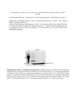
selected for further expression studies, Figure 1.
Expression of LigANI and LipL32 in P. pastoris
The coding sequences for the recombinant proteins LigANI (rLigANI) and LipL32 (rLipL32)
cloned in pPICZαB were under the control of the AOX1 promoter. In addition, pPICZαB
contains the α-factor signal sequence from S. cerevisiae, allowing secretion of the
recombinant protein. The concentration of rLigANI and rLipL32 in the culture supernatant
was found to increase with time, Figure 2A, and is related with a decrease in the intracellular
concentration of rLigANI, Figure 2B and C. In contrast, while the secretion of rLipL32
increased, so did the intracellular concentration, Figure 2D and E. Recombinant proteins of
the expected size were observed, rLigANI (61 kDa) and rLipL32 (32 kDa), yet there was
evidence of larger proteins, suggesting that the recombinant proteins had been glycosylated
by P. pastoris. Following 196 h induction at 28°C, the concentration of secreted protein
reached 0.93 g/L and 1.2 g/L for rLigANI and rLipL32, respectively. Large-scale (2 L
47
cultures) expression of rLigANI and rLipL32 resulted in yields of 276 mg/L and 285 mg/L,
respectively.
Recombinant protein purification and concentration
The supernatant containing the secreted rLigANI and rLipL32 was collected and
purified/concentrated using three alternative methods. In the first method, the proteins were
purified by ammonium sulphate precipitation. The optimal salt concentration for rLigANI
was 70-80%, while the precipitation of rLipL32 was similar under all concentrations tested.
The recombinant proteins were dialyzed to remove the ammonium sulphate and then
analysed by Western blotting, Figure 3A, B. Once again, there was evidence of posttranslation modification of the recombinant proteins. The yield for both rLigANI and
rLipL32 was similar, approximately 70 mg/L, corresponding to 24.5 and 27.6% of total
protein, respectively. In the second method, the supernatant was concentrated by
ultrafiltration which reduced the starting volume by 97%. The yield for rLigANI was 183
mg/L (66.3% total protein) compared to 106 mg/L (37.3% total protein) for rLipL32. The
samples were observed by 12% SDS-PAGE and compared to recombinant proteins expressed
and purified from E. coli (Figure 3 C). In the third method, the secreted proteins were
concentrated by lyophilisation. There was a 10-fold reduction in the initial sample volume
and the yield was 239 mg/L rLigANI and 224 mg/L rLipL32, equivalent to 86.7 and 70.7%
total protein, respectively.
Deglycosylation of LigANI and LipL32
In an analysis, using Vector NTI Advance 10.0 (Invitrogen) software, of the recombinant
protein amino acid sequences, LigANI was found to have seven potential N-glycosylation
sites, compared to one for LipL32. N-Glycosidase F (PNGase F) removes oligomannose,
48
hybrid, and complex N-glycans attached to asparagine, while Endoglycosidase H (Endo H)
releases oligomannose and hybrid N-glycans, but not complex N-glycans, and were used to
deglycosylate the recombinant proteins. Following deglycosylation, the larger molecular
weight species were no longer evident and the size of the rLigANI and rLipL32 corresponded
to the equivalent protein produced in E. coli, Figure 4. There did not appear to be any
difference in action between the two enzymes used.
Antigenicity of the recombinant LigANI and LipL32 proteins
The antigenicity of the purified proteins was evaluated by Western blotting with sera from
leptospirosis patients and with rabbit anti-Leptospira hyperimmune sera. The recombinant
proteins LigANI and LipL32 produced in E. coli were included as positive controls. Both
glycosylated and deglycosylated (Endo H and PNGase F treated) rLigANI were recognised
by the human and rabbit immune sera, Figure 5A, C and D, as were the glycosylated and
deglycosylated forms of rLipL32, Figure 5B, C and D.
Discussion
Previous studies have demonstrated the use of the Lig proteins and LipL32 in a range of
formats, including recombinant proteins [11-14], DNA vaccines [17, 22], microspheres and
liposomes [23, 24], fused to a cholera toxin subunit [25] or expressed in M. bovis bacille
Calmette-Guérin [16]. However, vaccine efficacy in the animal models has been highly
variable for these and other Leptospira proteins and they do not induce sterilizing immunity,
reviewed in [26]. Several reports suggest that the most likely explanation for the lack of a
consistent protective effect with recombinant proteins produced in E. coli is the failure of the
proteins to fold correctly [13, 22]. Structural modelling of Lig molecules predicted that the
49
bacterial immunoglobulin-like (Big) repeat domains have a highly folded β-immunoglobulin
sandwich structure [27]. E. coli expressed the full-length LigA at very low levels because of
its high toxicity, which resulted in a 50-fold decrease in viability of cells [28]. Furthermore,
expression of recombinant LigA in the E. coli pET expression system failed [14].
P. pastoris is an important host organism for the production of recombinant proteins
[19]. The large-scale production of recombinant proteins is necessary for pharmaceutical,
biomedical and biotechnological applications, therefore it is important to develop and to
optimize techniques for increased yield of the proteins of interest. In this work we cloned and
expressed a C-terminal fragment of LigA, LigANI, which includes six Big repeat domains of
the LigA protein, in the methylotrophic yeast P. pastoris. In addition, the full-length LipL32
protein was also expressed as a secreted protein. Previously we reported the expression of
recombinant LigANI in E. coli with a yield of 6-10 mg/L [13], while recombinant LipL32
was expressed at 40 mg/L [29]. In this study we report that large-scale expression in P.
pastoris resulted in yields of over 250 mg/L for both rLigANI and rLipL32, without the need
for subsequent solubilisation and/or re-folding steps. The strain used in this study, KM71H,
has a deletion in the AOX1 gene, which is partly replaced by ARG4 from S. cerevisiae and the
phenotype of these strains is MutS (Methanol utilization slow). The use of such strains is
advantageous as they do not require large amounts of methanol in large-scale cultures [1921].
Three low-cost purification strategies were evaluated, namely: i) ammonium
sulphate precipitation and desalting by dialysis, ii) ultrafiltration and iii) lyophilisation. The
most significant results in terms of yield were obtained using lyophilisation and ultrafiltration
to purify and/or concentrate the proteins. This is an important observation as these techniques
are applicable to large-scale cultures grown in bioreactors on an industrial scale. During
ultrafiltration the columns used had a cut-off of 30 kDa and our results demonstrated a
50
decreased yield of the rLipL32 protein, possibly due to the fact that the cut-off is very close
to the molecular weight of the recombinant protein. There was a significantly lower yield of
both rLigANI and rLipL32 when purified by ammonium sulphate precipitation.
LigANI and LipL32 were predicted to contain potential N-glycosylation sites and
treatment of the recombinant proteins with the enzymes Endo H and PNGase F confirmed
that post-translational modification had occurred during production and secretion in P.
pastoris, Figure 4. Deglycosylation removed the N-glycans attached to asparagine and when
analysed by SDS-PAGE and Western blotting, rLigANI and rLipL32 had similar molecular
weights as the corresponding proteins expressed in E. coli. N-glycosylation in yeast has a
composition of MannGlcNAc2 (Man: Mannose; GlcNAc: N-acetylglucosamine), where n is
the number of mannose oligosaccharides attached to the structure. This number has been
found to vary in P. pastoris from 3 to 17, depending on the expressed protein [30, 31]. The
attachment of a large number of mannose residues, known as hyperglycosylation, is rarely
observed in P. pastoris, compared to S. cerevisiae which hyperglycosylates the majority of
expressed proteins. Glycosylation can be influenced by some of the bioprocess parameters
used during growth and purification steps [32, 33]. Therefore, secreted proteins that are easily
recovered from the growth medium are likely to maintain the structure of the recombinant
protein. This may improve the protective immune response against leptospirosis when
rLigANI and rLipL32 are used as subunit vaccine candidates.
Conclusions
We believe that this is the first report of the use of P. pastoris to express pathogenic
Leptospira antigens. The aim of the study was to evaluate the large-scale expression of the
vaccine candidates LigA and LipL32 proteins in P. pastoris. The rLigANI and rLipL32
51
proteins described in this study were soluble and the purification step used simple and
inexpensive methods. Indeed, not only were the proteins expressed at a high level, but they
retained the antigenic characteristics of native the proteins. Furthermore, glycosylated
rLigANI and rLpiL32 were recognised by the antibodies presents in the sera of leptospirosis
patients and with antibodies raised against a heterologous Leptospira serovar.
Methods
Bacterial strains and growth conditions
L. interrogans serovar Copenhageni strain Fiocruz L1-130, originally isolated from a patient
with severe leptospirosis [34], was cultivated in Ellinghausen-McCullough-Johnson-Harris
(EMJH) medium supplemented with Leptospira Enrichment EMJH (Difco, USA) at 30 ºC. E.
coli strain TOP10 (Invitrogen) was grown in Luria-Bertani (LB) medium (1% tryptone, 0.5%
yeast extract, 0.5% NaCl and 2% agar) at 37 ºC with the addition of the zeocin 25 µg/mL.
P. pastoris strain KM71H (MutS, Invitrogen) was grown in Yeast extract peptone dextrose
(YPD) medium (1% yeast extract, 2% peptone and 2% D-glucose) supplemented with 100
µg/mL of zeocin at 28 ºC.
Cloning ligA and lipL32
We previously identified a C-terminal fragment of LigA, LigANI, as a vaccine candidate
[13]. Primers to amplify the DNA sequences coding for the LigANI polypeptide and the fulllength lipL32 gene were designed according the genome sequence of L. interrogans serovar
Copenhageni strain Fiocruz L1-130 [GenBank: AE016823]. The primer sequences (EcoRI
and
KpnI
sites
are
underlined)
used
in
this
CGGAATTCAATAATGTCTGATATTCTTACCGT,
TAGGTACCATGGCTCCGTTTTAATAGAG
study
were:
ligANI_F:
ligANI_R:
and
lipL32_F:
5'5'5'-
52
CGGAATTCTAGGTGGTCTGCCAA, lipL32_R: 5'-GGGGTACCACTTAGTCGCGTCA.
The PCR products were cloned in-frame into the pPICZαB vector (Invitrogen, Brazil). The
identity of the inserts was determined by DNA sequencing using the DYEnamic ET Dye
Terminator Cycle Sequencing Kit for MegaBACE DNA Analysis Systems – MegaBACE 500
(GE Healthcare, Brazil). Recombinant plasmids containing the LigANI coding sequence,
pPIC-LigANI, and lipL32, pPIC-LipL32, were propagated in E. coli TOP10, and the
plasmids isolated using the Perfectprep Plasmid Maxi kit (Eppendorf, USA). The plasmids
were linearized with restriction enzyme PmeI (New England BioLabs, USA). The linear
plasmid DNA was purified by phenol-chloroform extraction and DNA precipitation. P.
pastoris competent cells were transformed by electroporation (25 µF, 200 Ω, 2 kV) with 10
µg of linear plasmid DNA.
Screening for expression of recombinant LigANI and LipL32
Approximately 150 colonies of each plasmid construct were plated onto Buffered methanolcomplex medium (BMMY: 1% yeast extract, 2% peptone, 1.34% yeast nitrogen base,
0.00004% biotin, 0.5% methanol, 100 mM potassium phosphate and 2% agar, pH 6.0).
Following 24, 48 and 72 h incubation at 28ºC, expression of rLigANI and rLipL32 was
induced with 1% methanol and evaluated after 96 h. Expression of the recombinant proteins
was confirmed by colony immunoblotting [35]. Briefly, a nitrocellulose membrane (Hybond
ECL, GE Healthcare) was placed onto the surface of each petri dish and in direct contact with
the colonies for 3 h at 28°C. Any adherent matter was removed from the membrane by
washing with PBST (PBS, pH 7.4, 0.05% (v/v) Tween 20). After blocking (PBST, 5% nonfat dried milk), the membrane was incubated for 1 h at room temperature with anti-6×Hisperoxidase conjugate (Sigma-Aldrich, Brazil) at a dilution of 1:8,000 in PBS. After three
53
washes (5 min each) positive colonies were detected with 4-chloro-1-naphthol (SigmaAldrich).
The presence of the PCR products in the recombinant plasmids was also confirmed
by colony PCR. Crude genomic DNA extracts were prepared by boiling selected yeast
recombinant clones in water. PCR was performed as described above, using the crude
genomic DNA extracts as template. PCR products were analysed by horizontal gel
electrophoresis and visualized with GelRed (Uniscience, Brazil).
Expression of LigANI and LipL32 proteins in P. pastoris KM71H
A recombinant clone for each construct (rLigANI and rLipL32), positive for expression and
colony PCR, was selected and inoculated into a 1 L baffled flask containing 200 mL BMGY
broth (differs from BMMY in that the 1% methanol is replaced by 1% glycerol). The cultures
were incubated at 28°C, with shaking (250 rpm), for approximately 16–18 h until an OD600 of
2 to 6 was reached. The cells were harvested by centrifugation at 3,000 × g for 5 min and the
cell pellet suspended in the supernatant in 1/10 of the original volume (20 mL). The culture
was place in a 100 mL baffled flask and return to the incubator. Expression was induced by
the addition of methanol to a final concentration of 0.5%. Samples (supernatant and cells)
were collected at the following time points: 0, 24, 48, 72, 96, 120, 144, 168 and 196 h and
stored at –80°C. The cell pellets were suspended in breaking buffer (50 mM sodium
phosphate, 1 mM PMSF, 1 mM EDTA and 5% glycerol) and an equal volume of acidwashed glass beads (0.5 mm Ø). The samples were vortexed for 30 s followed by incubation
on ice for 30 s (8 cycles), centrifuged at 16,000 × g for 10 min at 4 ºC and the cleared
supernatant stored at –80°C.
The expression of the recombinant proteins were analysed by (12%) sodium dodecyl
sulphate-polyacrylamide gel electrophoresis (SDS–PAGE) and visualised by staining with
54
Coomassie Blue or Western blotting (WB). Samples were suspended in loading buffer (2%
SDS, 500 mM Tris pH 7.6, 1% bromophenol blue, 50% glycerol and 1% 2-mercaptoethanol)
and boiled for 10 min before separation by SDS-PAGE. For the WB assay the proteins were
electro transferred to a nitrocellulose membrane (Hybond ECL, GE Healthcare). After
blocking, PBS, 5% non-fat dried milk, overnight at 4ºC and three washes (5 min per wash) in
PBS-T, the membranes were incubated for 1 h with anti-LipL32 Mab (1:500 in PBS) or
mouse anti-LigANI polyclonal (1:500 in PBS), followed by 3 washes (5 min per wash) in
PBS-T. The rabbit anti-mouse IgG peroxidase conjugate (Sigma-Aldrich), diluted 1:6,000 in
PBS, was added and incubated for 1 h. The membranes were washed 5× in PBS-T and the
reactions were developed with 4-chloro-1-naphthol (Sigma-Aldrich).
LigANI and LipL32 were produced in large-scale using the P. pastoris MutS
secretory phenotype prior to purification, about the same conditions described above. Briefly,
P. pastoris was grown in BMGY medium (2 L) to an OD600 of 2 to 6, harvested by
centrifugation and suspended in BMMY expression medium in 1/10 of the original culture
volume (200 mL). The expression of the recombinant proteins was induced for 144 h with
methanol 0.5%. The supernatant containing the secreted recombinant proteins was cleared by
centrifugation, and stored at –80°C.
Purification and concentration of rLigANI and rLipL32
Three different strategies were used to purify and concentrate the secreted recombinant
proteins. The first strategy was based on ammonium sulphate precipitation: 85% ammonium
sulphate was added to the culture supernatant at 4°C, to final concentrations of: 25, 35, 45,
60, 70 and 80%. The precipitated proteins were collected by centrifugation at 10,000 × g for
15 min at 4°C and suspended in PBS and dialyzed in the same buffer for 48 h. Microcon YM30 Amicon Bioseparation filters (Millipore, USA), 30 kDa cut-off, were used to concentrate
55
the recombinant proteins expressed in the supernatant, following the manufacturer’s protocol.
Alternatively, proteins were concentrated by lyophilisation (Edwards Micro Modulyo) over
28 h and suspended in PBS, resulting in a concentration 10-fold of the initial sample. The
protein concentration in culture supernatants, concentrate and purified proteins samples were
determined by BCA Protein Assay Kit (Pierce, USA) with bovine serum albumin (BSA) as a
the standard.
Deglycosylation of rLigANI and rLipL32
Purified rLigANI and rLipL32 (1-20 µg) were incubated with 1× glycoprotein reaction buffer
at 100°C for 10 min to completely denature the glycoproteins. Deglycosylation was carried
out at 37°C for 1 h with 5× G5 (Endoglycosidase H) or 10× G7 (N-Glycosidase F) reaction
buffer and 1-5 µl of the relevant enzyme (Endoglycosidase H or N-Glycosidase F) according
to the manufacturer’s instructions (New England BioLabs).
Antigenicity of rLigANI and rLipL32
The ability of the recombinant proteins to interact specifically with products of the immune
response was determined by WB using sera collected from leptospirosis patients and
hyperimmune sera from infected rabbits. The use of subject sera for these experiments was
approved by the Internal Review Board of the Gonçalo Moniz Institute, Fiocruz-BA. A pool
of convalescent sera from severe leptospirosis patients was used at a dilution of 1:300 and an
anti-human IgG peroxidase conjugate at a 1:2,000 dilution. Rabbit anti-Leptospira
hyperimmune sera, specific to L. interrogans serovar Canicola strain Tande, was used at a
dilution of 1:500 and an anti-rabbit IgG peroxidase conjugate at a 1:3,000 dilution.
Competing interests
56
AJAM and OAD are inventors on a patent submission entitled: LigA and LigB proteins
(Leptospiral Ig-like (Lig) domains) for vaccination and diagnosis (Patent nos. BRPI0505529
and WO 2007070996). The other authors declare no competing interests.
Authors’ Contributions
DDH participated in the study design, performed the experiments and in the writing of the
manuscript. TLO performed the experiments. FKS participated in the construction of the
plasmids. KMF and CPH participated in the experiments on protein antigenicity and CR
participated in the protein purification steps. AJAM participated in the data analysis and the
writing of the manuscript. OAD coordinated the study and participated in the writing of the
manuscript. All authors read and approved the final manuscript.
Acknowledgements
This work was supported by the Brazilian National Research Council (CNPq), grant
475540/2008-5, the Research Support Foundation for the State of Bahia (FAPESB), grant
PES-0092/2008 (to AJAM) and the Oswaldo Cruz Foundation (to AJAM). DDH and KMF
received scholarships from CNPq. The funders had no role in study design, data collection
and analysis, decision to publish, or preparation of the manuscript.
57
References
1.
Bharti AR, Nally JE, Ricaldi JN, Matthias MA, Diaz MM, Lovett MA, Levett PN,
Gilman RH, Willig MR, Gotuzzo E et al: Leptospirosis: a zoonotic disease of global
importance. Lancet Infect Dis 2003, 3(12):757-771.
2.
Adler B, de la Pena Moctezuma A: Leptospira and leptospirosis. Vet Microbiol
2010, 140(3-4):287-296.
3.
Faine SB, Adler B, Bolin C, Perolat P: Leptospira and leptospirosis, 2nd edn.
Melbourne: MediSci; 1999.
4.
Reis RB, Ribeiro GS, Felzemburgh RD, Santana FS, Mohr S, Melendez AX, Queiroz
A, Santos AC, Ravines RR, Tassinari WS et al: Impact of environment and social
gradient on leptospira infection in urban slums. PLoS Negl Trop Dis 2008,
2(4):e228.
5.
McBride AJ, Athanazio DA, Reis MG, Ko AI: Leptospirosis. Curr Opin Infect Dis
2005, 18(5):376-386.
6.
Gouveia EL, Metcalfe J, de Carvalho AL, Aires TS, Villasboas-Bisneto JC, Queirroz
A, Santos AC, Salgado K, Reis MG, Ko AI: Leptospirosis-associated Severe
Pulmonary Hemorrhagic Syndrome, Salvador, Brazil. Emerg Infect Dis 2008,
14(3):505-508.
7.
Segura ER, Ganoza CA, Campos K, Ricaldi JN, Torres S, Silva H, Cespedes MJ,
Matthias MA, Swancutt MA, Lopez Linan R et al: Clinical spectrum of pulmonary
involvement in leptospirosis in a region of endemicity, with quantification of
leptospiral burden. Clin Infect Dis 2005, 40(3):343-351.
58
8.
Haake DA, Mazel MK, McCoy AM, Milward F, Chao G, Matsunaga J, Wagar EA:
Leptospiral outer membrane proteins OmpL1 and LipL41 exhibit synergistic
immunoprotection. Infect Immun 1999, 67(12):6572-6582.
9.
Yan W, Faisal SM, McDonough SP, Chang CF, Pan MJ, Akey B, Chang YF:
Identification and characterization of OmpA-like proteins as novel vaccine
candidates for Leptospirosis. Vaccine 2010, 28(11):2277-2283.
10.
Wang Z, Jin L, Wegrzyn A: Leptospirosis vaccines. Microb Cell Fact 2007, 6:39.
11.
Koizumi N, Watanabe H: Leptospiral immunoglobulin-like proteins elicit
protective immunity. Vaccine 2004, 22(11-12):1545-1552.
12.
Yan W, Faisal SM, McDonough SP, Divers TJ, Barr SC, Chang CF, Pan MJ, Chang
YF: Immunogenicity and protective efficacy of recombinant Leptospira
immunoglobulin-like protein B (rLigB) in a hamster challenge model. Microbes
Infect 2009, 11(2):230-237.
13.
Silva EF, Medeiros MA, McBride AJ, Matsunaga J, Esteves GS, Ramos JG, Santos
CS, Croda J, Homma A, Dellagostin OA et al: The terminal portion of leptospiral
immunoglobulin-like protein LigA confers protective immunity against lethal
infection in the hamster model of leptospirosis. Vaccine 2007, 25(33):6277-6286.
14.
Palaniappan RU, McDonough SP, Divers TJ, Chen CS, Pan MJ, Matsumoto M,
Chang YF: Immunoprotection of recombinant leptospiral immunoglobulin-like
protein A against Leptospira interrogans serovar Pomona infection. Infect Immun
2006, 74(3):1745-1750.
15.
Feng CY, Li QT, Zhang XY, Dong K, Hu BY, Guo XK: Immune strategies using
single-component LipL32 and multi-component recombinant LipL32-41-OmpL1
vaccines against leptospira. Braz J Med Biol Res 2009, 42(9):796-803.
59
16.
Seixas FK, da Silva EF, Hartwig DD, Cerqueira GM, Amaral M, Fagundes MQ,
Dossa RG, Dellagostin OA: Recombinant Mycobacterium bovis BCG expressing
the LipL32 antigen of Leptospira interrogans protects hamsters from challenge.
Vaccine 2007, 26(1):88-95.
17.
Branger C, Chatrenet B, Gauvrit A, Aviat F, Aubert A, Bach JM, Andre-Fontaine G:
Protection against Leptospira interrogans sensu lato challenge by DNA
immunization with the gene encoding hemolysin-associated protein 1. Infect
Immun 2005, 73(7):4062-4069.
18.
Sonrier C, Branger C, Michel V, Ruvoen-Clouet N, Ganiere JP, Andre-Fontaine G:
Evidence of cross-protection within Leptospira interrogans in an experimental
model. Vaccine 2000, 19(1):86-94.
19.
Sorensen HP: Towards universal systems for recombinant gene expression.
Microb Cell Fact 2010, 9:27.
20.
Cos O, Ramon R, Montesinos JL, Valero F: Operational strategies, monitoring and
control of heterologous protein production in the methylotrophic yeast Pichia
pastoris under different promoters: a review. Microb Cell Fact 2006, 5:17.
21.
Cregg JM, Cereghino JL, Shi J, Higgins DR: Recombinant protein expression in
Pichia pastoris. Mol Biotechnol 2000, 16(1):23-52.
22.
Faisal SM, Yan W, Chen CS, Palaniappan RU, McDonough SP, Chang YF:
Evaluation of protective immunity of Leptospira immunoglobulin like protein A
(LigA) DNA vaccine against challenge in hamsters. Vaccine 2008, 26(2):277-287.
23.
Faisal SM, Yan W, McDonough SP, Chang YF: Leptospira immunoglobulin-like
protein A variable region (LigAvar) incorporated in liposomes and PLGA
microspheres produces a robust immune response correlating to protective
immunity. Vaccine 2009, 27(3):378-387.
60
24.
Faisal SM, Yan W, McDonough SP, Chang CF, Pan MJ, Chang YF: Leptosomeentrapped leptospiral antigens conferred significant higher levels of protection
than those entrapped with PC-liposomes in a hamster model. Vaccine 2009,
27(47):6537-6545.
25.
Habarta A, Abreu PA, Olivera N, Hauk P, Cedola MT, Ferrer MF, Ho PL, Gomez
RM: Increased Immunogenicity to LipL32 of Leptospira interrogans when
Expressed as a Fusion Protein with the Cholera Toxin B Subunit. Curr Microbiol
2010, Aug 19. [Epub ahead of print]
26.
Ko AI, Goarant C, Picardeau M: Leptospira: the dawn of the molecular genetics
era for an emerging zoonotic pathogen. Nat Rev Microbiol 2009, 7(10):736-747.
27.
Matsunaga J, Barocchi MA, Croda J, Young TA, Sanchez Y, Siqueira I, Bolin CA,
Reis MG, Riley LW, Haake DA et al: Pathogenic Leptospira species express
surface-exposed
proteins
belonging
to
the
bacterial
immunoglobulin
superfamily. Mol Microbiol 2003, 49(4):929-945.
28.
Palaniappan RU, Chang YF, Jusuf SS, Artiushin S, Timoney JF, McDonough SP,
Barr SC, Divers TJ, Simpson KW, McDonough PL et al: Cloning and molecular
characterization of an immunogenic LigA protein of Leptospira interrogans.
Infect Immun 2002, 70(11):5924-5930.
29.
Seixas FK, Fernandes CH, Hartwig DD, Conceicao FR, Aleixo JA, Dellagostin OA:
Evaluation of different ways of presenting LipL32 to the immune system with
the aim of developing a recombinant vaccine against leptospirosis. Can J
Microbiol 2007, 53(4):472-479.
30.
Daly R, Hearn MT: Expression of heterologous proteins in Pichia pastoris: a
useful experimental tool in protein engineering and production. J Mol Recognit
2005, 18(2):119-138.
61
31.
Montesino R, Garcia R, Quintero O, Cremata JA: Variation in N-linked
oligosaccharide
structures
on
heterologous
proteins
secreted
by
the
methylotrophic yeast Pichia pastoris. Protein Expr Purif 1998, 14(2):197-207.
32.
Goochee CF, Monica T: Environmental effects on protein glycosylation.
Biotechnology (N Y) 1990, 8(5):421-427.
33.
Jenkins N, Parekh RB, James DC: Getting the glycosylation right: implications for
the biotechnology industry. Nat Biotechnol 1996, 14(8):975-981.
34.
Ko AI, Galvao Reis M, Ribeiro Dourado CM, Johnson WD, Jr., Riley LW: Urban
epidemic of severe leptospirosis in Brazil. Salvador Leptospirosis Study Group.
Lancet 1999, 354(9181):820-825.
35.
Sambrook J, Russell DW: Molecular Cloning: A Laboratory Manual, 3rd edn.
Cold Spring Harbor, NY: Cold Spring Harbor Laboratory Press; 2000.
62
Figure legends
Figure 1 - Screening for P. pastoris recombinant clones expressing rLigANI and
rLipL32.
Colony blot analysis of transformed P. pastoris strain KM71H with anti-6×His Mab. The tgD
recombinant protein expressed in P. pastoris KM71H was the positive control (+) and
untransformed P. pastoris KM71H was the negative control (–). Spots 1-7 are representative
rLigANI colonies and 8-12 are representative rLipL32 colonies. Arrows indicate the colonies
that were selected for large-scale expression studies.
Figure 2 - Expression of rLigANI and rLipL32 proteins in P. pastoris.
Time courses for the expression of secreted rLigANI and rLipL32 by P. pastoris induced for
up to 192 hours (8 days), (A) as determined by protein concentration (mg/mL). Western blot
analysis of the intracellular (pellet) and secreted (supernatant) expression of rLigANI (B and
C, respectively) and rLipL32 (D and E, respectively), using polyclonal anti-LigANI sera or
anti-LipL32 Mab. Samples (cells and supernatant) were collected at the various hourly time
points indicated. KM – negative control: untransformed P. pastoris KM71H culture.
Figure 3 - Purification of rLigANI and rLipL32 expressed in P. pastoris.
Recombinant proteins purified by precipitation with ammonium sulphate or by ultrafiltration.
Ammonium sulphate precipitated proteins were detected by Western blotting with (A)
polyclonal anti-LigANI sera or (B) an anti-LipL32 Mab. The effect of the various
concentrations of ammonium sulphate (expressed as percentage values) on the precipitation
of the recombinant proteins is displayed. (C) Affinity chromatography purified recombinant
LigANI (61 kDa) and LipL32 (32 kDa) produced in E. coli compared to purification by
63
ultrafiltration of rLigANI and rLipL32 secreted by P. pastoris. An equal volume (10 µL) of
both proteins was loaded on the gel.
Figure 4 - Deglycosylation of rLigANI and rLipL32 produced by P. pastoris.
To evaluate the post-translational modification of the rLigANI and rLipL32 proteins
produced and secreted by P. pastoris, the proteins were deglycosylated with PNGase F and
Endo H. The resultant proteins were visualized by (A) Western blotting with polyclonal antiLigANI sera and an anti-LipL32 Mab or by (B) SDS-PAGE stained with Coomassie blue.
The proteins were digested with PNGase F, Endo H or without enzyme (-). E-LigANI (61
kDa) and E-LipL32 (32 kDa) recombinant proteins were expressed and purified from E. coli.
Figure 5 - Antigenicity of the various forms of rLigANI and rLipL32.
Antigenicity was evaluated using rabbit anti-Leptospira sera (A) Lanes: 1 - rLigANI +
PNGase F; 2 - rLigANI + Endo H; 3 - glycosylated rLigANI and (B) Lanes: 4 - rLipL32 +
PNGase F; 5 - rLipL32 + Endo H; 6 – glycosylated rLipL32 or (C) convalescent sera from
leptospirosis patients, Lanes: 1 - glycosylated rLipL32; 2 - glycosylated rLigANI and (D) 3 –
rLipL32 + PNGAse F; 4 – LipL32 + Endo H; 5 - rLigANI + PNGAse F; 6 - rLigANI + Endo
H. E-LigANI (61 kDa) and E-LipL32 (32 kDa) recombinant proteins were expressed and
purified from E. coli.
64
Figure 1
(+)
1
2
(-)
3
4
5
6
8
9
10
11
7
12
65
Figure 2
A
B
0h
24h
48h
72h
96h
120h
144h
KM
61 kDa
C
0h
24h
48h
72h
96h
120h
144h
KM
61 kDa
D
0h
24h
48h
72h
96h
120h
144h
KM
32 kDa
E
0h
24h
48h
72h
96h
120h
144h
KM
32 kDa
66
Figure 3
A
E-LigANI
25%
35%
45%
60%
70%
80%
61 kDa
B
E-LipL32
25%
35%
45%
60%
70%
80%
32 kDa
E. coli
C
E-LigANI E-LipL32
P. pastoris
rLigANI
rLipL32
61 kDa
61 kDa
32 kDa
32 kDa
67
Figure 4
A
PNGase F Endo H
(-)
E-LigANI
PNGase F Endo H
(-)
E-LipL32
32 kDa
61 kDa
B
PNGase F Endo H
( - ) E-LigANI
PNGase F Endo H
(-)
E-LipL32
32 kDa
61 kDa
68
Figure 5
LigANI
A
1
2
LipL32
3
E-LigANI
B
4
5
6
E-LipL32
61 kDa
32 kDa
rLipL32
C
61 kDa
32 kDa
E-rLipL32 E-LigANI
1
2
D
3
rLigANI
4
5
6
61 kDa
32 kDa
69
4. ARTIGO 3
Immunoprotection by LigA and LipL32 produced in Pichia pastoris
and evaluated in the hamster model of lethal leptospirosis
(Artigo a ser submetido ao periódico Clinical and Vaccine Immunology)
70
Immunoprotection by LigA and LipL32 produced in Pichia pastoris and evaluated in
the hamster model of lethal leptospirosis
Running title: P. pastoris recombinant LigA and LipL32 protection in hamsters
Daiane D. Hartwig1, Karine M. Forster1, Thaís L. Oliveira1, Fabiana K. Seixas1, Marta
Amaral2, Alan J. A. McBride3 and Odir A. Dellagostin1*
1
Núcleo de Biotecnologia, Centro de Desenvolvimento Tecnológico, Universidade Federal de
Pelotas, Pelotas, RS, Brazil
2
Instituto de Biologia, Universidade Federal de Pelotas, Pelotas, RS, Brazil
3
Laboratório de Patologia e Biologia Molecular, Instituto Gonçalo Moniz, Fiocruz-BA,
Salvador, BA, Brazil
*Corresponding author: Odir A. Dellagostin, Centro de Biotecnologia, Universidade
Federal de Pelotas, Campus Universitário, Caixa Postal 354, CEP 96010-900, Pelotas, RS,
Brazil. Tel. +55 53 3275 7587; Fax +55 53 3275 7551
71
ABSTRACT
Leptospirosis is a widespread zoonosis of great importance to public health,
particularly in developing countries. A priority in research on leptospirosis is the
development of a vaccine able to elicit long-term immunity and to induce cross-protection
against the most common pathogenic Leptospira serovars. Several antigens of Leptospira
interrogans produced in Escherichia coli for use as subunit vaccines have demonstrated
promising results, but have presented with variable efficacy and low yield. The expression of
recombinant proteins in the methylotrophic yeast, Pichia pastoris, is relatively fast and
inexpensive. In addition, for purposes of vaccination, yeast recombinant proteins have been
shown to have natural adjuvant activity. In this study, we evaluated LigANI, which
corresponds to the six carboxy-terminal repeat domains of LigA, and the lipoprotein LipL32,
produced in P. pastoris. Both recombinant proteins induced significant immune humoral
responses in hamsters (P < 0.001), evaluated by ELISA. The LigANI vaccine preparation
significantly improved survival (P < 0.001) in hamsters challenged with 5×LD50 of L.
interrogans serovar Copenhageni. These results demonstrate that the large-scale production
of LigANI by P. pastoris shows potential as a potential vaccine candidate but requires further
development.
KEYWORDS: Leptospirosis, Vaccine, Pichia pastoris; Leptospira interrogans; LigA
protein; LipL32 protein; immunoprotection; hamster challenge model.
72
INTRODUCTION
Vaccination strategies remain the principal tool for the prevention of many infectious
diseases, including Leptospirosis. Pathogenic Leptospira spp. cause this zoonotic disease, one
of the most common in the world (1,2,29,39). In developed countries leptospirosis is an
emerging infectious disease associated with sporting events, tourism and recreational
activities, while in developing countries it is linked to poverty and poor sanitation. Globally,
more than 500,000 cases are reported each year (1,29). Severe infection is associated with a
mortality of 10-15% in cases of the traditional Weil’s disease and can be over 70% in cases
of severe pulmonary haemorrhage syndrome (SPHS) (15,29,35). In livestock, Leptospira
infection is associated with abortion, stillbirth, milk drop syndrome and occasionally death
(16).
However, an efficient vaccine with cross-protection against the different pathogenic
serovars remains a challenge. Efforts to develop recombinant leptospiral vaccines have
therefore focused on outer membrane proteins (OMP) (10,17,22). The Leptospiral
immunoglobulin-like (Lig) proteins, LigA, LigB and LigC, belong to a family of bacterial
immunoglobulin-like (Big) repeat domain proteins. They encode virulence determinants in
pathogenic strains (8,24,28,31), and are highly conserved (70–99 % identity) in
pathogenic Leptospira isolates (30). Several independent studies have evaluated these
proteins
as
potential
vaccine
candidates,
with
varying
degrees
of
efficacy
(12,14,21,32,37,40). The lipoprotein LipL32, also known as haemolysis-associated protein 1
(Hap1) (4), is described as the most abundant OMP exposed on the surface of the cell (11).
The lipL32 coding sequence is specific to the pathogenic Leptospira spp. (18). When this
protein was delivered as DNA vaccine, adenovirus mediated-vaccination or in a recombinant
Mycobacterium bovis BCG strain, it provided significant protection in gerbils or hamsters.
73
However, it failed to induce a protective immune response when used as recombinant subunit
vaccine (3,4,36).
Our group recently described the high yield expression of the LigANI (rLigANI) and
LipL32 (rLipL32) proteins by the methylotrophic yeast Pichia pastoris, using simple and
inexpensive expression and purification methods (20). This eukaryotic expression system is
used as a host for industrial production of recombinant proteins, has advantages such as high
and efficient expression of heterologous proteins that can be secreted into medium and
growth at high density in bioreactors (6,9,26). P. pastoris harbouring an expression vector
can efficiently secrete heterologous protein in the correctly folded, soluble and biologically
active form (34). The rLigANI and rLipL32 proteins secreted by P. pastoris were
glycosylated with the addition of mannose residues. In this study, we evaluated the specific
humoral immune response against rLigANI and rLipL32, expressed by P. pastoris, and their
efficacy as potential vaccine candidates in the hamster model of lethal leptospirosis.
MATERIALS AND METHODS
Bacterial strains and growth conditions. A virulent pathogenic L. interrogans serovar
Copenhageni strain FORSTER/CDTEC (unpublished data) was cultivated in EllinghausenMcCullough-Johnson-Harris (EMJH) medium supplemented with Leptospira Enrichment
EMJH (Difco, USA) at 30 ºC. Pichia pastoris strain KM71H MutS phenotype (Invitrogen,
Brazil) was grown in Yeast Extract Peptone Dextrose (YPD) medium (1% yeast extract, 2%
peptone and 2% D-glucose) at 30 ºC supplemented with 100 µg/ml of zeocin.
Experimental animals. Golden Syrian hamsters were housed at the animal facility of the
Biotechnology Centre, Federal University of Pelotas (UFPel). The animals were maintained
74
in accordance with the guidelines of the Ethics Committee in Animal Experimentation of
UFPel throughout the experimental period.
Production of the LigANI and LipL32 recombinant proteins in P. pastoris. The
expression of rLigANI and rLipL32 MutS secretory phenotype was performed using the
eukaryotic system based in P. pastoris, as described previously (20). Briefly, the recombinant
clones were grown in a baffled flask containing 2 L of BMGY broth (1% yeast extract, 2%
peptone, 1.34% yeast nitrogen base, 0.00004% biotin, 1% glycerol, 100 mM potassium
phosphate and 2% agar, pH 6.0). The cultures were incubated at 28°C until an OD600 of 2 to 6
was reached. The cells were harvested by centrifugation at 3,000 g for 5 min and the cell
pellet was resuspended in the BMMY broth (differs from BMGY in that the 1% glycerol is
replaced by 1% methanol) to 1/10 of the original volume (0.2 L). The culture was place in a 1
L baffled flask and return to the incubator. Maximum yield achieved by the addition of
methanol to a final concentration of 0.5% for 144 h. The Microcon YM-30 Amicon
Bioseparations (Millipore) were used to concentrate the recombinant proteins expressed in
the supernatant, following manufacturer’s protocol. The recombinant proteins were analysed
by (12%) sodium dodecyl sulphate-polyacrylamide gel electrophoresis (SDS–PAGE) and
visualized by staining with Coomassie Blue and Western blotting (WB). The LigANI and
LipL32 recombinant proteins concentration were determined by BCA Protein Assay Kit
(Pierce, USA) method using bovine serum albumin (BSA) as a standard.
Immunization and hamsters challenge experiments. Female hamsters, 4 - 6 weeks of age,
were used in all experiments. Animals were divided into groups of 12 animals, food and
water were provided ad libitum. Animals were immunized with rLigANI (80 µg) or rLipL32
(80 µg) together with 15% aluminium hydroxide adjuvant by intramuscular injection in the
75
hind legs twice, at 14 day intervals. Blood samples were collected through the retro-orbital
plexus before each immunization and challenge, the sera was collected and stored at −20ºC.
Twenty-eight days after the first dose all hamsters were challenged intraperitoneally with a
dose of 101 leptospires, equivalent to 5× LD50 of the L. interrogans serovar Copenhageni
strain FORSTER/CDTEC (unpublished data). The negative control group was inoculated
with phosphate buffered saline (PBS) + 15% aluminium hydroxide, the positive control group
was immunized with a bacterin vaccine consisting of 109 heat-killed whole-leptospires, as
previously described (36). Hamsters were monitored daily for clinical signs of leptospirosis
and euthanized when clinical signs of terminal disease appeared.
Evaluation of the humoral immune response. Antibody responses were monitored by an
indirect ELISA using rLigANI and rLipL32. ELISA plates (Polysorp Surface, Nunc) were
coated overnight at 4°C with 200 ng of recombinant protein per well, diluted in carbonatebicarbonate buffer pH 9.6. The plates were washed three times in PBS (pH 7.4) with 0.05%
(v/v) Tween 20 (PBST) and incubated with 200 µl of 5% blocking buffer, at 37ºC for 1 h.
The hamster sera, diluted 1:50, was added and the plate incubated for 1 h at 37ºC, followed
by three washes with PBST. Goat anti-hamster IgG peroxidase conjugate, 1:8,000 dilution,
(Serotec, USA) was added and incubated at 37ºC for 1 h, washed 5× with PBST and the
reaction visualized with o-phenylenediamine dihydrochloride (Sigma-Aldrich, Brazil) and
hydrogen peroxide. The reaction was stopped by the addition of 0.1 M sulphuric acid and
absorbance was determined at 492 nm using a Multiskan MCC/340 ELISA reader (Titertek
Instruments, USA). Mean values were calculated from sera samples assayed in triplicate.
Culture and histopathology analysis. Surviving hamsters on day 30 post challenge were
euthanized. Kidney and lung tissues were harvested post-euthanasia and studied for
76
histopathology. Sterilizing immunity was determined by culture isolation of leptospires from
kidney samples. From each organ 1 - 2 g of tissue was aseptically removed, processed and
transferred to 5 mL of EMJH medium (pH 7.2). Dark-field microscopy was performed during
8-week incubation period to identify positive cultures. For histopathological studies, kidney
and lung tissues samples were fixed in 10% formalin (pH 7.0) and embedded in paraffin. Six
sections of 5 - 6 µm thickness from organs were stained with haematoxylin and eosin and
examined by a qualified pathologist for evidence of interstitial nephritis or pulmonary
haemorrhage.
Imprint detection. The presence of leptospires in the kidneys of immunized hamster was
evaluated by the imprint method (7). Briefly, imprints were obtained by direct pressure of the
cut surface of the tissue sample onto poly-L-lysine-coated glass slides. Imprint slides were
dried at room temperature, fixed in methanol (10 min at 4ºC) and incubated for 30 min in a
dark humid chamber at 30°C. After three washes with 10% bovine foetal serum (BFS) diluted
in PBS, a Mab against LipL32 (1D9), diluted 1:100, was added and incubated in a dark
humid chamber at 30°C (1 h). The imprints were incubated (1 h) under the same conditions
with an anti-Leptospira FITC conjugate, diluted 1:100, after three washes with PBS + 10%
BSF. Nucleic acids were visualized by counterstaining with Hoestch, diluted 1:10, for 30 min
at 30oC in a dark humid chamber. Following five washes with PBS + 10% BSF, mounting
medium was added and a cover slip was sealed in place with acrylic. Staining was visualized
by fluorescence microscopy (Olympus) at an excitation wavelength of 450 nm.
Statistical analysis. Variance analysis was used to determine significant differences between
the assay results. The Fisher exact test and the Wilcoxon log-rank test were used to determine
significant differences for mortality and survival, respectively, using Epi Info 6 (Centres for
77
Disease Control, USA) and Prism 5 (Graphpad, USA), differences were considered
significant at P < 0.05.
RESULTS
Expression of rLigANI and rLipL32 in P. pastoris. The 61 kDa and 32 kDa bands which
corresponded to rLigANI and rLipL32, respectively, were secreted by P. pastoris after
methanol induction and visualized in SDS-PAGE (Fig. 1 A and B). There was evidence of
larger proteins, because the recombinant proteins had been glycosylated by P. pastoris. The
recombinant proteins accumulated in the culture medium during 144 h of methanol induction
and were purified by ultrafiltration for use in hamster immunoprotection assays.
Humoral immune response elicited by recombinant proteins in hamsters. Twenty-eight
days after the first immunization, the hamsters were challenged with a virulent strain of L.
interrogans serovar Copenhageni. Hamsters immunized with rLigANI, rLipL32 and
aluminium hydroxide gained weight during the 28 days post-immunization, reaching and
average of 92.24 ± 2.70 g. To quantify the specific antibody response, an ELISA was
performed with the sera collected on days 0, 14 and 28 post-immunization (pi) (Fig. 2).
Significant levels of circulating IgG antibodies were detected (P < 0.001). Fourteen days after
the first dose with rLipL32 there was a significant (P < 0.001) induction of IgG levels in the
hamster immunized compared to the negative control group (PBS/aluminium hydroxide).
However, the IgG level in those hamsters immunized with rLigANI did not differ from the
negative control group at\ 14 days. In the twenty-eight days after the first dose was observed
difference (P < 0.001) between the three groups (rLigANI, rLipL32 and aluminium
hydroxide), Fig. 2.
78
Immunoprotection of hamsters immunized with rLigANI and rLipL32. Female hamsters
were challenged with 101 leptospires (5 × LD50 for female hamsters) 28 days after the first
immunization and mortality was determined for up to 30 days post-challenge. Of the
hamsters immunized with 2× 80 µg of rLigANI in aluminium hydroxide, 25% (3/12) were
protected against lethal challenge (Table 1) however, this was not significant, P = 0.22, in
terms of mortality (Fisher exact test). Nonetheless, immunization with rLigANI significantly
improved survival (Wilcoxon Log-rank test) compared to the negative control group, P <
0.001. Median survival in the negative control group was nine days compared to 17.5 days in
the rLigANI group (Fig. 3). Immunization with rLipL32 failed to induce protection in the
hamster model (Table 1 and Fig. 3). However, survival in hamsters immunized with rLipL32
was significant compared to the control group, P < 0.001 (Wilcoxon Log-rank test). Median
survival in the negative control group was nine days compared to 12 days in the rLipL32
group. All hamsters in the positive control group (Table 1, Fig. 3) were protected against
mortality, P < 0.001 (Fisher exact test).
Histopathological analysis. Thirty days after the challenge with a virulent leptospiral strain
the surviving hamsters were necropsied. During this 30-day follow-up period there was no
clinical evidence of infection, but of the surviving hamsters immunized with LigANI one
present macroscopic evidence of pulmonary haemorrhage and two hamsters present icteric
kidneys. Severe pathological lesions were found in animals vaccinated with rLigANI, which
survived during the course of the experiment, as prominent interstitial infiltrate of
lymphocytes and plasma cells, hemorrhage and necrosis. In the lung was evidenced
hemorrhage and edema, including in animals immunized with bacterin.
79
Detection of leptospires in surviving hamsters post-challenge. Among the rLigANI
immunized hamsters which survived, leptospires were isolated in kidney tissues cultures. The
imprint evaluation confirmed these results, in the three hamsters that survived challenge,
leptospires were detected in the kidney samples, Fig. 4 A and B. In contrast, none of the
hamsters in the positive control group (bacterin vaccine) had evidence of leptospires (culture
or imprint), indicating that sterilizing immunity was induced, Fig. 4 C and D.
DISCUSSION
Our previous work described the cloning and expression of the vaccine candidates
LigANI and LipL32 from L. interrogans in P. pastoris. We showed the soluble expression of
these proteins in methylotrophic yeast, resulting in a significant increase in yield compared to
expression in E. coli. In addition, the proteins were glycosylated and retained the antigenic
characteristics of the native proteins (20). In the present study the main objective was to
assess the potential of rLigANI and rLipL32 secreted in a mannosylated form by P. pastoris
as vaccine candidates against leptospirosis, using the standard hamster model. LigA and the
LipL32 were evaluated in previous studies as vaccine candidates however, efficacy varied
considerably (3,12-14,19,21,32,36). A potential problem with subunit vaccines is the
incorrect folding of the recombinant protein by the E. coli host. In a previous study, LigANI
required solubilisation in urea and extensive dialysis to maintain the protein in solution prior
to vaccine preparation (37). There were further reports of inefficient expression in E. coli,
possibly due to toxicity of the recombinant proteins (31,32). In contrast, the rLigANI and
rLipL32 expressed in P. pastoris were soluble and easily purified. Another possible
explanation for the variable efficacies reported in the previous studies is the challenge strain,
the majority used poorly virulent strains that required a high lethal dose, 106 - 108 leptospires,
and even then failed to cause 100% mortality in the control groups (3,12,32). The challenge
80
strain used in this study was highly virulent, the challenge dose was 10 leptospires, equivalent
to 5× the LD50.
We previously expressed LigANI (a polypeptide fragment corresponding to the six
carboxy-terminal Ig-like repeat domains of LigA) in an E. coli-based expression system and
this antigen significantly protected immunized hamsters (37). However, the vaccine
preparation included Freund’s adjuvant and this is incompatible for human and animal use. In
a study using aluminium hydroxide adjuvant, LigA significantly protected hamsters against
lethal challenge. In addition, anti-LigA antibodies were protective but there were extensive
kidney lesions present in the surviving hamsters (32). When evaluated as DNA vaccine, LigA
was immunoprotective but there was extensive kidney damage and the negative control
groups demonstrated a high survival rate (12). In the current study, we used a highly virulent
strain of L. interrogans serovar Copenhageni, which a low challenge dose that reproducibly
induced characteristic disease manifestations and death in all negative control animals. The
rLigANI evaluated in the present study demonstrated an efficacy of only 25%, furthermore
all of the surviving animals were culture positive and contained lesions typical of severe
leptospirosis. However, rLigANI significantly increased survival compared to the control
group. This result once again demonstrates the variable efficacy associated with the Lig
vaccine candidates. As the rLigANI polypeptide used in this study was expressed in P.
pastoris it is likely that the glycosylated recombinant protein varied significantly from the
native protein. This finding is further supported by the observation that rLigANI induced a
significant IgG response in the immunized hamsters, but one that was not protective (Fig. 2).
Although immunization with rLipL32 elicited a significant IgG response (Fig. 2), it
failed to protect against lethal challenge. Yet, as seen with rLigANI, immunization with
rLipL32 significantly increased survival. These results are in agreement with previous studies
using recombinant LipL32 expressed in E. coli (3). However, this antigen when expressed in
81
an adenovirus construct, recombinant M. bovis BCG or used as DNA vaccine demonstrated a
protective effect (3,36). These observations indicate that the efficacy of the antigens is
correlated to the form in which they are presented to the host immune system (27).
Adjuvant/antigen delivery vehicles including microspheres and liposomes were evaluated and
demonstrated significantly higher protection in hamsters compared to aluminium hydroxide
adjuvant (14).
The rLigANI and rLipL32 proteins evaluated in this study were predicted to contain
potential N-glycosylation sites and this was shown experimentally (20). Glycosylation is
known to play a critical role in antigen recognition and immunity (33). N-linked
oligosaccharides in P. pastoris have been reported to consist of eight to 14 mannose residues,
are branched and exclusively found as the terminal sugar of the secreted proteins by P.
pastoris strains (5,38). Thus, the variability in glycan structure may have an impact on the
ability of the mannosylated protein to bind at mannose receptors and influence its
presentation to immune system. As the mannosylated proteins are recognized by the mannose
receptors (MR) on macrophages and dendritic cells, these antigens are presented by class I
and II major histocompatibility complexes (MHC), resulting in the activation of CD4+ (23)
and CD8+ T cells (25).
The current study demonstrated that the rLigANI and rLipL32 proteins expressed in
P. pastoris induced significant antibody levels in immunized hamsters, nevertheless, they
failed to protect against lethal challenge. Although the recombinant proteins increased
survival in immunized hamsters it appears that the glycosylation or folding of LigANI by P.
pastoris is not compatible with that of the native protein. It is possible that glycosylation of
the recombinant proteins evaluated in this study failed to stimulate a protective immune
response. The major benefit of the P. pastoris expression system in the production of subunit
82
vaccine is the high yields reported. However, this is of no benefit if the efficacy of the
vaccine preparation is poor.
83
REFERENCES
1. Adler, B. and M. A. de la Pena. 2010. Leptospira and leptospirosis. Vet.Microbiol.
140:287-296. doi:S0378-1135(09)00116-3 [pii];10.1016/j.vetmic.2009.03.012 [doi].
2. Bharti, A. R., J. E. Nally, J. N. Ricaldi, M. A. Matthias, M. M. Diaz, M. A. Lovett, P.
N. Levett, R. H. Gilman, M. R. Willig, E. Gotuzzo, and J. M. Vinetz. 2003.
Leptospirosis: a zoonotic disease of global importance. Lancet Infect.Dis. 3:757-771.
3. Branger, C., B. Chatrenet, A. Gauvrit, F. Aviat, A. Aubert, J. M. Bach, and G.
Andre-Fontaine. 2005. Protection against Leptospira interrogans sensu lato challenge by
DNA immunization with
the gene encoding hemolysin-associated
protein
1.
Infect.Immun. 73:4062-4069.
4. Branger, C., C. Sonrier, B. Chatrenet, B. Klonjkowski, N. Ruvoen-Clouet, A. Aubert,
G. Andre-Fontaine, and M. Eloit. 2001. Identification of the hemolysis-associated
protein 1 as a cross-protective immunogen of Leptospira interrogans by adenovirusmediated vaccination. Infect.Immun. 69:6831-6838.
5. Bretthauer, R. K. and F. J. Castellino. 1999. Glycosylation of Pichia pastoris-derived
proteins. Biotechnol.Appl.Biochem. 30 ( Pt 3):193-200.
6. Cereghino, G. P., J. L. Cereghino, C. Ilgen, and J. M. Cregg. 2002. Production of
recombinant
proteins
in
fermenter
cultures
of
the
yeast
Pichia
pastoris.
Curr.Opin.Biotechnol. 13:329-332. doi:S0958166902003300 [pii].
7. Chagas-Junior, A. D., A. J. McBride, D. A. Athanazio, C. P. Figueira, M. A.
Medeiros, M. G. Reis, A. I. Ko, and F. W. McBride. 2009. An imprint method for
detecting leptospires in the hamster model of vaccine-mediated immunity for leptospirosis.
J.Med.Microbiol.
[doi].
58:1632-1637.
doi:jmm.0.014050-0
[pii];10.1099/jmm.0.014050-0
84
8. Choy, H. A., M. M. Kelley, T. L. Chen, A. K. Moller, J. Matsunaga, and D. A. Haake.
2007. Physiological osmotic induction of Leptospira interrogans adhesion: LigA and LigB
bind extracellular matrix proteins and fibrinogen. Infect.Immun. 75:2441-2450.
doi:IAI.01635-06 [pii];10.1128/IAI.01635-06 [doi].
9. Cregg, J. M., J. L. Cereghino, J. Shi, and D. R. Higgins. 2000. Recombinant protein
expression
in
Pichia
pastoris.
Mol.Biotechnol.
16:23-52.
doi:MB:16:1:23
[pii];10.1385/MB:16:1:23 [doi].
10. Cullen, P. A., D. A. Haake, and B. Adler. 2004. Outer membrane proteins of pathogenic
spirochetes. FEMS Microbiol.Rev. 28:291-318.
11. Cullen, P. A., X. Xu, J. Matsunaga, Y. Sanchez, A. I. Ko, D. A. Haake, and B. Adler.
2005. Surfaceome of Leptospira spp. Infect.Immun. 73:4853-4863.
12. Faisal, S. M., W. Yan, C. S. Chen, R. U. Palaniappan, S. P. McDonough, and Y. F.
Chang. 2008. Evaluation of protective immunity of Leptospira immunoglobulin like
protein A (LigA) DNA vaccine against challenge in hamsters. Vaccine 26:277-287.
doi:S0264-410X(07)01189-9 [pii];10.1016/j.vaccine.2007.10.029 [doi].
13. Faisal, S. M., W. Yan, S. P. McDonough, C. F. Chang, M. J. Pan, and Y. F. Chang.
2009. Leptosome-entrapped leptospiral antigens conferred significant higher levels of
protection than those entrapped with PC-liposomes in a hamster model. Vaccine 27:65376545. doi:S0264-410X(09)01236-5 [pii];10.1016/j.vaccine.2009.08.051 [doi].
14. Faisal, S. M., W. Yan, S. P. McDonough, and Y. F. Chang. 2009. Leptospira
immunoglobulin-like protein A variable region (LigAvar) incorporated in liposomes and
PLGA microspheres produces a robust immune response correlating to protective
immunity.
Vaccine
27:378-387.
[pii];10.1016/j.vaccine.2008.10.089 [doi].
doi:S0264-410X(08)01504-1
85
15. Gouveia, E. L., J. Metcalfe, A. L. de Carvalho, T. S. Aires, J. C. Villasboas-Bisneto,
A. Queirroz, A. C. Santos, K. Salgado, M. G. Reis, and A. I. Ko. 2008. Leptospirosisassociated severe pulmonary hemorrhagic syndrome, Salvador, Brazil. Emerg.Infect.Dis.
14:505-508.
16. Guitian, J., M. C. Thurmond, and S. K. Hietala. 1999. Infertility and abortion among
first-lactation dairy cows seropositive or seronegative for Leptospira interrogans serovar
hardjo. J.Am.Vet.Med.Assoc. 215:515-518.
17. Haake, D. A., M. K. Mazel, A. M. McCoy, F. Milward, G. Chao, J. Matsunaga, and
E. A. Wagar. 1999. Leptospiral outer membrane proteins OmpL1 and LipL41 exhibit
synergistic immunoprotection. Infect.Immun. 67:6572-6582.
18. Haake, D. A., M. A. Suchard, M. M. Kelley, M. Dundoo, D. P. Alt, and R. L.
Zuerner. 2004. Molecular evolution and mosaicism of leptospiral outer membrane
proteins involves horizontal DNA transfer. J.Bacteriol. 186:2818-2828.
19. Habarta, A., P. A. Abreu, N. Olivera, P. Hauk, M. T. Cedola, M. F. Ferrer, P. L.
Ho, and R. M. Gomez. 2010. Increased Immunogenicity to LipL32 of Leptospira
interrogans when Expressed as a Fusion Protein with the Cholera Toxin B Subunit.
Curr.Microbiol. doi:10.1007/s00284-010-9739-6 [doi].
20. Hartwig, D. D., T. L. Oliveira, F. K. Seixas, K. M. Forster, C. Rizzi, C. P. Hartleben,
A. J. McBride, and O. A. Dellagostin. 2010. High yield expression of leptospirosis
vaccine candidates LigA and LipL32 in the methylotrophic yeast Pichia pastoris.
Microb.Cell Fact. 9:98. doi:1475-2859-9-98 [pii];10.1186/1475-2859-9-98 [doi].
21. Koizumi, N. and H. Watanabe. 2004. Leptospiral immunoglobulin-like proteins elicit
protective immunity. Vaccine 22:1545-1552.
22. Koizumi, N. and H. Watanabe. 2005. Leptospirosis vaccines: past, present, and future.
J.Postgrad.Med. 51:210-214.
86
23. Lam, J. S., M. K. Mansour, C. A. Specht, and S. M. Levitz. 2005. A model vaccine
exploiting fungal mannosylation to increase antigen immunogenicity. J.Immunol.
175:7496-7503. doi:175/11/7496 [pii].
24. Lin, Y. P. and Y. F. Chang. 2007. A domain of the Leptospira LigB contributes to high
affinity
binding
of
fibronectin.
Biochem.Biophys.Res.Commun.
362:443-448.
doi:S0006-291X(07)01696-8 [pii];10.1016/j.bbrc.2007.07.196 [doi].
25. Luong, M., J. S. Lam, J. Chen, and S. M. Levitz. 2007. Effects of fungal N- and Olinked mannosylation on the immunogenicity of model vaccines. Vaccine 25:4340-4344.
doi:S0264-410X(07)00337-4 [pii];10.1016/j.vaccine.2007.03.027 [doi].
26. Macauley-Patrick, S., M. L. Fazenda, B. McNeil, and L. M. Harvey. 2005.
Heterologous protein production using the Pichia pastoris expression system. Yeast
22:249-270. doi:10.1002/yea.1208 [doi].
27. Marciani, D. J. 2003. Vaccine adjuvants: role and mechanisms of action in vaccine
immunogenicity. Drug Discov.Today 8:934-943. doi:S1359644603028642 [pii].
28. Matsunaga, J., M. A. Barocchi, J. Croda, T. A. Young, Y. Sanchez, I. Siqueira, C.
A. Bolin, M. G. Reis, L. W. Riley, D. A. Haake, and A. I. Ko. 2003. Pathogenic
Leptospira species express surface-exposed proteins belonging to the bacterial
immunoglobulin superfamily. Mol.Microbiol. 49:929-945.
29. McBride, A. J., D. A. Athanazio, M. G. Reis, and A. I. Ko. 2005. Leptospirosis.
Curr.Opin.Infect.Dis. 18:376-386.
30. McBride, A. J., G. M. Cerqueira, M. A. Suchard, A. N. Moreira, R. L. Zuerner, M.
G. Reis, D. A. Haake, A. I. Ko, and O. A. Dellagostin. 2009. Genetic diversity of the
Leptospiral
immunoglobulin-like
Infect.Genet.Evol.
(Lig)
9:196-205.
[pii];10.1016/j.meegid.2008.10.012 [doi].
genes
in
pathogenic
Leptospira
spp.
doi:S1567-1348(08)00219-0
87
31. Palaniappan, R. U., Y. F. Chang, S. S. Jusuf, S. Artiushin, J. F. Timoney, S. P.
McDonough, S. C. Barr, T. J. Divers, K. W. Simpson, P. L. McDonough, and H. O.
Mohammed. 2002. Cloning and molecular characterization of an immunogenic LigA
protein of Leptospira interrogans. Infect.Immun. 70:5924-5930.
32. Palaniappan, R. U., S. P. McDonough, T. J. Divers, C. S. Chen, M. J. Pan, M.
Matsumoto, and Y. F. Chang. 2006. Immunoprotection of recombinant leptospiral
immunoglobulin-like protein A against Leptospira interrogans serovar Pomona
infection. Infect.Immun. 74:1745-1750.
33. Rudd, P. M., T. Elliott, P. Cresswell, I. A. Wilson, and R. A. Dwek. 2001.
Glycosylation and the immune system. Science 291:2370-2376.
34. Sagt, C. M., B. Kleizen, R. Verwaal, M. D. de Jong, W. H. Muller, A. Smits, C.
Visser, J. Boonstra, A. J. Verkleij, and C. T. Verrips. 2000. Introduction of an Nglycosylation
site
increases
secretion
of
heterologous
proteins
in
yeasts.
Appl.Environ.Microbiol. 66:4940-4944.
35. Segura, E. R., C. A. Ganoza, K. Campos, J. N. Ricaldi, S. Torres, H. Silva, M. J.
Cespedes, M. A. Matthias, M. A. Swancutt, L. R. Lopez, E. Gotuzzo, H. Guerra, R.
H. Gilman, and J. M. Vinetz. 2005. Clinical spectrum of pulmonary involvement in
leptospirosis in a region of endemicity, with quantification of leptospiral burden.
Clin.Infect.Dis. 40:343-351.
36. Seixas, F. K., E. F. da Silva, D. D. Hartwig, G. M. Cerqueira, M. Amaral, M. Q.
Fagundes, R. G. Dossa, and O. A. Dellagostin. 2007. Recombinant Mycobacterium
bovis BCG expressing the LipL32 antigen of Leptospira interrogans protects hamsters
from challenge. Vaccine 26:88-95.
37. Silva, E. F., M. A. Medeiros, A. J. McBride, J. Matsunaga, G. S. Esteves, J. G.
Ramos, C. S. Santos, J. Croda, A. Homma, O. A. Dellagostin, D. A. Haake, M. G.
88
Reis, and A. I. Ko. 2007. The terminal portion of leptospiral immunoglobulin-like
protein LigA confers protective immunity against lethal infection in the hamster model
of leptospirosis. Vaccine.
38. Trimble, R. B., C. Lubowski, C. R. Hauer, III, R. Stack, L. McNaughton, T. R.
Gemmill, and S. A. Kumar. 2004. Characterization of N- and O-linked glycosylation of
recombinant human bile salt-stimulated lipase secreted by Pichia pastoris. Glycobiology
14:265-274. doi:10.1093/glycob/cwh036 [doi];cwh036 [pii].
39. Vinetz, J. M. 2001. Leptospirosis. Curr.Opin.Infect.Dis. 14:527-538.
40. Yan, W., S. M. Faisal, S. P. McDonough, T. J. Divers, S. C. Barr, C. F. Chang, M. J.
Pan, and Y. F. Chang. 2009. Immunogenicity and protective efficacy of recombinant
Leptospira immunoglobulin-like protein B (rLigB) in a hamster challenge model.
Microbes.Infect.
11:230-237.
3.[pii];10.1016/j.micinf.2008.11.008 [doi].
doi:S1286-4579(08)00335-
89
Table 1. Prophylactic efficacy of rLigANI and rLipL32 vaccines in hamsters challenged with
a virulent leptospiral strain.
Treatment group
No. of animals
% Survival
rLigANI
12
25% (3)
rLipL32
12
0% (0)
aluminium hydroxide
12
0% (0)
killed whole-leptospires
6
100% (6)
90
Figure 1. SDS-page of the rLigANI and rLipL32 expressed by P. pastoris KM71H. The
recombinant proteins were concentrated and purified by ultrafiltration directly from the
culture supernatant. Lanes: rLigANI (61 kDa) and rLipL32 (32 kDa): recombinant proteins
expressed in P. pastoris.
Figure 2. Systemic antibodies absorbencies of hamsters inoculated with rLigANI, rLipL32
and aluminium hydroxide (control). Recombinant LigANI and rLipL32 expressed by P.
pastoris was used as antigen in the ELISA. Results are expressed as the mean absorbance. *P
< 0.001 in comparison to the control group. (IM) Intramuscular immunized animals. (C)
Intraperitoneally challenged. Each point corresponds to absorbance mean of each animal.
Figure 3. Survival of hamsters challenged with virulent L. interrogans after immunization
with recombinant proteins. The Wilcoxon Log-rank test was used to determine significant
differences for survival between the immunized group with rLigANI and rLipL32 or with
aluminium hydroxide and killed whole-leptospires control groups (P < 0.001).
Figure 4. Direct observation of leptospires in imprint samples. (A and B) Leptospires were
detected in kidney from hamsters immunized with rLigANI and challenge with a virulent
strain of L. interrogans serovar Copenhageni. (B and C) Absence of leptospires in kidney
samples of hamsters immunized with 109 killed whole-leptospires and challenge with a
virulent strain of L. interrogans serovar Copenhageni. Magnification_1000. Samples were
treated with FITC or Hoestch for LipL32 protein or DNA visualization, respectively.
91
Figure 1
rLigANI
rLipL32
61 kDa
32 kDa
92
Figure 2
IM
IM
C
*
*
*
*
93
Percent survival
Figure 3
100
90
80
70
60
50
40
30
20
10
0
aluminum hydroxide
rLigANI
rLipL32
killed whole-leptospires
0 2 4 6 8 10 12 14 16 18 20 22 24 26 28 30
Days
94
Figure 4
FITC
A
Hoestch
B
FITC
C
Hoestch
D
95
5. CONCLUSÕES
A) As proteínas recombinantes rLigAni e rLipL32 de L. interrogans foram expressas
com sucesso na forma secretória em P. pastoris, utilizando o vetor pPICZαB;
B) O rendimento das proteínas recombinantes expressas neste sistema eucarioto foi
consideravelmente mais alto que o reportado em estudos prévio, onde foi utilizada a
bactéria E. coli;
C) As proteínas recombinantes foram glicosiladas e mantiveram-se antigênicas;
D) Além disso, na forma de vacina de subunidade recombinante as proteínas
glicosiladas foram capazes de induzir resposta imune humoral específica em
hamsters;
E) rLigANI e rLipL32 não demonstraram potencial imunoprotetor no referido modelo
animal, desafiado com cepa virulenta de L. interrogans sorovar Copenhageni. No
entanto, conferiram uma maior sobrevida aos animais imunizados com estas
proteínas, em comparação ao grupo controle negativo.
96
6. REFERÊNCIAS
ALT DP, ZUERNER RL, and BOLIN CA. Evaluation of antibiotics for treatment of
cattle infected with Leptospira borgpetersenii serovar hardjo. Journal of the American
Veterinary Medical Association, 219: 636-639, 2001.
ANDRE-FONTAINE G, BRANGER C, GRAY AW, and KLAASEN HL. Comparison of
the efficacy of three commercial bacterins in preventing canine leptospirosis.
Vet.Rec., 153: 165-169, 2003.
BHARADWAJ R. Leptospirosis--a reemerging disease? Indian J.Med.Res., 120: 136138, 2004.
BHARTI AR, NALLY JE, RICALDI JN, MATTHIAS MA, DIAZ MM, LOVETT MA,
LEVETT PN, GILMAN RH, WILLIG MR, GOTUZZO E, and VINETZ JM.
Leptospirosis: a zoonotic disease of global importance. Lancet Infect.Dis., 3: 757771, 2003.
BRANGER C, CHATRENET B, GAUVRIT A, AVIAT F, AUBERT A, BACH JM, and
ANDRE-FONTAINE G. Protection against Leptospira interrogans sensu lato
challenge by DNA immunization with the gene encoding hemolysin-associated
protein 1. Infect.Immun., 73: 4062-4069, 2005.
BRANGER C, SONRIER C, CHATRENET B, KLONJKOWSKI B, RUVOEN-CLOUET
N, AUBERT A, ANDRE-FONTAINE G, and ELOIT M. Identification of the hemolysisassociated protein 1 as a cross-protective immunogen of Leptospira interrogans by
adenovirus-mediated vaccination. Infect.Immun., 69: 6831-6838, 2001.
BULACH DM, KALAMBAHETI T, PENA-MOCTEZUMA A, and ADLER B.
Lipopolysaccharide biosynthesis in Leptospira. J.Mol.Microbiol.Biotechnol., 2: 375380, 2000.
BULACH DM, ZUERNER RL, WILSON P, SEEMANN T, MCGRATH A, CULLEN PA,
DAVIS J, JOHNSON M, KUCZEK E, ALT DP, PETERSON-BURCH B, COPPEL RL,
97
ROOD JI, DAVIES JK, and ADLER B. Genome reduction in Leptospira
borgpetersenii reflects limited transmission potential. Proc.Natl.Acad.Sci.U.S.A, 103:
14560-14565, 2006.
CEREGHINO GP, CEREGHINO JL, ILGEN C, and CREGG JM. Production of
recombinant
proteins
in
fermenter
cultures
of
the
yeast
Pichia
pastoris.
Curr.Opin.Biotechnol., 13: 329-332, 2002.
CEREGHINO JL and CREGG JM. Heterologous protein expression in the
methylotrophic yeast Pichia pastoris. FEMS Microbiol.Rev., 24: 45-66, 2000.
CHOY HA, KELLEY MM, CHEN TL, MOLLER AK, MATSUNAGA J, and HAAKE DA.
Physiological osmotic induction of Leptospira interrogans adhesion: LigA and LigB
bind extracellular matrix proteins and fibrinogen. Infect.Immun., 75: 2441-2450, 2007.
CLARK TG and CASSIDY-HANLEY D. Recombinant subunit vaccines: potentials
and constraints. Dev.Biol.(Basel), 121: 153-163, 2005.
CULLEN PA, XU X, MATSUNAGA J, SANCHEZ Y, KO AI, HAAKE DA, and ADLER
B. Surfaceome of Leptospira spp. Infect.Immun., 73: 4853-4863, 2005.
DALY R and HEARN MT. Expression of the human activin type I and II receptor
extracellular domains in Pichia pastoris. Protein Expr.Purif., 46: 456-467, 2006.
DESAI S, VAN TU, LIERZ M, ESPELAGE W, ZOTA L, SARBU A, CZERWINSKI M,
SADKOWSKA-TODYS M, AVDICOVA M, REETZ J, LUGE E, GUERRA B,
NOCKLER K, and JANSEN A. Resurgence of field fever in a temperate country: an
epidemic of leptospirosis among seasonal strawberry harvesters in Germany in 2007.
Clin.Infect.Dis., 48: 691-697, 2009.
98
ELLIS T, IMRIE A, KATZ AR, and EFFLER PV. Underrecognition of leptospirosis
during a dengue fever outbreak in Hawaii, 2001-2002. Vector.Borne.Zoonotic.Dis., 8:
541-547, 2008.
FAINE SB, ADLER B, BOLIN C, and PEROLAT P. Leptospira and Leptospirosis.
1999.
FAISAL SM, YAN W, CHEN CS, PALANIAPPAN RU, MCDONOUGH SP, and
CHANG YF. Evaluation of protective immunity of Leptospira immunoglobulin like
protein A (LigA) DNA vaccine against challenge in hamsters. Vaccine, 26: 277-287,
2008.
FAISAL
SM,
YAN
W,
MCDONOUGH
SP,
and
CHANG
YF.
Leptospira
immunoglobulin-like protein A variable region (LigAvar) incorporated in liposomes
and PLGA microspheres produces a robust immune response correlating to
protective immunity. Vaccine, 27: 378-387, 2009.
FLANNERY B, COSTA D, CARVALHO FP, GUERREIRO H, MATSUNAGA J, DA
SILVA ED, FERREIRA AG, RILEY LW, REIS MG, HAAKE DA, and KO AI.
Evaluation of recombinant Leptospira antigen-based enzyme-linked immunosorbent
assays for the serodiagnosis of leptospirosis. J.Clin.Microbiol., 39: 3303-3310, 2001.
GELLISSEN
G.
Heterologous
protein
production
in
methylotrophic
yeasts.
Appl.Microbiol.Biotechnol., 54: 741-750, 2000.
GOUVEIA EL, METCALFE J, DE CARVALHO AL, AIRES TS, VILLASBOASBISNETO JC, QUEIRROZ A, SANTOS AC, SALGADO K, REIS MG, and KO AI.
Leptospirosis-associated severe pulmonary hemorrhagic syndrome, Salvador, Brazil.
Emerg.Infect.Dis., 14: 505-508, 2008.
HAAKE DA. Hamster model of leptospirosis. Curr.Protoc.Microbiol., Chapter 12: Unit,
2006.
99
HAAKE DA, CHAO G, ZUERNER RL, BARNETT JK, BARNETT D, MAZEL M,
MATSUNAGA J, LEVETT PN, and BOLIN CA. The leptospiral major outer membrane
protein LipL32 is a lipoprotein expressed during mammalian infection. Infect.Immun.,
68: 2276-2285, 2000.
HAAKE DA, SUCHARD MA, KELLEY MM, DUNDOO M, ALT DP, and ZUERNER
RL. Molecular evolution and mosaicism of leptospiral outer membrane proteins
involves horizontal DNA transfer. J.Bacteriol., 186: 2818-2828, 2004.
HAMBURGER ZA, BROWN MS, ISBERG RR, and BJORKMAN PJ. Crystal structure
of invasin: a bacterial integrin-binding protein. Science, 286: 291-295, 1999.
HAUK P, MACEDO F, ROMERO EC, VASCONCELLOS SA, DE MORAIS ZM,
BARBOSA AS, and HO PL. In LipL32, the major leptospiral lipoprotein, the C
terminus is the primary immunogenic domain and mediates interaction with collagen
IV and plasma fibronectin. Infect.Immun., 76: 2642-2650, 2008.
JENKINS N, PAREKH RB, and JAMES DC. Getting the glycosylation right:
implications for the biotechnology industry. Nat.Biotechnol., 14: 975-981, 1996.
KOIZUMI N and WATANABE H. Leptospirosis vaccines: past, present, and future.
J.Postgrad.Med., 51: 210-214, 2005a.
KOIZUMI N and WATANABE H. Leptospirosis vaccines: past, present, and future.
J.Postgrad.Med., 51: 210-214, 2005b.
LEVETT PN. Leptospirosis. Clin.Microbiol.Rev., 14: 296-326, 2001.
LIN YP, LEE DW, MCDONOUGH SP, NICHOLSON LK, SHARMA Y, and CHANG
YF. Repeated domains of leptospira immunoglobulin-like proteins interact with elastin
and tropoelastin. J.Biol.Chem., 284: 19380-19391, 2009.
100
LUO Y, FREY EA, PFUETZNER RA, CREAGH AL, KNOECHEL DG, HAYNES CA,
FINLAY BB, and STRYNADKA NC. Crystal structure of enteropathogenic
Escherichia coli intimin-receptor complex. Nature, 405: 1073-1077, 2000.
MACAULEY-PATRICK
S,
FAZENDA
ML,
MCNEIL
B,
and
HARVEY
LM.
Heterologous protein production using the Pichia pastoris expression system. Yeast,
22: 249-270, 2005.
MARTINEZ R, PEREZ A, QUINONES MC, CRUZ R, ALVAREZ A, ARMESTO M,
FERNANDEZ C, MENENDEZ J, RODRIGUEZ I, BARO M, DIAZ M, RODRIGUEZ J,
SIERRA G, OBREGON AM, TOLEDO ME, and FERNANDEZ N. Efficacy and safety
of a vaccine against human leptospirosis in Cuba. Rev.Panam.Salud Publica, 15:
249-255, 2004.
MATSUNAGA J, BAROCCHI MA, CRODA J, YOUNG TA, SANCHEZ Y, SIQUEIRA
I, BOLIN CA, REIS MG, RILEY LW, HAAKE DA, and KO AI. Pathogenic Leptospira
species express surface-exposed proteins belonging to the bacterial immunoglobulin
superfamily. Mol.Microbiol., 49: 929-945, 2003.
MCBRIDE
AJ,
ATHANAZIO
DA,
REIS
MG,
and
KO
AI.
Leptospirosis.
Curr.Opin.Infect.Dis., 18: 376-386, 2005.
MELDGAARD M and SVENDSEN I. Different effects of N-glycosylation on the
thermostability of highly homologous bacterial (1,3-1,4)-beta-glucanases secreted
from yeast. Microbiology, 140 ( Pt 1): 159-166, 1994.
PALANIAPPAN RU, CHANG YF, HASSAN F, MCDONOUGH SP, POUGH M, BARR
SC, SIMPSON KW, MOHAMMED HO, SHIN S, MCDONOUGH P, ZUERNER RL,
QU J, and ROE B. Expression of leptospiral immunoglobulin-like protein by
Leptospira interrogans and evaluation of its diagnostic potential in a kinetic ELISA.
J.Med.Microbiol., 53: 975-984, 2004.
101
PALANIAPPAN RU, CHANG YF, JUSUF SS, ARTIUSHIN S, TIMONEY JF,
MCDONOUGH SP, BARR SC, DIVERS TJ, SIMPSON KW, MCDONOUGH PL, and
MOHAMMED HO. Cloning and molecular characterization of an immunogenic LigA
protein of Leptospira interrogans. Infect.Immun., 70: 5924-5930, 2002.
PETERSEN AM, BOYE K, BLOM J, SCHLICHTING P, and KROGFELT KA. First
isolation of Leptospira fainei serovar Hurstbridge from two human patients with Weil's
syndrome. J.Med.Microbiol., 50: 96-100, 2001.
REIS RB, RIBEIRO GS, FELZEMBURGH RD, SANTANA FS, MOHR S, MELENDEZ
AX, QUEIROZ A, SANTOS AC, RAVINES RR, TASSINARI WS, CARVALHO MS,
REIS MG, and KO AI. Impact of environment and social gradient on Leptospira
infection in urban slums. PLoS.Negl.Trop.Dis., 2: e228, 2008.
SEGURA ER, GANOZA CA, CAMPOS K, RICALDI JN, TORRES S, SILVA H,
CESPEDES MJ, MATTHIAS MA, SWANCUTT MA, LOPEZ LR, GOTUZZO E,
GUERRA H, GILMAN RH, and VINETZ JM. Clinical spectrum of pulmonary
involvement in leptospirosis in a region of endemicity, with quantification of leptospiral
burden. Clin.Infect.Dis., 40: 343-351, 2005.
SEIXAS FK, DA SILVA EF, HARTWIG DD, CERQUEIRA GM, AMARAL M,
FAGUNDES
MQ,
DOSSA
RG,
and
DELLAGOSTIN
OA.
Recombinant
Mycobacterium bovis BCG expressing the LipL32 antigen of Leptospira interrogans
protects hamsters from challenge. Vaccine, 26: 88-95, 2007.
SILVA EF, MEDEIROS MA, MCBRIDE AJ, MATSUNAGA J, ESTEVES GS, RAMOS
JG, SANTOS CS, CRODA J, HOMMA A, DELLAGOSTIN OA, HAAKE DA, REIS
MG, and KO AI. The terminal portion of leptospiral immunoglobulin-like protein LigA
confers protective immunity against lethal infection in the hamster model of
leptospirosis. Vaccine, 2007.
102
SONRIER C, BRANGER C, MICHEL V, RUVOEN-CLOUET N, GANIERE JP, and
ANDRE-FONTAINE G. Evidence of cross-protection within Leptospira interrogans in
an experimental model. Vaccine, 19: 86-94, 2000.
VERNEL-PAUILLAC F and GOARANT C. Differential cytokine gene expression
according to outcome in a hamster model of leptospirosis. PLoS.Negl.Trop.Dis., 4:
e582, 2010.
VINETZ JM. Leptospirosis. Curr.Opin.Infect.Dis., 14: 527-538, 2001.
WANG Z, JIN L, and WEGRZYN A. Leptospirosis vaccines. Microb.Cell Fact., 6: 39,
2007.
ZHUO JT, WANG SS, and LAN WL. A discussion on setting up target age group for
immunization against leptospirosis. Zhonghua Liu Xing.Bing.Xue.Za Zhi., 16: 228230, 1995.
103
7. ANEXOS
Artigo 2: Publicado no periódico Microbial Cell Factories
Hartwig et al. Microbial Cell Factories 2010, 9:98
http://www.microbialcellfactories.com/content/9/1/98
RESEARCH
Open Access
High yield expression of leptospirosis vaccine
candidates LigA and LipL32 in the
methylotrophic yeast Pichia pastoris
Daiane D Hartwig1, Thaís L Oliveira1, Fabiana K Seixas1, Karine M Forster1, Caroline Rizzi1, Cláudia P Hartleben1,
Alan JA McBride2, Odir A Dellagostin1*
Abstract
Background: Leptospirosis, a zoonosis caused by Leptospira spp., is recognized as an emergent infectious disease.
Due to the lack of adequate diagnostic tools, vaccines are an attractive intervention strategy. Recombinant proteins
produced in Escherichia coli have demonstrated promising results, albeit with variable efficacy. Pichia pastoris is an
alternative host with several advantages for the production of recombinant proteins.
Results: The vaccine candidates LigANI and LipL32 were cloned and expressed in P. pastoris as secreted proteins.
Large-scale expression resulted in a yield of 276 mg/L for LigANI and 285 mg/L for LipL32. The recombinant
proteins were glycosylated and were recognized by antibodies present in the sera of patients with severe
leptospirosis.
Conclusions: The expression of LigANI and LipL32 in P. pastoris resulted in a significant increase in yield compared
to expression in E. coli. In addition, the proteins were secreted, allowing for easy purification, and retained the
antigenic characteristics of the native proteins, demonstrating their potential application as subunit vaccine
candidates.
Background
Leptospira interrogans sensu lato is the causative agent
of Leptospirosis, one of the most widespread zoonotic
diseases in the world [1-3]. In Brazil alone there are
over 10,000 cases of leptospirosis reported annually during the epidemics that affect the poor communities in
the major urban centres of Brazil [4]. Mortality ranges
from 10-15% in cases of the traditional Weil’s disease
and can be over 70% in cases of severe pulmonary haemorrhage syndrome (SPHS) and, even with aggressive
intervention strategies, mortality remains high [5-7].
Due to the lack of adequate tools leptospirosis is underdiagnosed, therefore vaccination remains a viable alternative for the management of this disease. Several
groups, including our own, have demonstrated the use
of subunit vaccines against leptospirosis, albeit with
varying degrees of efficacy [8-10], in particular the use
* Correspondence: [email protected]
1
Núcleo de Biotecnologia, Centro de Desenvolvimento Tecnológico,
Universidade Federal de Pelotas, Pelotas, RS, Brazil
Full list of author information is available at the end of the article
of the Leptospiral immunoglobulin-like (Lig) proteins,
LigA and LigB [11-14], and the immunodominant lipoprotein, LipL32 [15-18].
Escherichia coli has been used extensively as a host for
heterologous protein expression, but potential limitations include the yield, folding and post-translational
modifications of the recombinant protein. An alternative
host to E. coli is the methylotrophic yeast, Pichia pastoris. This yeast strain has emerged as a powerful and
inexpensive expression system for the heterologous production of recombinant proteins with the following
characteristics: (i) techniques for genetic modifications
are available; (ii) proteins may be secreted; (iii) posttranslational modification and (iv) high yield, reviewed
in [19-21].
We previously expressed the Lig polypeptides, LigANI,
LigBNI and LigBrep, in several E. coli-based expression
systems. To date the recombinant proteins were insoluble, required extensive dialysis during purification and
the yield was poor [13]. In this work we describe the
use of the methylotrophic yeast P. pastoris for the
© 2010 Hartwig et al; licensee BioMed Central Ltd. This is an Open Access article distributed under the terms of the Creative Commons
Attribution License (http://creativecommons.org/licenses/by/2.0), which permits unrestricted use, distribution, and reproduction in
any medium, provided the original work is properly cited.
Hartwig et al. Microbial Cell Factories 2010, 9:98
http://www.microbialcellfactories.com/content/9/1/98
Page 2 of 7
cloning, expression, purification and antigenic characterization of the leptospiral vaccine candidates LigANI and
LipL32.
A
Results
Plasmid construction and sequence analysis
The DNA sequences that encode for the LigA polypeptide, LigANI, (1800 bp) and LipL32 (766 bp) were
amplified by PCR and cloned into the P. pastoris expression vector pPICZaB. Of the 150 P. pastoris colonies
screened for expression of each recombinant protein, 30
colonies were strongly recognised by a monoclonal antibody (Mab) specific to the 6×His tag at the C-terminus
of the recombinant proteins. Colony PCR was used to
confirm the presence of the insert in the expression vector and clones exhibiting the highest expression levels
were selected for further expression studies, Figure 1.
The coding sequences for the recombinant proteins
LigANI (rLigANI) and LipL32 (rLipL32) cloned in pPICZaB were under the control of the AOX1 promoter. In
addition, pPICZaB contains the a-factor signal sequence
from S. cerevisiae, allowing secretion of the recombinant
protein. The concentration of rLigANI and rLipL32 in
the culture supernatant was found to increase with time,
Figure 2A, and is related with a decrease in the intracellular concentration of rLigANI, Figure 2B and 2C. In
contrast, while the secretion of rLipL32 increased, so
did the intracellular concentration, Figure 2D and 2E.
Recombinant proteins of the expected size were
observed, rLigANI (61 kDa) and rLipL32 (32 kDa), yet
there was evidence of larger proteins, suggesting that
the recombinant proteins had been glycosylated by P.
pastoris. Following 196 h induction at 28°C, the
1
2
(-)
3
4
5
6
8
9
10
11
0h
24h
48h
72h
96h
120h
144h
KM
61 kDa
C
Expression of LigANI and LipL32 in P. pastoris
(+)
B
0h
24h
48h
72h
96h
120h
144h
KM
61 kDa
D
0h
24h
48h
72h
96h
120h
144h
KM
32 kDa
E
0h
24h
48h
72h
96h
120h
144h
KM
32 kDa
Figure 2 Expression of rLigANI and rLipL32 proteins in
P. pastoris. Time courses for the expression of secreted rLigANI and
rLipL32 by P. pastoris induced for up to 192 hours (8 days), (A) as
determined by protein concentration (mg/mL). Western blot
analysis of the intracellular (pellet) and secreted (supernatant)
expression of rLigANI (B and C, respectively) and rLipL32 (D and E,
respectively), using polyclonal anti-LigANI sera or anti-LipL32 Mab.
Samples (cells and supernatant) were collected at the various hourly
time points indicated. KM - negative control: untransformed
P. pastoris KM71 H culture.
concentration of secreted protein reached 0.93 g/L and
1.2 g/L for rLigANI and rLipL32, respectively. Largescale (2 L cultures) expression of rLigANI and rLipL32
resulted in yields of 276 mg/L and 285 mg/L,
respectively.
7
12
Figure 1 Screening for P. pastoris recombinant clones
expressing rLigANI and rLipL32. Colony blot analysis of
transformed P. pastoris strain KM71 H with anti-6×His Mab. The tgD
recombinant protein expressed in P. pastoris KM71 H was the
positive control (+) and untransformed P. pastoris KM71 H was the
negative control (-). Spots 1-7 are representative rLigANI colonies
and 8-12 are representative rLipL32 colonies. Arrows indicate the
colonies that were selected for large-scale expression studies.
Recombinant protein purification and concentration
The supernatant containing the secreted rLigANI and
rLipL32 was collected and purified/concentrated using
three alternative methods. In the first method, the proteins were purified by ammonium sulphate precipitation.
The optimal salt concentration for rLigANI was 70-80%,
while the precipitation of rLipL32 was similar under all
concentrations tested. The recombinant proteins were
dialyzed to remove the ammonium sulphate and then
analysed by Western blotting, Figure 3A, B. Once again,
Hartwig et al. Microbial Cell Factories 2010, 9:98
http://www.microbialcellfactories.com/content/9/1/98
A
E-LigANI
25%
35%
45%
60%
Page 3 of 7
70%
N-glycosylation sites, compared to one for LipL32.
N-Glycosidase F (PNGase F) removes oligomannose,
hybrid, and complex N-glycans attached to asparagine,
while Endoglycosidase H (Endo H) releases oligomannose and hybrid N-glycans, but not complex N-glycans,
and were used to deglycosylate the recombinant proteins. Following deglycosylation, the larger molecular
weight species were no longer evident and the size of
the rLigANI and rLipL32 corresponded to the equivalent protein produced in E. coli, Figure 4. There did not
appear to be any difference in action between the two
enzymes used.
80%
61 kDa
B
E-LipL32
25%
35%
45%
60%
70%
80%
32 kDa
P. pastoris
E. coli
C
E-LigANI E-LipL32
rLigANI
rLipL32
Antigenicity of the recombinant LigANI and LipL32
proteins
61 kDa
61 kDa
32 kDa
32 kDa
Figure 3 Purification of rLigANI and rLipL32 expressed in P.
pastoris. Recombinant proteins purified by precipitation with
ammonium sulphate or by ultrafiltration. Ammonium sulphate
precipitated proteins were detected by Western blotting with (A)
polyclonal anti-LigANI sera or (B) an anti-LipL32 Mab. The effect of
the various concentrations of ammonium sulphate (expressed as
percentage values) on the precipitation of the recombinant proteins
is displayed. (C) Affinity chromatography purified recombinant
LigANI (61 kDa) and LipL32 (32 kDa) produced in E. coli compared
to purification by ultrafiltration of rLigANI and rLipL32 secreted by P.
pastoris. An equal volume (10 μL) of both proteins was loaded on
the gel.
there was evidence of post-translation modification of
the recombinant proteins. The yield for both rLigANI
and rLipL32 was similar, approximately 70 mg/L, corresponding to 24.5 and 27.6% of total protein, respectively.
In the second method, the supernatant was concentrated
by ultrafiltration which reduced the starting volume by
97%. The yield for rLigANI was 183 mg/L (66.3% total
protein) compared to 106 mg/L (37.3% total protein) for
rLipL32. The samples were observed by 12% SDS-PAGE
and compared to recombinant proteins expressed and
purified from E. coli (Figure 3C). In the third method,
the secreted proteins were concentrated by lyophilisation. There was a 10-fold reduction in the initial sample
volume and the yield was 239 mg/L rLigANI and 224
mg/L rLipL32, equivalent to 86.7 and 70.7% total protein, respectively.
Deglycosylation of LigANI and LipL32
In an analysis, using Vector NTI Advance 10.0 (Invitrogen) software, of the recombinant protein amino acid
sequences, LigANI was found to have seven potential
The antigenicity of the purified proteins was evaluated
by Western blotting with sera from leptospirosis
patients and with rabbit anti-Leptospira hyperimmune
sera. The recombinant proteins LigANI and LipL32 produced in E. coli were included as positive controls. Both
glycosylated and deglycosylated (Endo H and PNGase F
treated) rLigANI were recognised by the human and
rabbit immune sera, Figure 5A, C and 5D, as were the
glycosylated and deglycosylated forms of rLipL32, Figure
5B, C and 5D.
Discussion
Previous studies have demonstrated the use of the Lig
proteins and LipL32 in a range of formats, including
recombinant proteins [11-14], DNA vaccines [17,22],
microspheres and liposomes [23,24], fused to a cholera
toxin subunit [25] or expressed in M. bovis bacille
Calmette-Guérin [16]. However, vaccine efficacy in the
animal models has been highly variable for these and
A
PNGase F Endo H
(-)
E-LigANI
PNGase F Endo H
(-)
E-LipL32
32 kDa
61 kDa
B
PNGase F Endo H
( - ) E-LigANI
PNGase F Endo H
(-)
E-LipL32
32 kDa
61 kDa
Figure 4 Deglycosylation of rLigANI and rLipL32 produced by
P pastoris. To evaluate the post-translational modification of the
rLigANI and rLipL32 proteins produced and secreted by P. pastoris,
the proteins were deglycosylated with PNGase F and Endo H. The
resultant proteins were visualized by (A) Western blotting with
polyclonal anti-LigANI sera and an anti-LipL32 Mab or by (B) SDSPAGE stained with Coomassie blue. The proteins were digested with
PNGase F, Endo H or without enzyme (-). E-LigANI (61 kDa) and ELipL32 (32 kDa) recombinant proteins were expressed and purified
from E. coli.
Hartwig et al. Microbial Cell Factories 2010, 9:98
http://www.microbialcellfactories.com/content/9/1/98
LigANI
A
1
2
Page 4 of 7
LipL32
3
E-LigANI
B
4
5
6
E-LipL32
61 kDa
32 kDa
rLipL32
C
61 kDa
32 kDa
E-rLipL32 E-LigANI
1
2
D
3
rLigANI
4
5
6
61 kDa
32 kDa
Figure 5 Antigenicity of the various forms of rLigANI and
rLipL32. Antigenicity was evaluated using rabbit anti-Leptospira sera
(A) Lanes: 1 - rLigANI + PNGase F; 2 - rLigANI + Endo H; 3 glycosylated rLigANI and (B) Lanes: 4 - rLipL32 + PNGase F; 5 rLipL32 + Endo H; 6 - glycosylated rLipL32 or (C) convalescent sera
from leptospirosis patients, Lanes: 1 - glycosylated rLipL32; 2 glycosylated rLigANI and (D) 3 - rLipL32 + PNGAse F; 4 - LipL32 +
Endo H; 5 - rLigANI + PNGAse F; 6 - rLigANI + Endo H. E-LigANI (61
kDa) and E-LipL32 (32 kDa) recombinant proteins were expressed
and purified from E. coli.
other Leptospira proteins and they do not induce sterilizing immunity, reviewed in [26]. Several reports suggest that the most likely explanation for the lack of a
consistent protective effect with recombinant proteins
produced in E. coli is the failure of the proteins to fold
correctly [13,22]. Structural modelling of Lig molecules
predicted that the bacterial immunoglobulin-like (Big)
repeat domains have a highly folded b-immunoglobulin
sandwich structure [27]. E. coli expressed the full-length
LigA at very low levels because of its high toxicity,
which resulted in a 50-fold decrease in viability of cells
[28]. Furthermore, expression of recombinant LigA in
the E. coli pET expression system failed [14].
P. pastoris is an important host organism for the production of recombinant proteins [19]. The large-scale
production of recombinant proteins is necessary for
pharmaceutical, biomedical and biotechnological applications, therefore it is important to develop and to optimize techniques for increased yield of the proteins of
interest. In this work we cloned and expressed a Cterminal fragment of LigA, LigANI, which includes six
Big repeat domains of the LigA protein, in the methylotrophic yeast P. pastoris. In addition, the full-length
LipL32 protein was also expressed as a secreted protein.
Previously we reported the expression of recombinant
LigANI in E. coli with a yield of 6-10 mg/L [13], while
recombinant LipL32 was expressed at 40 mg/L [29]. In
this study we report that large-scale expression in P.
pastoris resulted in yields of over 250 mg/L for both rLigANI and rLipL32, without the need for subsequent
solubilisation and/or re-folding steps. The strain used in
this study, KM71 H, has a deletion in the AOX1 gene,
which is partly replaced by ARG4 from S. cerevisiae and
the phenotype of these strains is MutS (Methanol utilization slow). The use of such strains is advantageous as
they do not require large amounts of methanol in largescale cultures [19-21].
Three low-cost purification strategies were evaluated,
namely: i) ammonium sulphate precipitation and
desalting by dialysis, ii) ultrafiltration and iii) lyophilisation. The most significant results in terms of yield
were obtained using lyophilisation and ultrafiltration to
purify and/or concentrate the proteins. This is an
important observation as these techniques are applicable to large-scale cultures grown in bioreactors on an
industrial scale. During ultrafiltration the columns
used had a cut-off of 30 kDa and our results demonstrated a decreased yield of the rLipL32 protein, possibly due to the fact that the cut-off is very close to the
molecular weight of the recombinant protein. There
was a significantly lower yield of both rLigANI and
rLipL32 when purified by ammonium sulphate
precipitation.
LigANI and LipL32 were predicted to contain potential N-glycosylation sites and treatment of the recombinant proteins with the enzymes Endo H and PNGase F
confirmed that post-translational modification had
occurred during production and secretion in P. pastoris,
Figure 4. Deglycosylation removed the N-glycans
attached to asparagine and when analysed by SDSPAGE and Western blotting, rLigANI and rLipL32 had
similar molecular weights as the corresponding proteins
expressed in E. coli. N-glycosylation in yeast has a composition of Man n GlcNAc 2 (Man: Mannose; GlcNAc:
N-acetylglucosamine), where n is the number of mannose oligosaccharides attached to the structure. This
number has been found to vary in P. pastoris from 3 to
17, depending on the expressed protein [30,31]. The
attachment of a large number of mannose residues,
known as hyperglycosylation, is rarely observed in P.
pastoris, compared to S. cerevisiae which hyperglycosylates the majority of expressed proteins. Glycosylation
can be influenced by some of the bioprocess parameters
used during growth and purification steps [32,33].
Therefore, secreted proteins that are easily recovered
from the growth medium are likely to maintain the
structure of the recombinant protein. This may improve
the protective immune response against leptospirosis
when rLigANI and rLipL32 are used as subunit vaccine
candidates.
Conclusions
We believe that this is the first report of the use of
P. pastoris to express pathogenic Leptospira antigens.
The aim of the study was to evaluate the large-scale
expression of the vaccine candidates LigA and LipL32
proteins in P. pastoris. The rLigANI and rLipL32
Hartwig et al. Microbial Cell Factories 2010, 9:98
http://www.microbialcellfactories.com/content/9/1/98
proteins described in this study were soluble and the
purification step used simple and inexpensive methods.
Indeed, not only were the proteins expressed at a high
level, but they retained the antigenic characteristics of
native the proteins. Furthermore, glycosylated rLigANI
and rLpiL32 were recognised by the antibodies presents
in the sera of leptospirosis patients and with antibodies
raised against a heterologous Leptospira serovar.
Methods
Bacterial strains and growth conditions
L. interrogans serovar Copenhageni strain Fiocruz L1130, originally isolated from a patient with severe leptospirosis [34], was cultivated in Ellinghausen-McCulloughJohnson-Harris (EMJH) medium supplemented with
Leptospira Enrichment EMJH (Difco, USA) at 30°C. E.
coli strain TOP10 (Invitrogen) was grown in Luria-Bertani (LB) medium (1% tryptone, 0.5% yeast extract, 0.5%
NaCl and 2% agar) at 37°C with the addition of zeocin
to 25 μg/mL. P. pastoris strain KM71 H (MutS, Invitrogen) was grown in Yeast extract peptone dextrose
(YPD) medium (1% yeast extract, 2% peptone and 2%
D-glucose) supplemented with 100 μg/mL of zeocin at
28°C.
Cloning ligA and lipL32
We previously identified a C-terminal fragment of LigA,
LigANI, as a vaccine candidate [13]. Primers to amplify
the DNA sequences coding for the LigANI polypeptide
and the full-length lipL32 gene were designed according
the genome sequence of L. interrogans serovar Copenhageni strain Fiocruz L1-130 [GenBank: AE016823]. The
primer sequences (EcoRI and KpnI sites are underlined)
used in this study were: ligANI_F: 5’-CGGAATTCAATAATGTCTGATATTCTTACCGT, ligANI_R: 5’TAGGTACCATGGCTCCGTTTTAATAGAG and
lipL32_F: 5’-CGGAATTCTAGGTGGTCTGCCAA,
lipL32_R: 5’-GGGGTACCACTTAGTCGCGTCA. The
PCR products were cloned in-frame into the pPICZaB
vector (Invitrogen, Brazil). The identity of the inserts
was determined by DNA sequencing using the DYEnamic ET Dye Terminator Cycle Sequencing Kit for
MegaBACE DNA Analysis Systems - MegaBACE 500
(GE Healthcare, Brazil). Recombinant plasmids containing the LigANI coding sequence, pPIC-LigANI, and
lipL32, pPIC-LipL32, were propagated in E. coli TOP10,
and the plasmids isolated using the Perfectprep Plasmid
Maxi kit (Eppendorf, USA). The plasmids were linearized with restriction enzyme PmeI (New England BioLabs, USA). The linear plasmid DNA was purified by
phenol-chloroform extraction and DNA precipitation.
P. pastoris competent cells were transformed by electroporation (25 μF, 200 Ω, 2 kV) with 10 μg of linear plasmid DNA.
Page 5 of 7
Screening for expression of recombinant LigANI and
LipL32
Approximately 150 colonies of each plasmid construct
were plated onto Buffered methanol-complex medium
(BMMY: 1% yeast extract, 2% peptone, 1.34% yeast
nitrogen base, 0.00004% biotin, 0.5% methanol, 100 mM
potassium phosphate and 2% agar, pH 6.0). Following
24, 48 and 72 h incubation at 28°C, expression of rLigANI and rLipL32 was induced with 1% methanol and
evaluated after 96 h. Expression of the recombinant proteins was confirmed by colony immunoblotting [35].
Briefly, a nitrocellulose membrane (Hybond ECL, GE
Healthcare) was placed onto the surface of each petri
dish and in direct contact with the colonies for 3 h at
28°C. Any adherent matter was removed from the membrane by washing with PBST (PBS, pH 7.4, 0.05% (v/v)
Tween 20). After blocking (PBST, 5% non-fat dried
milk), the membrane was incubated for 1 h at room
temperature with anti-6×His-peroxidase conjugate
(Sigma-Aldrich, Brazil) at a dilution of 1:8,000 in PBS.
After three washes (5 min each) positive colonies were
detected with 4-chloro-1-naphthol (Sigma-Aldrich).
The presence of the PCR products in the recombinant
plasmids was also confirmed by colony PCR. Crude
genomic DNA extracts were prepared by boiling
selected yeast recombinant clones in water. PCR was
performed as described above, using the crude genomic
DNA extracts as template. PCR products were analysed
by horizontal gel electrophoresis and visualized with
GelRed (Uniscience, Brazil).
Expression of LigANI and LipL32 proteins in P. pastoris
KM71H
A recombinant clone for each construct (rLigANI and
rLipL32), positive for expression and colony PCR, was
selected and inoculated into a 1 L baffled flask containing 200 mL BMGY broth (differs from BMMY in that
the 1% methanol is replaced by 1% glycerol). The cultures were incubated at 28°C, with shaking (250 rpm),
for approximately 16-18 h until an OD600 of 2 to 6 was
reached. The cells were harvested by centrifugation at
3,000 × g for 5 min and the cell pellet resuspended in
the supernatant equivalent to 1/10 of the original
volume (20 mL). The culture was place in a 100 mL
baffled flask and return to the incubator. Expression was
induced by the addition of methanol to a final concentration of 0.5%. Samples (supernatant and cells) were
collected at the following time points: 0, 24, 48, 72, 96,
120, 144, 168 and 196 h and stored at -80°C. The cell
pellets were suspended in breaking buffer (50 mM
sodium phosphate, 1 mM PMSF, 1 mM EDTA and 5%
glycerol) and an equal volume of acid-washed glass
beads (0.5 mm Ø). The samples were vortexed for 30 s
followed by incubation on ice for 30 s (8 cycles),
Hartwig et al. Microbial Cell Factories 2010, 9:98
http://www.microbialcellfactories.com/content/9/1/98
centrifuged at 16,000 × g for 10 min at 4°C and the
cleared supernatant stored at -80°C.
The expression of the recombinant proteins were analysed by (12%) sodium dodecyl sulphate-polyacrylamide
gel electrophoresis (SDS-PAGE) and visualised by staining with Coomassie Blue or Western blotting (WB).
Samples were suspended in loading buffer (2% SDS, 500
mM Tris pH 7.6, 1% bromophenol blue, 50% glycerol
and 1% 2-mercaptoethanol) and boiled for 10 min
before separation by SDS-PAGE. For the WB assay the
proteins were electro transferred to a nitrocellulose
membrane (Hybond ECL, GE Healthcare). After blocking, PBS, 5% non-fat dried milk, overnight at 4°C and
three washes (5 min per wash) in PBST, the membranes
were incubated for 1 h with anti-LipL32 Mab (1:500 in
PBS) or mouse anti-LigANI polyclonal (1:500 in PBS),
followed by 3 washes (5 min per wash) in PBST. The
rabbit anti-mouse IgG peroxidase conjugate (SigmaAldrich), diluted 1:6,000 in PBS, was added and incubated for 1 h. The membranes were washed 5× in PBST
and the reactions were developed with 4-chloro-1naphthol (Sigma-Aldrich).
LigANI and LipL32 were produced in large-scale using
the P. pastoris Mut S secretory phenotype, under the
same conditions described above. Briefly, P. pastoris was
grown in BMGY medium (2 L) to an OD600 of 2 to 6,
harvested by centrifugation and suspended in 200 mL
BMMY expression medium (1/10 of the original culture
volume). The expression of the recombinant proteins
was induced for 144 h by the addition of methanol to
0.5%. The supernatant containing the secreted recombinant proteins was cleared by centrifugation, and stored
at -80°C.
Purification and concentration of rLigANI and rLipL32
Three different strategies were used to purify and concentrate the secreted recombinant proteins. The first
strategy was based on ammonium sulphate precipitation:
85% ammonium sulphate was added to the culture
supernatant at 4°C, to final concentrations of: 25, 35, 45,
60, 70 and 80%. The precipitated proteins were collected
by centrifugation at 10,000 × g for 15 min at 4°C, suspended in PBS and dialyzed in the same buffer for 48 h.
Microcon YM-30 Amicon Bioseparation filters (Millipore, USA), 30 kDa cut-off, were used to concentrate
the recombinant proteins expressed in the supernatant,
following the manufacturer’s protocol. Alternatively,
proteins were concentrated by lyophilisation (Edwards
Micro Modulyo) over 28 h and suspended in PBS,
resulting in a 10-fold concentration of the initial sample.
The protein concentration in culture supernatants, concentrates and purified protein samples were determined
using the BCA Protein Assay Kit (Pierce, USA) with
bovine serum albumin (BSA) as a the standard.
Page 6 of 7
Deglycosylation of rLigANI and rLipL32
Purified rLigANI and rLipL32 (1-20 μg) were incubated
with 1× glycoprotein reaction buffer at 100°C for 10
min to completely denature the glycoproteins. Deglycosylation was carried out at 37°C for 1 h with 5× G5
(Endoglycosidase H) or 10× G7 (N-Glycosidase F) reaction buffer and 1-5 μl of the relevant enzyme (Endoglycosidase H or N-Glycosidase F) according to the
manufacturer’s instructions (New England BioLabs).
Antigenicity of rLigANI and rLipL32
The ability of the recombinant proteins to interact specifically with products of the immune response was
determined by WB using sera collected from leptospirosis patients and hyperimmune sera from infected rabbits.
The use of patient sera for these experiments was
approved by the Internal Review Board of the Gonçalo
Moniz Institute, Fiocruz-BA. A pool of convalescent
sera from severe leptospirosis patients was used at a
dilution of 1:300 and an anti-human IgG peroxidase
conjugate at a 1:2,000 dilution. Rabbit anti-Leptospira
hyperimmune sera, specific to L. interrogans serovar
Canicola strain Tande, was used at a dilution of 1:500
and an anti-rabbit IgG peroxidase conjugate at a 1:3,000
dilution.
Acknowledgements
This work was supported by the Brazilian National Research Council (CNPq),
grant 475540/2008-5, the Research Support Foundation for the State of
Bahia (FAPESB), grant PES-0092/2008 (to AJAM) and the Oswaldo Cruz
Foundation (to AJAM). DDH and KMF received scholarships from CNPq. The
funders had no role in study design, data collection and analysis, decision to
publish, or preparation of the manuscript.
Author details
1
Núcleo de Biotecnologia, Centro de Desenvolvimento Tecnológico,
Universidade Federal de Pelotas, Pelotas, RS, Brazil. 2Laboratório de Patologia
e Biologia Molecular, Instituto Gonçalo Moniz, Fiocruz-BA, Salvador, BA, Brazil.
Authors’ contributions
DDH participated in the study design, performed the experiments and in
the writing of the manuscript. TLO performed the experiments. FKS
participated in the construction of the plasmids. KMF and CPH participated
in the experiments on protein antigenicity and CR participated in the
protein purification steps. AJAM participated in the data analysis and the
writing of the manuscript. OAD coordinated the study and participated in
the writing of the manuscript. All authors read and approved the final
manuscript.
Competing interests
AJAM and OAD are inventors on a patent submission entitled: LigA and LigB
proteins (Leptospiral Ig-like (Lig) domains) for vaccination and diagnosis
(Patent nos. BRPI0505529 and WO 2007070996). The other authors declare
no competing interests.
Received: 25 October 2010 Accepted: 6 December 2010
Published: 6 December 2010
References
1. Bharti AR, Nally JE, Ricaldi JN, Matthias MA, Diaz MM, Lovett MA, Levett PN,
Gilman RH, Willig MR, Gotuzzo E, et al: Leptospirosis: a zoonotic disease of
global importance. Lancet Infect Dis 2003, 3(12):757-771.
Hartwig et al. Microbial Cell Factories 2010, 9:98
http://www.microbialcellfactories.com/content/9/1/98
2.
3.
4.
5.
6.
7.
8.
9.
10.
11.
12.
13.
14.
15.
16.
17.
18.
19.
20.
21.
22.
23.
Adler B, de la Pena Moctezuma A: Leptospira and leptospirosis. Vet
Microbiol 2010, 140(3-4):287-296.
Faine SB, Adler B, Bolin C, Perolat P: Leptospira and leptospirosis.
Melbourne: MediSci;, 2 1999.
Reis RB, Ribeiro GS, Felzemburgh RD, Santana FS, Mohr S, Melendez AX,
Queiroz A, Santos AC, Ravines RR, Tassinari WS, et al: Impact of
environment and social gradient on leptospira infection in urban slums.
PLoS Negl Trop Dis 2008, 2(4):e228.
McBride AJ, Athanazio DA, Reis MG, Ko AI: Leptospirosis. Curr Opin Infect
Dis 2005, 18(5):376-386.
Gouveia EL, Metcalfe J, de Carvalho AL, Aires TS, Villasboas-Bisneto JC,
Queirroz A, Santos AC, Salgado K, Reis MG, Ko AI: Leptospirosis-associated
Severe Pulmonary Hemorrhagic Syndrome, Salvador, Brazil. Emerg Infect
Dis 2008, 14(3):505-508.
Segura ER, Ganoza CA, Campos K, Ricaldi JN, Torres S, Silva H, Cespedes MJ,
Matthias MA, Swancutt MA, Lopez Linan R, et al: Clinical spectrum of
pulmonary involvement in leptospirosis in a region of endemicity, with
quantification of leptospiral burden. Clin Infect Dis 2005, 40(3):343-351.
Haake DA, Mazel MK, McCoy AM, Milward F, Chao G, Matsunaga J,
Wagar EA: Leptospiral outer membrane proteins OmpL1 and LipL41
exhibit synergistic immunoprotection. Infect Immun 1999,
67(12):6572-6582.
Yan W, Faisal SM, McDonough SP, Chang CF, Pan MJ, Akey B, Chang YF:
Identification and characterization of OmpA-like proteins as novel
vaccine candidates for Leptospirosis. Vaccine 2010, 28(11):2277-2283.
Wang Z, Jin L, Wegrzyn A: Leptospirosis vaccines. Microb Cell Fact 2007,
6:39.
Koizumi N, Watanabe H: Leptospiral immunoglobulin-like proteins elicit
protective immunity. Vaccine 2004, 22(11-12):1545-1552.
Yan W, Faisal SM, McDonough SP, Divers TJ, Barr SC, Chang CF, Pan MJ,
Chang YF: Immunogenicity and protective efficacy of recombinant
Leptospira immunoglobulin-like protein B (rLigB) in a hamster challenge
model. Microbes Infect 2009, 11(2):230-237.
Silva EF, Medeiros MA, McBride AJ, Matsunaga J, Esteves GS, Ramos JG,
Santos CS, Croda J, Homma A, Dellagostin OA, et al: The terminal portion
of leptospiral immunoglobulin-like protein LigA confers protective
immunity against lethal infection in the hamster model of leptospirosis.
Vaccine 2007, 25(33):6277-6286.
Palaniappan RU, McDonough SP, Divers TJ, Chen CS, Pan MJ, Matsumoto M,
Chang YF: Immunoprotection of recombinant leptospiral
immunoglobulin-like protein A against Leptospira interrogans serovar
Pomona infection. Infect Immun 2006, 74(3):1745-1750.
Feng CY, Li QT, Zhang XY, Dong K, Hu BY, Guo XK: Immune strategies
using single-component LipL32 and multi-component recombinant
LipL32-41-OmpL1 vaccines against leptospira. Braz J Med Biol Res 2009,
42(9):796-803.
Seixas FK, da Silva EF, Hartwig DD, Cerqueira GM, Amaral M, Fagundes MQ,
Dossa RG, Dellagostin OA: Recombinant Mycobacterium bovis BCG
expressing the LipL32 antigen of Leptospira interrogans protects
hamsters from challenge. Vaccine 2007, 26(1):88-95.
Branger C, Chatrenet B, Gauvrit A, Aviat F, Aubert A, Bach JM, AndreFontaine G: Protection against Leptospira interrogans sensu lato
challenge by DNA immunization with the gene encoding hemolysinassociated protein 1. Infect Immun 2005, 73(7):4062-4069.
Sonrier C, Branger C, Michel V, Ruvoen-Clouet N, Ganiere JP, AndreFontaine G: Evidence of cross-protection within Leptospira interrogans in
an experimental model. Vaccine 2000, 19(1):86-94.
Sorensen HP: Towards universal systems for recombinant gene
expression. Microb Cell Fact 2010, 9:27.
Cos O, Ramon R, Montesinos JL, Valero F: Operational strategies,
monitoring and control of heterologous protein production in the
methylotrophic yeast Pichia pastoris under different promoters: a
review. Microb Cell Fact 2006, 5:17.
Cregg JM, Cereghino JL, Shi J, Higgins DR: Recombinant protein
expression in Pichia pastoris. Mol Biotechnol 2000, 16(1):23-52.
Faisal SM, Yan W, Chen CS, Palaniappan RU, McDonough SP, Chang YF:
Evaluation of protective immunity of Leptospira immunoglobulin like
protein A (LigA) DNA vaccine against challenge in hamsters. Vaccine
2008, 26(2):277-287.
Faisal SM, Yan W, McDonough SP, Chang YF: Leptospira immunoglobulinlike protein A variable region (LigAvar) incorporated in liposomes and
Page 7 of 7
24.
25.
26.
27.
28.
29.
30.
31.
32.
33.
34.
35.
PLGA microspheres produces a robust immune response correlating to
protective immunity. Vaccine 2009, 27(3):378-387.
Faisal SM, Yan W, McDonough SP, Chang CF, Pan MJ, Chang YF:
Leptosome-entrapped leptospiral antigens conferred significant higher
levels of protection than those entrapped with PC-liposomes in a
hamster model. Vaccine 2009, 27(47):6537-6545.
Habarta A, Abreu PA, Olivera N, Hauk P, Cedola MT, Ferrer MF, Ho PL,
Gomez RM: Increased Immunogenicity to LipL32 of Leptospira
interrogans when Expressed as a Fusion Protein with the Cholera Toxin
B Subunit. Curr Microbiol 2010.
Ko AI, Goarant C, Picardeau M: Leptospira: the dawn of the molecular
genetics era for an emerging zoonotic pathogen. Nat Rev Microbiol 2009,
7(10):736-747.
Matsunaga J, Barocchi MA, Croda J, Young TA, Sanchez Y, Siqueira I,
Bolin CA, Reis MG, Riley LW, Haake DA, et al: Pathogenic Leptospira
species express surface-exposed proteins belonging to the bacterial
immunoglobulin superfamily. Mol Microbiol 2003, 49(4):929-945.
Palaniappan RU, Chang YF, Jusuf SS, Artiushin S, Timoney JF,
McDonough SP, Barr SC, Divers TJ, Simpson KW, McDonough PL, et al:
Cloning and molecular characterization of an immunogenic LigA protein
of Leptospira interrogans. Infect Immun 2002, 70(11):5924-5930.
Seixas FK, Fernandes CH, Hartwig DD, Conceicao FR, Aleixo JA,
Dellagostin OA: Evaluation of different ways of presenting LipL32 to the
immune system with the aim of developing a recombinant vaccine
against leptospirosis. Can J Microbiol 2007, 53(4):472-479.
Daly R, Hearn MT: Expression of heterologous proteins in Pichia pastoris:
a useful experimental tool in protein engineering and production. J Mol
Recognit 2005, 18(2):119-138.
Montesino R, Garcia R, Quintero O, Cremata JA: Variation in N-linked
oligosaccharide structures on heterologous proteins secreted by the
methylotrophic yeast Pichia pastoris. Protein Expr Purif 1998, 14(2):197-207.
Goochee CF, Monica T: Environmental effects on protein glycosylation.
Biotechnology (NY) 1990, 8(5):421-427.
Jenkins N, Parekh RB, James DC: Getting the glycosylation right:
implications for the biotechnology industry. Nat Biotechnol 1996,
14(8):975-981.
Ko AI, Galvao Reis M, Ribeiro Dourado CM, Johnson WD, Riley LW: Urban
epidemic of severe leptospirosis in Brazil. Salvador Leptospirosis Study
Group. Lancet 1999, 354(9181):820-825.
Sambrook J, Russell DW: Molecular Cloning: A Laboratory Manual. Cold
Spring Harbor, NY: Cold Spring Harbor Laboratory Press;, 3 2000.
doi:10.1186/1475-2859-9-98
Cite this article as: Hartwig et al.: High yield expression of leptospirosis
vaccine candidates LigA and LipL32 in the methylotrophic yeast Pichia
pastoris. Microbial Cell Factories 2010 9:98.
Submit your next manuscript to BioMed Central
and take full advantage of:
• Convenient online submission
• Thorough peer review
• No space constraints or color figure charges
• Immediate publication on acceptance
• Inclusion in PubMed, CAS, Scopus and Google Scholar
• Research which is freely available for redistribution
Submit your manuscript at
www.biomedcentral.com/submit
Download