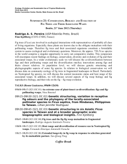
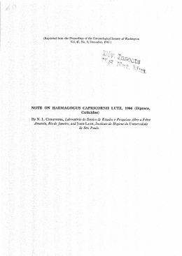
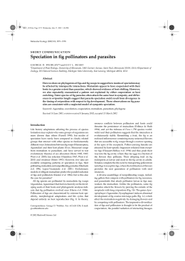
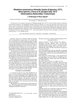
Available online at www.sciencedirect.com
CERAMICS
INTERNATIONAL
Ceramics International 41 (2015) 3069–3074
www.elsevier.com/locate/ceramint
A theoretical investigation of the structural and electronic properties
of orthorhombic CaZrO3
I.L.V. Rosaa, M.C. Oliveiraa,b, M. Assisa, M. Ferrera, R.S. Andréa, E. Longob, M.F.C. Gurgelc,n
a
LIEC–Department of Chemistry, Universidade Federal de São Carlos, Via Washington Luiz, Km 235, P.O. Box 676, 13565-905 São Carlos, São Paulo, Brazil
b
Institute of Chemistry, Universidade Estadual Paulista—Unesp, P.O. Box 355, 14801-907 Araraquara, São Paulo, Brazil
c
Department of Chemistry, Universidade Federal de Goiás, Regional de Catalão, Av. Dr. Lamartine Pinto de Avelar, 75704-020 Catalão, Goiás, Brazil
Received 30 July 2014; received in revised form 21 October 2014; accepted 25 October 2014
Available online 6 November 2014
Abstract
A CaZrO3 (CZO) powder was prepared by the soft chemical, polymeric precursor method (PPM). The CZO crystalline structure was
investigated by powder X-ray diffraction (XDR), Retvield Refinament data, Raman spectra and ultraviolet–visible absorption spectroscopy. A
theoretical study was performed using a periodic quantum mechanical calculation (CRYSTAL09 program). The periodic model built for the
crystalline CZO structure was consistent with the experimental data obtained from structural and electronic properties. These results show that the
material has an orthorhombic structure with experimental and theoretical gap values of 5.7 eV and 6.2 eV, respectively. In this article, we discuss
the hybridization process of the oxygen p-orbitals and of the zirconium d-orbitals and analyze their band structures and density of states (partial
and total).
& 2014 Elsevier Ltd and Techna Group S.r.l. All rights reserved.
Keywords: Perovskite; First principles; Electronic properties; CZO
1. Introduction
ABO3 perovskites (where A ¼ Ca, Sr, Pb or Ba and B ¼ Ti
or Zr) are oxides that have been extensively investigated due to
their significance in fundamental research and in various
technological applications [1–3]. These perovskite-type structures, where A and B are cations that correspond to oxygen
ions, can have various symmetries (e.g. cubic unit cell,
tetragon and distorted) depending on processing conditions
and structural order [4]. Interest is increasing in these materials
because of their physical and chemical properties that are
optimal for diverse applications. Many studies have proposed
low-cost preparation methods. The literature reports several
synthesis methods such as a solid-state reaction [5], propionic
acid routes [6], malate [7], co-precipitation [8], sol-gel [9],
modified Pechini [10] and citrate [11].
n
Corresponding author. Tel.: þ55 64 34415334; fax: þ 5564 34415300.
E-mail address: [email protected] (M.F.C. Gurgel).
http://dx.doi.org/10.1016/j.ceramint.2014.10.149
0272-8842/& 2014 Elsevier Ltd and Techna Group S.r.l. All rights reserved.
Calcium Zirconate (CZO) has excellent chemical and physical properties including chemical stability [12–14]. CaZrO3 is an
orthorhombic perovskite (Pcnm space group) at ambient temperatures. This structure consists of small deformations in the
ZrO6 octahedron with Zr-O bond lengths from 2.091(1) to
2.101(1) Å and O-Zr-O bond angles from 88:0(1)1 to 99:0(1)1.
CaZrO3 can be cubic with Pbnm (at low temperature) and
Pm3m (after high temperature heat treatment) space groups [15].
Brik et al. [16] used the DFT method to investigate the
structural, electronic, and energy properties of the surface
(0 0 1) of the CZO cubic terminations (CaO and ZrO). They
observed that the CaO surface termination had lower energy
and calculated 3.283 eV (LDA) and 3.315 eV (GGA) for the
CZO cubic structure.
Hou [17] used the pseudopotential method for plane wave
calculations to analyze the structural and electronic properties,
and the elastic constant of the cubic CZO and concluded that
the material is mechanically stable with a band gap of 3.30 eV.
Stoch et al. [18] showed that CZO obtained through a solidstate reaction has a similar orthorhombic structure. The full
3070
I.L.V. Rosa et al. / Ceramics International 41 (2015) 3069–3074
potential linear plane wave method (FLAPW) showed the gap
values of 4.1 eV.
Our group used periodic ab intio analysis to investigate
correlations between experimental and theoretical studies on
perovskites [19–24]. In the present study, we present XDR,
UV–vis and Raman spectroscopy of crystalline orthorhombic
CZO prepared by PPM. We built a periodic model based on
Rietveld refinement data and associated the results with the
quantum mechanical theoretical study to investigate the structure and electronic properties of this important material.
2. Experimental details
2.1. Synthesis of pure CZO
Pure CZO was obtained by dissolving ZrOCl2.8H2O
(0.0102 mol) into 40.0 mL of deionized water (60 1C)
while stirring. Then, C6H8O7(0.1224 mol) and CaCl2.2H2O
(0.0102 mol) were added. Next, ethyleneglycol was added and
the temperature increased to 80 1C, promoting there actional
polymerization. This process converted the material into a
resin. The resin was then heated at 350 1C for 4 h causing
pyrolysis. Finally, the material was heated again at 900 1C for
2 h, resulting in the crystallized CZO material [22].
laser providing excitation light (1064 nm), at a spectral
resolution of 4 cm_1. The UV–vis absorption spectra of the
CZO powders were measured with a Cary 5 G instrument.
2.3. CZO structure and periodic model details
Periodic quantum mechanical calculations (ab initio) were
performed using the CRYSTAL09 [29] program and showed
the orthorrombic structure with Pcnm space group of the CZO
material. A package within the framework of the Density
Functional Theory and a gradient-corrected correlation function by Lee, Yang and Parr, combined with the Becke3
exchange functional (B3LYP) were employed. The atomic
centers were described by all-electron basis sets for the Ca, Zr,
Ti and O atoms [30]. The XCrySDen program was used to
design the periodic model and band structure and to outline the
density of states diagrams [31]. We built the periodic model
2.2. Rietveld refinements and characterization
The CZO crystalline powder was analyzed by X-ray
diffraction (XRD). The diffraction patterns of the crystalline
powders were recorded by a Rigaku RINT2000 diffractometer
in step scan mode (D2 h¼ 0.02_, 20_ 6 2 h 6 130_) using
copper radiation (kKa1¼ 1.5406 A˚, kKa2 ¼ 1.5444 A1, Ka1/
Ka2 ¼ 0.5) monochromatized by a graphite crystal. The tube
power was 42 kV 120 mA, with a divergence slit of 0.5 mm
and receiving slit of 0.30 mm. Rietveld refinements [25] were
performed with the GSAS program [26]. The peak profile
function was modeled using the Thompson-Cox-Hastings
pseudo-Voigt (pV-TCH) convolution with the asymmetry
function described by Finger et al. [27]. Strain anisotropy
broadening was corrected by the phenomenological model
described by Stephens [28]. Raman spectra were recorded on
an RFS/ 100/S Bruker FT-Raman spectrometer with Nd:YAG
Fig. 1. XRD patterns for crystalline CZO powder annealed at 900 1C for 2 h.
Table 1
Rietveld refinement data for pure CZO.
Space group¼ Pnmc
Lattice parameter (Å)
Centralatoms
Calcium
Zirconium
Oxygen 1
Oxygen 2
Agreement indicies
Rwp 8.02%
a¼ 5.59444
b ¼8.02115
Atomic coordinates
c¼ 5.76111
X
Y
Z
0.0125
0.0
0.61301
0.29986
0.25
0.0
0.25
0.05664
0.04798
0.5
0.04238
0.3011
R2F 3.02%
χ2 1.301
Fig. 2. Representation of the unit cell to periodic model CZO.
I.L.V. Rosa et al. / Ceramics International 41 (2015) 3069–3074
based on the Rietveld refinement results (Table 1) for the CZO
crystalline structure of using a unit cell to represent the
periodic models.
3071
3. Results and discussion
3.1. X-ray diffraction
Fig. 1 illustrates the XRD patterns of the CZO powders
calcined at 900 1C for 2 h. The pattern is typical of the ordered
orthorhombic perovskite phase (n1 35–0790)
3.2. Rietveld refinement
The Rietveld refinement results (Table 1) were used to
construct the periodic pattern of the CZO (Fig. 2). This periodic
model consists of 4 zirconium atoms, 4 calcium atoms and 12
oxygen atoms, resulting in an orthorhombic structure (Pcmn).
3.3. Raman spectroscopy
Fig. 3. Raman spectrum of the CZO material, heat-treated at 900 1 C for 2 h.
Raman spectroscopy measurements were performed to
identify the material at short range. Fig. 3 shows the Raman
spectrum for the ceramic CZO powder annealed at 900 1C/ for
2 h.
Fig. 3 shows the Raman spectrum of CZO heat-treated at
900 1C for 2 h. This spectrum has peaks that are characteristic
of a material with a short-range orthorhombic structure. The
frequencies of the CZO Raman active modes (Table 2) are in
Table 2
Frequencies (cm 1) of the Raman active modes for CZO powders heat-treated at 900 1C for 2 h.
Rights
CZO
M. Tarrida, et al. [33] (Ca,Sr)ZrO3 and
obtained Ca(Sn,Zr)O3) (Pnma) symmetry group.
(λ cm 1)
H. Zheng, et. al.[34] CaZrO3–
V.M. Orera, et al. [35]
CaTiO3 (Pnma) symmetry group. CaZrO3 (Pcmn) symmetry
group.
(Ag) Network modes 141
(Ca-ZrO3)
145
P. McMillan, et al.[36]
CaZrO3 (Pbnm) symmetry
group.
143
(B2g) bonding
modes (flexion)
(O-Zr-O)
185
151
172
185
(B1g) bonding
modes (flexion)
(O-Zr-O)
(B1g) bonding
modes (flexion)
(O-Zr-O)
206
193
213
190
212
190
212
189
212
228
229
227
227
224
235
263
233
262
234
262,5
233
260
287
286
286,5
285
305
358
302
338
356
(Ag) bonding modes 257
(flexion) (O-Zr-O)
(Ag) bonding modes 281
(flexion) (O-Zr-O)
(Ag) torsional modes 354
(Zr-O3)
(Ag þ B1g) torsional 433
modes (Zr-O3)
(B2g) stretching
modes
(Ag þ B2g)
stretching modes
464
358
C.H. Perry, et al.[37]
CaZrO3 Orthorhombic
153
182
357
186
228
340
423
437
418
370
408
423
439
470
439
439
469
437
468
515
545
551
537
543
548
377
418
543
547
3072
I.L.V. Rosa et al. / Ceramics International 41 (2015) 3069–3074
agreement with the literature [32–37]. The overall half-width at
half-height of the Raman spectrum bands is proportional to the
inverse of the grain size. The frequencies at 141 cm 1 and
185 cm 1 correspond to the A1g vibrational modes and the
Zr-O (B2g) bending modes, respectively. The frequencies at
206 and 228 cm 1 are ascribed to the Zr -O bending modes
(B1g). The 257 and 281 cm 1 frequencies are ascribed to the
Zr -O bending (A1g) modes, 354 cm 1 to the torsional mode
(A1g) and 433 cm 1 to the torsional modes (A1g þ B1g). The
Raman band at the stretching modes Zr-O (A1g þ B2g) is also
apparent.
3.4. UV–vis spectroscopy
Fig. 4 shows the optical absorption curve as a function of
photon energy for the pure CZO samples.
The experimental Egap value for this sample was calculated
by the Wood and Tauc method, which relates sample
absorbance with photon energy to obtain the optical energy
gap (Egap) [38].
The UV–vis absorbance spectrum (Fig. 4) shows that the
Gap Energy is 5.7 eV. This experimental gap value is in good
agreement with the theoretical Egap of 6.23 eV.
3.5. Band structure
K-point sampling was conducted at 40 points within the
irreducible part of the Brillouin zone. The theoretical results were
represented in terms of band structures. Fig. 5(a-c) illustrate the
Brillouin zone, band structure and atomic orbital contributions
related to the oxygen, zirconium and calcium atoms. Fig. 5(a)
illustrates that the Brillouin zone for the unit cell is determined by
the CZO kpoints: Ѓ¼ (0,0,0), X¼ (1/2,0,0) Z¼ (1/2,0,0); U¼
(0,0,1/2); T¼ (0,1/2,1/2), Ѓ¼ (0,0,0). The gap was evaluated as
6.23 eV, which is considered a direct gap from (0,0,0) to (0,0,0)
kpoints. Fig.5(b) represents the structure band. Fig. 5(c) shows the
valence band (VB) from 0 to -0.62 eV where the oxygen orbitals
(px, py and pz) are located. Fig. 5(d) shows the conduction band
(CV) of the zirconium atomic orbitals 4d2z, 4d2xy, 4dxy, 4dxz, 4dyz,
situated from 6.23 to 8.11 eV. Slight contributions from the
atomic orbitals of the Ca atoms are also visible.
Several researchers have used computational methods and
theoretical models to interpret the structural and electronic
Fig. 4. Absorption Spectrum as a function of energy (eV) for the CZO powder properties of calcium zirconate with cubic and orthorhombic
heat-treated at 900 1C for 2 h.
structures [16–18]. Our article discusses an experimental and
Fig. 5. Representation of the CZO band structure (a) brillouin zone, (b) band structure, (c) p atomic orbitals in the VB and ( d) d and s atomic orbitals in the CB.
I.L.V. Rosa et al. / Ceramics International 41 (2015) 3069–3074
theoretical study of orthorhombic CZO. We synthesized the
material by PPM and carried out computations using the
CRYSTAL 09 program [29]. In the case of CZO synthesized
by the MPP procedure both experimental and theoretical Egap
values are 5.7 and 6.23 eV, respectively
3.6. Partial DOS
Fig. 6(a-d) shows total DOS of CZO from the atomic
orbitals of zirconium, calcium and oxygen, respectively. Some
of the atoms in this structure contribute equally given the
3073
structural symmetry of CZO. A predominant contribution of d
and p atomic orbitals from the Zirconium and Oxygen atoms in
the conduction band region (CB) and in the valence band
region (BV), respectively, was noticed. The s contributions of
the atomic orbitals from the Calcium atoms are also present in
the CB and BV bands being, however, less intense. Fig. 6(a)
illustrates the equivalent contributions to total DOS from the
atomic orbitals (4d) of the four zirconium atoms (Zr1-4). Fig. 6
(b) shows the equivalent contributions from the atomic orbitals
of the four calcium atoms (Ca1-4). The CZO structure is
composed of twelve oxygen atoms with contributions from the
px, py and pz orbitals. DOS from the oxygen atoms was
divided into two equivalent oxygen groups (O1-4 and O5-12)
(Fig. 6c-d).
Fig. 7(a) shows the CZO unit cell and the diagonal plane
designated by the red dots among the Zr-O-Ca-O-Zr atoms.
Fig. 7(b) illustrates the electron density map of the Ca, Zr and
O atoms and shows contour lines indicating hybridization
among the atoms in the diagonal plane. A homogeneous
borderline between the Zr-O and Ca-O atoms representing
covalent and ionic bonds, respectively [39].
4. Conclusions
Fig. 6. Total DOS from the pure CZO: (a) 4 zirconium atoms (Zr1-4) (b) 4 calcium
atoms (Ca1-4), (c) 4 oxygen atoms (O1-4) and (d) 8 oxygen atoms (O5-12).
Crystalline CZO was synthesized by PPM. XRD patterns,
Rietveld refinement and Raman spectroscopy confirmed the
orthorhombic structure of this material, whereas UVvis absorption allowed calculation of the value gap Egap (5.7 eV). The
periodic quantum mechanical calculation (PQMC) through
CRYSTAL09 was used to determine the first-principle calculations needed to study the structure and electronic properties of
CZO. Our theoretical band structure and DOS results show
hybridization between Zr and O atoms resulting from strong
covalent bonding between zirconium d-states of and oxygen
p-states. CZO has a direct band gap with Egap ¼ 6.23 eV.
Fig. 7. Representation of the Zr-Ca-O diagonal plane from CZO (a) unit cell and (b) electron density map.
3074
I.L.V. Rosa et al. / Ceramics International 41 (2015) 3069–3074
Acknowledgments
The financial support of this research project by the
Brazilian research funding agencies CNPq, CAPES and
FAPESP is gratefully acknowledged
[20]
References
[21]
[1] V.V. Bannikov, I.R. Shein, V.L. Kozhevnikov, A.L. Ivanovskii, Magnetism without magnetic ions in non-magnetic perovskites SrTiO3, SrZrO3
and SrSnO3, J. Magn. Magn. Mater. 320 (2008) 936–942.
[2] M.F.C. Gurgel, M.L. Moreira, E.C. Paris, J.W.M. Espinosa, P.S. Pizani,
J.A. Varela, E. Longo, BaZrO3 photoluminescence property: an Ab initio
analysis of structural deformation and symmetry changes, Int. J. Quantum
Chem. 111 (2011) 694–701.
[3] H. Pinto, A. Stashans, Computational study of self-trapped hole polarons
in tetragonal BaTiO3, Phys. Rev. B 65 (2002) 1343041–1343046.
[4] P.S. Pizani, E.R. Leite, F.M. Pontes, E.C. Paris, J.H. Rangel, E.J.H. Lee,
E. Longo, P. Delega, J.A. Varela, Photoluminescence of disordered ABO
(3) perovskites, Appl. Phys. Lett. 77 (2000) 824–826.
[5] K.R. Kambale, A.R. Kulkarni, N. Venkataramani, Grain growth kinetics
of barium titanate synthesized using conventional solid state reaction
route, Ceram. Int. 40 (2014) 667–673.
[6] T. Horikawa, N. Mikami, T. Makita, J. Tanimura, M. Kataoka, K. Sato,
M. Nunoshita, Dielectric-properties of (Ba,Sr)TiO3 thin-films deposited
by rf-sputtering, Jpn. J. Appl. Phys. 1 (32) (1993) 4126–4130.
[7] C. Marcilly, P. Courty, B. Delmon, Preparation of highly dispersed mixed
oxides and oxide solid solutions by pyrolysis of amorphous organic
precursors, J. Am. Ceram. Soc. 53 (1970) 56–57.
[8] A.V. Murugan, V. Samuel, S.C. Navale, V. Ravi, Phase evolution of
NiTiO3 prepared by coprecipitation method, Mater. Lett. 60 (2006)
1791–1792.
[9] I. Chilibon, J.N. Marat-Mendes, Ferroelectric ceramics by sol-gel
methods and applications: a review, J. Sol-Gel Sci.Techol. 64 (2012)
571–611.
[10] Y.J. Lin, Y.H. Chang, W.D. Yang, B.S. Tsai, Synthesis and characterization of ilmenite NiTiO3 and CoTiO3 prepared by a modified Pechini
method, J. Non-Cryst. Sol. 352 (2006) 789–794.
[11] H. Taguchi, D. Matsuda, M. Nagao, K. Tanihata, Y. Miyamoto, Synthesis
of perovskite-type (La1-XSrx)MnO3 (O-less-than-or-equal-to-X-less-thanor-equal-to-0.3) at low-temperature, J. Am. Ceram. Soc. 75 (1992)
201–202.
[12] Y. Du, Z.P. Jin, P.Y. Huang, Thermodynamic calculation of the zirconia
calcia system, J. Am. Ceram. Soc. 75 (1992) 3040–3048.
[13] G. Rog, M. Dudek, A. Kozlowska-Rog, M. Bucko, Calcium zirconate:
preparation, solid oxide properties and application to the galvanic cells,
Electrochim. Acta 47 (2002) 4523–4529.
[14] J. Szczerba, Z. Pedzich, The effect of natural dolomite admixtures on
calcium zirconate-periclase materials microstructure evolution, Ceram.
Int. 36 (2010) 535–547.
[15] H.J.A. Koopmans, G.M.H. Vandevelde, P.J. Gellings, Powder neutrondiffraction study of the perovskites CaTiO3 and CaZrO3, Acta Crystallogr. C 39 (1983) 1323–1325.
[16] M.G. Brik, C.G. Ma, V. Krasnenko, First-principles calculations of the
structural and electronic properties of the cubic CaZrO3 (001) surfaces,
Surf. Sci. 608 (2013) 146–153.
[17] Z.F. Hou, Ab initio calculations of elastic modulus and electronic
structures of cubic CaZrO3, Phys. B –Condens. Matter 403 (2008)
2624–2628.
[18] P. Stoch, J. Szczerba, J. Lis, D. Madej, Z. Pedzich, Crystal structure and
ab initio calculations of CaZrO3, J. Am. Ceram. Soc 32 (2012) 665–670.
[19] M.S.C. Camara, M.F.C. Gurgel, S.R. Lazaro, T.M. Boschi, P.S. Pizani,
E.R. Leite, A. Beltran, E. Longo, Room temperature photoluminescence
[22]
[23]
[24]
[25]
[26]
[27]
[28]
[29]
[30]
[31]
[32]
[33]
[34]
[35]
[36]
[37]
[38]
[39]
of the Li2ZnTi3O8 spinel: Experimental and theoretical study, Int.
J. Quantum Chem. 103 (2005) 580–587.
L.S. Cavalcante, N.C. Batista, T. Badapanda, M.G.S. Costa, M.S. Li,
W. Avansi, V.R. Mastelaro, E. Longo, J.W.M. Espinosa, M.F.C. Gurgel,
Local electronic structure, optical bandgap and photoluminescence (PL)
properties of Ba(Zr0.75Ti0.25)O3 powders, Mater. Sci. Semicon. Proc. 16
(2013) 1035–1045.
V.M. Longo, A.T. de Figueiredo, S. de Lazaro, M.F. Gurgel,
M.G.S. Costa, C.O. Paiva-Santos, J.A. Varela, E. Longo,
V.R. Mastelaro, F.S. De Vicente, A.C. Hernandes, R.W.A. Franco,
Structural conditions that leads to photoluminescence emission in SrTiO3:
An experimental and theoretical approach, J. Appl. Phys. 104 (2008)
0235151–02351511.
J.M.A. Nunes, J.W.M. Espinosa, M.F.C. Gurgel, P.S. Pizani, S.H. Leal,
M. Santos, E. Longo, Photoluminescent properties of lead zirconate
powders obtained by the polymeric precursor method, Ceram. Int. 38
(2012) 4593–4599.
E.C. Paris, M.F.C. Gurgel, M.R. Joya, G.P. Casali, C.O. Paiva-Santos,
T.M. Boschi, P.S. Pizani, J.A. Varela, E. Longo, Structural deformation
monitored by vibrational properties and orbital modeling in (Pb,Sm)TiO3
systems, J. Phys. Chem. Solids 71 (2010) 12–17.
J.R. Sambrano, E. Orhan, M.F.C. Gurgel, A.B. Campos, M.S. Goes,
C.O. Paiva-Santos, J.A. Varela, E. Longo, Theoretical analysis of the
structural deformation in Mn-doped BaTiO3, Chem. Phys. Lett. 402
(2005) 491–496.
H.M. Rietveld, A profile refinement method for nuclear and magnetic
structures, J. Appl. Crystallogr 2 (1969) 65.
A.C. Larson, R.B. Von Dreele, General Structure Analysis System
(GSAS) Program, Rep. N1 (LAUR 86–748), Los Alamos National
Laboratory, University of California, Los Alamos, NM, 2004.
L.W. Finger, D.E. Cox, A.P. Jephcoat, A correction for powder
diffraction peak asymmetry due to axial divergence, J. Appl. Crystallogr
27 (1994) 892–900.
P.W. Stephens, Phenomenological model of anisotropic peak broadening
in powder diffraction, J. Appl. Crystallogr. 32 (1999) 281–289.
R. Dovesi, V.R. Saunders, C. Roetti, R. Orlando, C.M. Zicovich-Wilson,
F. Pascale, B. Civalleri, K. Doll, N.M. Harrison, I.J. Bush, P.D.' Arco,
M. Llunell, CRYSTAL09 User's Manual, University of Torino, Torino,
2009.
CRYSTAL Basis Sets Library. 〈http://www.crystal.unito.it/Basis_Sets/
Ptable.html〉, 2014 (accessed 04.07.14).
A. Kokalj, XCrySDen - a new program for displaying crystalline
structures and electron densities, J. Mol.Graph. Model. 17 (1999)
176–179.
I. Levin, E. Cockayne, M.W. Lufaso, J.C. Woicik, J.E. Maslar, Local
structures and Raman spectra in the Ca(Zr,Ti)O3 perovskite solid
solutions, Chem. Mater. 18 (2006) 854–860.
P. McMillan, N. Ross, The raman-spectra of several orthorhombic
calcium-oxide perovskites, Phys. Chem. Miner. 16 (1988) 21–28.
V.M. Orera, C. Pecharroman, J.I. Pena, R.I. Merino, C.J. Serna, Vibrational spectroscopy of CaZrO3 single crystals, J. Phys.-Condens. Matter
10 (1998) 7501–7510.
C.H. Perry, D.J. McCarthy, G. Rupprech, Dielectric dispersion of some
perovskite zirconates, Phys. Rev. 138 (1965) 1537–1538.
M. Tarrida, H. Larguem, M. Madon, Structural investigations of (Ca,Sr)
ZrO3 and Ca(Sn,Zr)O3 perovskite compounds, Phys. Chem. Miner. 36
(2009) 403–413.
H. Zheng, I.M. Reaney, G. de Gyorgyfalva, R. Ubic, J. Yarwood,
M.P. Seabra, V.M. Ferreira Raman, spectroscopy of CaTiO3-based
perovskite solid solutions, J. Mater. Res. 19 (2004) 488–495.
D.L. Wood, J. Tauc, Weak absorption tails in amorphous semiconductors,
Phys. Rev. B 5 (1972) 3144.
D.F. Shriver, P.W. Atkins, Inorganic Chemistry, fourth ed., Oxford
University, United States, 2006.
Download