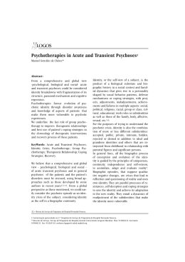
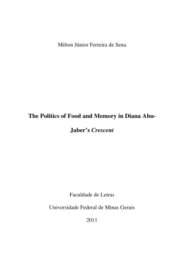
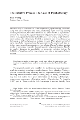
This article appeared in a journal published by Elsevier. The attached
copy is furnished to the author for internal non-commercial research
and education use, including for instruction at the authors institution
and sharing with colleagues.
Other uses, including reproduction and distribution, or selling or
licensing copies, or posting to personal, institutional or third party
websites are prohibited.
In most cases authors are permitted to post their version of the
article (e.g. in Word or Tex form) to their personal website or
institutional repository. Authors requiring further information
regarding Elsevier’s archiving and manuscript policies are
encouraged to visit:
http://www.elsevier.com/copyright
Author's personal copy
Journal of Psychiatric Research 45 (2011) 727e734
Contents lists available at ScienceDirect
Journal of Psychiatric Research
journal homepage: www.elsevier.com/locate/psychires
Police officers under attack: Resilience implications of an fMRI study
Julio F.P. Peres a, *, Bernd Foerster b, Leandro G. Santana c, Mauricio Domingues Fereira c,
Antonia G. Nasello d, Mariângela Savoia d, Alexander Moreira-Almeida e, Henrique Lederman a
a
Department of Diagnostic Imaging – Radiologia Clinica Universidade Federal de Sao Paulo, Escola Paulista de Medicina, SP, Brazil
Philips Medical Systems, USA
c
Sao Paulo State Militarized Police Force, Brazil
d
Faculty of Medical Sciences, Santa Casa de Sao Paulo, Brazil
e
School of Medicine, Universidade Federal de Juiz de Fora, Minas Gerais, Brazil
b
a r t i c l e i n f o
a b s t r a c t
Article history:
Received 30 July 2010
Received in revised form
22 September 2010
Accepted 2 November 2010
Objective: Crime is now a top-priority public-health issue in many urban areas. Sao Paulo’s state police
force was the target of gunfire attack on an unprecedented scale. Several officers were killed or wounded,
and many more were affected by psychological trauma. We investigated the brain activity underlying
trauma, the coping effect of psychotherapy, and resilience in a highly homogenous sample that experienced the same traumatic event. The design applied was a between-group comparison of cerebral
blood-oxygenation-level-dependent signals and symptom scores of police officers with and without
partial Posttraumatic Stress Disorder (pPTSD).
Method: We used functional magnetic resonance imaging (fMRI) to investigate the retrieval of traumatic
memories of 36 volunteers divided in three groups: (1) pPTSD policemen submitted to psychotherapy;
(2) pPTSD policemen on the wait list; and (3) symptom-free (resilient) policemen. All participants were
given a baseline fMRI scan and a follow-up scan some 40 days later. Not given psychotherapy, groups 2
and 3 were controls.
Results: Group 1 showed 37% fewer PTSD symptoms post-psychotherapy and their scores and neural
expressions were comparable to Group 3 resilient policemen. A marked increased in medial prefrontal
cortex (mPFC) activity was concomitant with decreased amygdala activity during traumatic memory
retrieval in both resilient and pPTSD participants (after psychotherapy) and these findings were associated with symptom attenuation.
Conclusions: Our results provide neurophysiological evidence of resilience in a high-risk group for PTSD.
Psychotherapy may help to build narratives and resilient integrated translations of fragmented traumatic
memories via mPFC, and thus weaken their sensory content while strengthening them cognitively.
! 2010 Elsevier Ltd. All rights reserved.
Keywords:
PTSD
Police
Psychotherapy
Resilience
Neuroimaging
fMRI
1. Introduction
Most of us have dealt with a traumatic event of some kind such as
loss, accident, or illness, or will be dealing with one at some point in
our lives. Psychological trauma is closely related to the development
of posttraumatic stress disorder (PTSD), involving three sets of
symptoms: (i) reliving trauma (traumatic memories, nightmares,
intrusive thoughts); (ii) emotional avoidance/numbness (affective
distance, emotional anaesthesia); and (iii) increased arousal (irritability, insomnia and hypervigilance). Lifetime prevalence of PTSDtriggering traumatic events may be as much as 50e90%, and actual
* Corresponding author. Rua Maestro Cardim 887, Sao Paulo e SP, Brazil, Postal
Code 01323-001, fax: þ55 11 3284 8929.
E-mail address: [email protected] (J.F.P. Peres).
0022-3956/$ e see front matter ! 2010 Elsevier Ltd. All rights reserved.
doi:10.1016/j.jpsychires.2010.11.004
prevalence in the general population is about 8% (Kessler et al.,1995;
Vieweg et al., 2006), while partial PTSD (pPTSD) in an at-risk group
exposed to combat such as Vietnam veterans has been estimated at
approximately 30% (Weiss et al., 1992). After noting that individuals
who do not meet the full set of diagnostic criteria for PTSD may
suffer from clinically significant symptoms of PTSD (Weiss et al.,
1992), the concept of pPTSD or sub-threshold PTSD was introduced to describe subsyndromal forms of PTSD (Blanchard et al.,
1995; Stein et al., 1997).
Crime is now one of the most frequent causes of death in many
countries. Coping with stressful and life-threatening situations is
part of policing so officers are routinely exposed to critical-incident
stressors in the line of duty. Although active policemen are understudied relative to other traumatized samples, this population may
be critical to our understanding of responses to traumatic stressors
Author's personal copy
J.F.P. Peres et al. / Journal of Psychiatric Research 45 (2011) 727e734
(Kessler et al., 1995; Vieweg et al., 2006; Weiss et al., 1992). A
criminal organization attacked the State of Sao Paulo Militarized
Police Force on an unprecedented scale in the period May 12e23,
2006, and many officers were killed or wounded in firefights. Some
were traumatized, while others remained symptom free.
Whether an event is traumatic or not will depend on an individual’s perceptual neural-circuitry processing and underlying
resilience, which is the ability to cope effectively and adapt in the
face of loss, hardship or adversity (Bonanno, 2004; Block and
Kremen, 1996). Resilient individuals reported fewer posttraumatic
symptoms after combat and showed greater ability to optimize
emotional functioning through the use of alternative cognitive
strategies (Bonanno, 2004; Florian et al., 1995). Neuroscientists
have yet to comprehensively research this field. Examining neural
mechanisms underlying psychological trauma or resilience is
difficult given the heterogeneous symptoms and peculiarities of
traumatic memories (key symptoms of PTSD). There are several
methodological challenges and complex factors to control such as:
(i) traumatized individuals typically present various comorbidities
(e.g. major depression, substance abuse, etc.), (ii) traumatic events
of different kinds (violence, accidents, loss, etc.) involve distinct
sensory levels and modalities of memories (visual, tactile, olfactory,
auditory, affective), (iii) different PTSD symptoms and emotions
may accompany specific neural interactions during retrieval of
traumatic memories (e.g. dissociative experiences are psychoneurophysiologically different from hyperarousal experiences), (iv)
the heterogeneous nature of trauma may pose difficulties when
inducing reproducible responses in patients, or comparable activations in healthy control subjects, (v) the recency of the memories
being studied is often different (memory expression may be
modified over time, causing changes in the neural substrates
involved).
In the last ten years, however, neuroimaging research has yielded important information on heightened amygdala responsivity in
PTSD patients during symptomatic states, and has found that
medial prefrontal cortex (mPFC) responsivity is inversely associated
with PTSD-symptom severity (Shin et al., 2006). Nevertheless, the
directionality of the PFC to amygdalaeactivity correlation has been
inconsistent: negative in PTSD cases but positive in controls, suggesting coupling only in psychopathology (Shin et al., 2005; Peres
et al., 2008).
Two basic issues yet to be addressed are the reasons for most
trauma survivors not developing PTSD and the predictors of
positive outcomes in traumatized victims (Shin et al., 2005, 2006).
All the police officers in our sample had come under fire and seen
colleagues being shot, so our study offered a unique opportunity
to investigate victims who were all affected by the same traumatic
event, which they reported as watching wounded colleagues
dying after pleading for help. The study controlled all five variables listed above and was conducted 3 months after the criminal
attacks, so there was sufficient time for a confident diagnosis of
PTSD. In order to address the object of the study e brain activity
underlying trauma, psychotherapy effect and resilience e we used
functional magnetic resonance imaging (fMRI) to detect alterations in brain activity related to overcoming trauma after
psychotherapy.
Since we had screened for startle response and hypervigilance
(Fig. 1) as prevalent symptoms, we hypothesized that pPTSD
policemen would present an exaggerated amygdala response
pattern (Bremner, 2003) during traumatic memory retrieval prior to
psychotherapy. On the other hand, we predicted that resilient
policemen and pPTSD policemen would show increased PFC activation after psychotherapy. Based on studies that have suggested
that lower cortisol levels in traumatized victims and combat veterans
(Yehuda et al., 1995) pose a risk factor for PTSD, we hypothesized that
Arrival
Rest
Post-fMRI
150
Cortisol ng/dL
728
100
50
0
Psychotherapy
Wait List
Healthy
Fig. 1. Samples of saliva cortisol were collected using the non-invasive Salivette-Sarstedt method on three occasions: (i) on arrival at the neuroimaging center (ii) after
30 min in repose, and (iii) 15 min after concluding the fMRI scan. All salivary samples
were obtained from 6pm to 8pm.
pPTSD policemen would present lower cortisol levels than healthy
(symptom-free) policemen.
2. Methods
2.1. Participants
We initially examined 97 police officers, all targeted in the wave of
gunfire attacks prior to the study, of whom 29.6% developed pPTSD.
Twenty-four of them with pPTSD and 12 healthy (resilient) men were
randomly selected for the study. All 36 policemen studied (mean age
28.2, SD ¼ 3.2) were free of comorbidities; there were no differences
in recency of the stressor and all had experienced the same event,
which caused trauma for some, but not others (Table 1). According to
Blanchard et al. (1995) and Stein et al. (1997) we considered pPTSD
subjects as those who presented fewer than the required number of
DSM-IV criterion B, or C or D symptoms for full PTSD. The pPTSD
individuals selected for the study did not present symptoms in the
Table 1
Demographic characteristics of the 36 policemen.
Gender (male/female)
Cerebral dominance
(right/non-right)
Marital status
(single/married)
Age subjects
(years/months)
(mean/SD/range)
Comorbidities (with/
without)
Psychotropic medication
(with/without)
Age of memories
(months/days)
Education (secondary
school/university)
Type of trauma (criminal
attack/others)
Previous traumatic events
scoring over 10 (Impact
Event Scale)
pPTSD
Psychotherapy
group (n ¼ 12)
pPTSD
Wait-list
group (n ¼ 12)
12/0
12/0
12/0
12/0
8/4
5/7
6/6
31.2/5.8/
24e36
27.6/3.9/
23e31
28.2/7.8/
25e38
0/12
0/12
0/12
0/12
0/12
0/12
3.5
3.10
3.15
12/0
12/0
12/0
12/0
12/0
12/0
0
0
0
12/0
12/0
Healthy group
(n ¼ 12)
Summary of demographic characteristics of all pPTSD subjects (24) and healthy
subjects (12). SD, Standard deviation.
Author's personal copy
J.F.P. Peres et al. / Journal of Psychiatric Research 45 (2011) 727e734
avoidance and numbing cluster of the Clinician Administered PTSD
Scale for DSM-IV (CAPS; Blake et al., 1990), but did present re-experiencing and hyperarousal clusters. In the present study specifically,
pPTSD subjects had the same recurrent traumatic memories (criterion B), hypervigilance and exaggerated startle response (criterion D)
as prevalent symptoms, but presented sub-threshold symptoms for
criterion C e numbing of general responsiveness e thus not meeting
full DSM-IV criteria for PTSD. Police officers with dissociative
expression (criterion C) e which is an important confounding variable found in neuroimaging studies of PTSD patients (Shin et al., 2005,
2006; Peres et al., 2008) e were excluded. Moreover, to test whether
volunteers’ responses were dissociative or hypervigilant, and to
ensure sample homogeneity (Griffin et al., 1997; Lanius et al., 2002,
2006), we monitored heart rate before the study and during fMRI
scans, as well as saliva cortisol expression, since its measures differed
significantly in its relationship to dissociation versus hypervigilant
PTSD subjects (Simeon et al., 2008) (Fig. 1). Samples of saliva cortisol
were collected using the non-invasive Salivette-Sarstedt method
(Kirschbaum and Hellhammer, 1994; Poll et al., 2007). Only hyperresponsive volunteers were selected for the study. After giving
informed written consent, subjects were divided in three groups: (1)
pPTSD policemen subjected to Exposure and Cognitive Restructuring
Therapy (ECRT; Marks et al., 1998), (2) pPTSD policemen not yet
subjected to psychotherapy (wait-list), and (3) healthy (resilient)
policemen.
2.2. Symptom measures
Two blinded researchers examined the three groups before the
first and after the second fMRI scan. The Structured Clinical Interview from DSM IV (SCID; First et al., 1995) and the ClinicianAdministered PTSD Scale (CAPS; Blake et al., 1990) were applied to
all participants. In addition, the following standardized evaluations
were self-administered after each fMRI scan: the Beck Depression
Inventory (Beck et al., 1961), the Beck Anxiety Inventory (Beck et al.,
1988), and Impact of Event Scale (Horowitz et al., 1979), Dissociative
Experiences Scale (Fiszman et al., 2004) and Resilience Quotient Test
(Reivich and Shatte, 2002/validated by Barbosa, 2006). Because
most participants reported that spirituality/religiousness was an
important factor in their lives we also used the Brief Religious
Coping (RCOPE; Pargament et al., 2000) and Duke Religious Index
(DUREL; Koenig et al., 1997).
2.3. Sensory modalities of traumatic memories
The Traumatic Memory Inventory (TMI; Hopper and van der
Kolk, 2001) was used to evaluate and classify the basic characteristics of traumatic, pleasant and neutral memories. This inventory
evaluates the intensity and vivacity of five modalities of traumatic
memory (visual, affective, tactile, olfactory, auditory, and narrative),
and may be used to assess their evolution over time. Thus, pPTSD
subjects underwent the same type of assessment prior to treatment
as a baseline control condition, and following ECRT as a post-treatment condition. Policemen in control groups 2 and 3 were administered the TMI twice in forty days.
2.4. Standardizing the stressor event and activation paradigm
The policemen taking part were all officers involved in deadly
shooting incidents during the concerted wave of attacks, for whom
this was the stressor event of major emotional impact (Table 1).
When interviewed, they unanimously referred to the particular
events they described as the most traumatic: seeing wounded
colleagues pleading for help, or even dying, during the firefights. A
real-life recording of policemen under fire followed by death was
729
used as paradigm to evoke the traumatic memory. The policemen
selected for the study were those who, on listening to this stimulus,
presented faster heart rates (adrenergic response) rather than lower
ones (dissociative response) varying 20% or more from their baseline rates. All the policemen had a repertoire of pleasant memories
on a beach and neutral memories of an off-the-air television. White
noise (like an off-the-air television) and the sound of waves on
a beach with similar frequencies to the traumatic-event recording
(500e100,000 Hz) were used as neutral and pleasant stimuli
respectively. The frequencies of all auditory paradigms (neutral,
pleasant and traumatic) were also matched. All three memories
were from the same period as the traumatic episode which occurred
3 months prior to the study.
2.5. Psychological rehabilitation program
Exposure-based therapy and cognitive restructuring (ETCR) led
to PTSD patients showing marked improvements that were stable
over time (Marks et al., 1998; Peres et al., 2007; Bryant et al., 2008).
Exposure-based therapy in particular is often indicated as the
psychological treatment of choice for patients with traumatic
memories (Weiss et al., 1992; Bonanno, 2004; Peres et al., 2008).
Our research team had applied the ETCR procedure as standard
treatment for trauma victims, which helped them attain psychological growth on the basis of their negative experiences (Peres
et al., 2007). The police force’s Social and Legal Assistance Center
issued mental health rehabilitation guidelines for policemen
involved in life-threatening incidents based on psychological
assessments of officers involved in any incident that causes or may
cause psychological trauma. Its 28-day multidisciplinary psychological rehabilitation program involves ETCR groups (Marks et al.,
1998; Peres et al., 2007), art therapy and ecological walks.
2.6. Neuroimaging design
Three months after the traumatic event, we assessed the neural
mechanisms underpinning the psychological process related to
coping with trauma, as well as predictors of resilience, using fMRI
adapted for an acoustic-cue paradigm, with two scans interspaced
40-days. Recall or retrieval was cued by three types of memories:
(a) pleasant (soothing ocean-beach noise); (b) neutral (white noise
from an off-the-air TV station); and (c) traumatic (real-life
recording of gunfire during the wave of attacks). Group 1 was given
its baseline fMRI scan with a follow-up scan after the ETCR. Control
groups 2 and 3 were given baseline and follow-up scans but not
therapeutic procedures. Altered hemodynamic responses e bloodoxygenation-level-dependent (BOLD) e were measured while
reliving the traumatic memory recall (TR), the pleasant memory
(PL) and the neutral noise (NT). Conditions TR, PL and NT were
combined three times in an acquisition series. Participants were
subjected to a single period of acquisition totaling 6 min and 45 s.
Each series included three 30-s blocks. During both fMRI scans,
participants were asked to evoke their traumatic, pleasant and
neutral memories while three recordings were played through ear
phones. They were asked to lie down, be still, breathe through the
nose, and recall sensations and emotions associated with the
traumatic and pleasant memories as vividly as possible. The block
sequence was organized in random order and balanced across
participants. Blocks were separated by a 15-s period as a distinct
cognitive task for emotional separation from the next memory task,
while participants listened and mentally ran through a countdown
from 15 to 1 (Condition C). This design enabled us to examine BOLD
signals related to traumatic memory in both traumatized and
resilient police officers, and to discuss the clinical implications of
the findings for resilience.
Author's personal copy
730
J.F.P. Peres et al. / Journal of Psychiatric Research 45 (2011) 727e734
CAPS versus Amygdala
0.5
0.4
r^2=0.97
0.3
Amygdala
0.2
0.1
0
-0.1 10
20
30
40
50
60
-0.2
CAPS
-0.3
Pre-treatment
-0.4
Pos-treatment
-0.5
CAPS versus mPFC
0.6
r^2=0.97
0.4
mPFC
0.2
0
-0.2
-0.4
-0.6
10
20
30
Pre-treatment
Pos-treatment
40
50
60
CAPS
Author's personal copy
J.F.P. Peres et al. / Journal of Psychiatric Research 45 (2011) 727e734
2.7. fMRI procedures and data analysis
FMRI data were obtained using a clinical 1.5 T MR scanner, Achieva
Pulsar, Philips Medical Systems. Images were acquired with fat
saturated Echo Planar Imaging (TE/TR ¼ 50/3000 ms) in oblique axial
direction (29 slices, 4 mm slice thickness, 0.5 mm gap, 96 # 96 matrix
size with a quadratic field of view of 230 mm resulting in 2.4 mm inplane resolution). T1-weighted anatomic images were obtained with
a 3D TFE technique (TE/TR ¼ 5.1/25 ms, 0.76 # 0.76 # 1.6 mm3 spatial
resolution). BOLD data were preprocessed and statistically analyzed
using the statistical parametric mapping package SPM2, Welcome
Department of Cognitive Neurology. Images were realigned for
motion correction, normalized to Talairach coordinates and spatially
smoothed using a 5-mm Gaussian kernel. A voxel-by-voxel statistical
analysis was applied to the data, using a general linear model and
block designs. Analysis included a temporal highpass filter (128 s cutoff period), a voxel threshold of P ¼ 0.005 and a cluster threshold
(corrected for multiple comparisons) Pcor ¼ 0.05; only clusters larger
than 600 mm3 were considered significant. Multiple comparison
correction was performed by taking corrected p-values obtained from
analysis using the SPM2 software package, which implements
random-field-theory based algorithm correction (Nichols and
Hayasaka, 2003). Activation patterns were overlaid on the subject’s
T1 structure. We used region-of-interest (ROI) analyses for orbitofrontal and prefrontal cortex, anterior cingulate cortex, insula, thalamus, amygdala, hippocampus, and parietal lobes to test our a priori
hypotheses. ROIs were defined by the automated anatomical labeling
masks (Tzourio-Mazoyer et al., 2002). The statistical thresholds
employed were p < 0.05 (small volume corrected) for ROI analyses
and p < 0.001 for whole-brain analyses. BOLD signal change was
correlated with change in total CAPS score from before to after
treatment, and the statistical threshold for this analysis was p < 0.01.
2.8. Correlation analysis
Pearson correlations were generated to assess the association
between variations in BOLD signals of the group subjected to
psychotherapy. Significance tests for the correlations were limited
to the ROI structures, since these were the areas that would most
likely interact with each other during the pleasant, neutral and
traumatic memory retrievals. Given the sample size, all results
were confirmed using Spearman Rank correlations, which were
also performed between the CAPS score and percentage change in
BOLD responses for relevant ROI in the psychotherapy group. Since
results for both methods were similar, only Pearson correlations
have been shown. In order to calculate the sample correlation
coefficient (Pearson productemoment correlation coefficient) we
used the equation below:
731
P
P
X* Y
X*Y $
ffiffiffiffiffiffiffiffiffiffiffiffiffiffiffiffiffiffiffiffiffiffiffiP
ffiffiffiffiffiffiffiffiffiffiffiffiffi#
ffiffiffiffiffiffi"
ffiffiffiffiffiN
ffiffiffiffiffiffiffiffiffiffiffiffiffiffiffiffiffiffiP
ffiffiffiffiffiffiffiffiffiffiffiffiffi#
ffiffiffi
r ¼ sffi"
P 2 ð YÞ2
P 2 ð XÞ2
*
Y $
X $
N
N
P
3. Results
Overall, results showed that pPTSD participants (Group 1 and 2)
had markedly higher levels of left-amygdala activity (Talairach
$17/-6/-23, T-score ¼ 3.4, Pcorr<0.001, voxels ¼ 681) and decreased
activity in mPFC ('10/36/-15, T-score ¼ $4.8, Pcorr<0.001,
voxels ¼ 1945) during traumatic memory retrieval in the first fMRI
(Fig. 2). Again, increased left-amygdala activity ($17/-6/-23, Tscore ¼ 3.4, Pcorr<0.001, voxels ¼ 576) (Fig. 2) and decreased mPFC
activity ('10/36/-15, T-score ¼ $4.8, Pcorr<0.001, voxels ¼ 1623)
were observed in the second fMRI of Group 2. In contrast, the mPFC
BOLD response to traumatic memory retrieval was significantly
greater for Group 1 than for Group 2 in the second fMRI (*P < 0.05,
two-sample t test corrected for multiple comparisons), and Group-1
activation was significantly lower (more so after psychotherapy
than before it) in the left amygdala ($17/-6/-23, T-score ¼ 3.4,
Pcorr<0.001, voxels ¼ 92). In the second fMRI, Groups 1 and 3 both
showed less left-amygdala activity ($17/-6/-23, T-score ¼ 3.4, Pcorr
<0.001, voxels ¼ 232) and increased mPFC activity ('10/36/-15, Tscore ¼ -4.8, Pcorr <0.001, voxels ¼ 1852). Differences between
Group 30 s first and second scans did not reach significance, which
was to be expected for the resilient control group. In the same ROI,
we found that all three groups activated the mPFC when retrieving
pleasant memories in the first and second scans.
Whole-brain analysis and other ROIs such as anterior cingulate and
insula showed no statistically significant differences. In all three
groups (2 pPTSD and 1symptom-free), we observed normal release of
cortisol (92.7 ng/dL, SD ¼ 4.2) as expected for their age group and time
of day. For all conditions (exept induced relaxation e condition II) the
normal reference value ranges from 70 ng/dL to 170 ng/dL for the time
of day when salivary samples were obtained e 6pm to 8pm (Fig. 1).
Groups 1 and 2 showed similar symptom scores on initial
measures [48 ' 3.62 and 46 ' 2.70 (CAPS), 38 ' 2.63 and 35 ' 3.12
(IES), 13 ' 5.35 and 15 ' 6.45 (BDI) and 28 ' 2.54 and 31 ' 3.44
(BAI)]. Only the Group 1 subjects showed a fall of 37% or more in
total CAPS score post-psychotherapy, and we also observed
significant improvements in symptoms on their second measures
[19 ' 5.03, p ¼ 0.03 (CAPS), 10 ' 2.97, p ¼ 0.04 (IES), and 11 ' 2.03,
p ¼ 0.05 (BAI)] compared with their first, whereas Group 2 showed
no significant changes in its scores on the second set of measures
compared with the first [46 ' 2.70 and 49 ' 4.82, p ¼ 0.19 (CAPS),
Fig. 2. Correlation between changes in BOLD and changes in total severity of posttraumatic stress disorder (Clinician-Administered PTSD Scale, or CAPS) following ECRT. The
functional maps display the areas where changes in BOLD activity in medial prefrontal cortex (mPFC) and amygdala correlated with changes in total CAPS score. The scatter plots
display the direction of these correlations (increase in total CAPS on the horizontal axis, extent of BOLD activity on the vertical axis).
Author's personal copy
732
J.F.P. Peres et al. / Journal of Psychiatric Research 45 (2011) 727e734
Table 2
Inventory scores of psychotherapy and control groups.
pPTSD psychotherapy
Pre-psychotherapy
CAPS
48 ' 3.62
IES
38 ' 2.63
BDI
13 ' 5.35
BAI
28 ' 2.54
pPTSD wait-list group
1st Measures
CAPS
43 ' 4.82
IES
32 ' 5.64
BDI
16 ' 5.61
BAI
29 ' 5.10
Healthy control group
1st Measures
CAPS
12 ' 6.83
IES
9 ' 4.47
BDI
6 ' 6.15
BAI
12 ' 3.11
Post-psychotherapy
19 ' 5.03
10 ' 2.97
7 ' 3.09
11 ' 2.03
Significance
0.03
0.04
0.18
0.05
2nd Measures
46 ' 2.70
35 ' 3.12
15 ' 6.45
31 ' 3.44
Significance
0.19
0.16
0.21
0.09
2nd Measures
14 ' 5.71
7 ' 7.92
7 ' 5.33
10 ' 4.41
Significance
0.23
0.32
0.29
0.25
Psychotherapy treatment response characteristics (mean ' S.D.) (paired t-tests) and
control groups characteristics (mean ' S.D.) (paired t-tests) interspaced 40 days for
the Clinician Administered PTSD Scale (CAPS), the Impact of Event Scale (IES), the
Beck Depression Inventory (BDI), and the Beck Anxiety Inventory (BAI).
35 ' 3.12 and 37 ' 5.64, p ¼ 0.16 (IES), 31 ' 3.44 and 32 ' 5.10,
p ¼ 0.09 (BAI)]. After psychotherapy, Group 1 and 3 symptom scores
were comparable [19 ' 5.03 and 14 ' 5.71 (CAPS), 10 ' 2.97 and
7 ' 7.92 (IES), 11 ' 2.03 and 10 ' 4.41 (BAI)]. Group 3 scores did not
show significant changes compared to the first and second
measurements [12 ' 6.83 and 14 ' 5.71, p ¼ 0.23 (CAPS), 9 ' 4.47
and 7 ' 7.92, p ¼ 0.32 (IES), 12 ' 3.11 and 10 ' 4.41, p ¼ 0.25 (BAI)].
Group 1’s post-psychotherapy IES scores (10 ' 2.97) showed the
traumatic event causing less impact than previously (38 ' 2.63),
and coincided approximately with Group 3 (healthy) IES scores
(9 ' 4.47) (Table 2).
Group 30 s most salient resilience traits were self-efficacy
(p ¼ 0.05), empathy (p ¼ 0.04) and optimism (p ¼ 0.05). In relation to
religiosity, the resilient group’s scores showed significant prominence of intrinsic religiosity (p ¼ 0.02) as posited by DUREL, and two
main factors of positive religious coping with major life stressors
shown by the RCOPE: (1) “I sought God’s love and care” (seeking
spiritual support) (p ¼ 0.05) and (2) “I tried to put my plans into
action together with God” (collaborative religious coping) (p ¼ 0.04).
Pearson’s correlation coefficients were used to test correlation
between mPFC/amygdala activities and symptom scores. All correlations are reported for two-tailed p < 0.05. Only Group 1 showed
a positive correlation between change in total CAPS score and
change in mPFC activity ('10/36/-15) from pre- to post-psychotherapy (r ¼ 0.82, p ¼ 0.02). There was also a significant positive
correlation between mPFC and post-psychotherapy narrative TMI
scores (r ¼ 0.81, p ¼ 0.03). A negative correlation was found between
left-amygdala activation ($17/-6/-23) and change in total CAPS
score from pre- to post-psychotherapy (r ¼ 0.86, p ¼ 0.04).
4. Discussion
For the first time, it was possible to examine the neurofunctional
reciprocities of a homogeneous set of traumatized individuals
through control of complex variables (free of comorbidities and
medications, no need for washout, same age of traumatic memory,
same traumatic event also experienced by resilient individuals) in
relation to coping (Group 1), continuity (Group 2) and spontaneous
resilience to trauma (Group 3). After psychotherapy, Group 1 was
comparable to Group 3 resilient policemen in terms of symptom
scores and neural expressions related to traumatic memory
retrieval. These findings underline the importance of psychotherapy
for shortening the period of suffering and/or avoiding symptoms
becoming chronic e since Group 2 pPTSD policemen (not subjected
to psychotherapy) continued to present the same symptoms with
signs of worsening, whereas all those subjected to psychotherapy
presented a reduction of at least 37% in total CAPS scores.
Evidence from neuroimaging research indicates that the PFC
underlies many cognitive skills (Wood and Grafman, 2003). Current
and previous findings related to mPFC deactivation report that
pPTSD and PTSD patients experience difficulty in activating this
area, which is related to cognitive categorization and labeling of
internal states (Peres et al., 2007; Shin et al., 2006). Higher brain
regions such as the mPFC fail to diminish exaggerated arousal and
distress symptoms mediated via the amygdala, and this may be
related to the pathological responses found in psychologically
traumatized victims (Peres et al., 2008). The hypothesis that primary
pathology in PTSD may be amygdala hyper-responsivity rather than
deficient mPFC suggests ‘bottom-up’ activation of the amygdala on
100
90
80
70
60
50
40
30
20
10
0
Visual
Tactile
pPTSD Pre-psychoth
pPTSD Post-psychoth
Olfactory
Auditory
pPTSD 1st measures
pPTSD 2nd measures
Affective
Narrative
Healthy 1st measures
Healthy 2nd measures
Fig. 3. Memory modality and intensity scores of traumatic memory obtained after both fMRI scans for Group 1 (red), 2 (green) and 3 (blue). Traumatic memory was affectively and
sensorially less intense, and narrative scores were higher for Group 1 after psychotherapy. The sensory, affective and narrative modalities of traumatic memory remained similar for
Group 2 on first and second measures. Group 3 showed a well-defined narrative structure and low scores for sensory modalities of traumatic memory on both measures.
Author's personal copy
J.F.P. Peres et al. / Journal of Psychiatric Research 45 (2011) 727e734
the mPFC (Gilboa et al., 2004). Most neuroimaging studies of PTSD
show reduced mPFC activity (Peres et al., 2005, 2007; Lanius et al.,
2001), and some find increased amygdala activity during threat
processing (Peres et al., 2008; Shin et al., 2006).
Integrating sensory traces of memories into structured therapeutic narratives is one of the main challenges for psychotherapies
applied to trauma victims (Peres et al., 2005, 2008; Shin et al., 2006),
and pPTSD individuals require the same level of care (Carlier and
Gersons, 1995). Neural correlations with post-psychotherapy
improvement were quite marked: as CAPS and narrative TMI scores
improved, mPFC activation increased and amygdala activation
decreased. Group 1’s increased mPFC activation correlated with postpsychotherapy symptom improvement, which suggests that more
active cognitive mPFC processing affected the resilience of pPTSD
subjects. Because the PFC plays a major role in integrating cortical
functioning and mediating perception and storage of memories in the
cortical system, this region may be particularly important for processing traumatic memories and the subsequent development of
PTSD symptoms (McFarlane et al., 2002).
Research has pointed to the nonverbal nature of traumatic
memory recall in PTSD subjects, compared to a more verbal pattern
in healthy subjects (Lanius et al., 2004). Psychotherapy may help to
build narratives and resilient integrated translations of fragmented
traumatic memories via mPFC, and thus weaken their sensory
content while strengthening them cognitively (Fig. 3). We found
that all three groups activated the mPFC while retrieving pleasant
and neutral memories in the first and second scans, which suggests
preservation of the declarative memory system in pPTSD subjects
for non-traumatic events (Lanius et al., 2004; Peres et al., 2008). On
the basis of our results for Group 1 and 2, we would postulate that
diminished mPFC activity when processing stressor information
during periods of intense emotional arousal heightens the probability of the amygdala being activated. It was interesting to note
that increased mPFC activity was concomitant with less amygdala
activity for a traumatic memory in both the "resilient" and "pPTSD
after psychotherapy" groups.
The TMI scores showed that retrieval of memory of traumatic
events was emotionally and sensorially less intense for Group 1
after psychotherapy. They were able to communicate their
memories in a more structured narrative, like Group 3, which
showed a well-defined narrative structure and low scores for
sensory modalities of traumatic memory on both TMI measures
(Fig. 3). Unlike the psychotherapy group, in the second set of
symptom measurements, Group 2 did not show significantly
better scores in terms of psychological improvement (Table 2) and
the sensory modalities of traumatic memory remained similar
(Fig. 3).
Previous research on correlations between CAPS and BOLD signals
show that improvements in patients’ symptoms were related to
higher levels of PFC activity and less amygdala activity (Peres et al.,
2008; Shin et al., 2006). The higher TMI narrative scores (Fig. 3) for
the traumatic memory after psychotherapy were also correlated with
higher levels of mPFC activity, strengthening the evidence for
involvement of this region in the psychotherapy applied. The therapeutic effects may be largely due to extinction learning (Charney
2004; Phelps et al., 2004), which builds a new response hierarchy
and gradually replaces the previous association with fear. The similarities between Group 1 post-psychotherapy and Group 3 in relation
to neural expression and symptom scores show that resilience can be
developed and psychotherapy can affect this learning process.
Emotional flexibility is a critical mechanism underlying the
ability of resilient people to successfully adapt to ever-changing
environments (Bonanno, 2004; Block and Kremen, 1996 Charney
2004). Resilient police officers scored high on religiosity and two
indicators of resilient coping were observed: seeking spiritual
733
support and collaborative religious coping. This cognitive reserve
related to supportive feelings may have influenced their resilient
processing. Fear extinction is also mediated by inhibitory control of
the mPFC over amygdala-based fear processes (Phelps et al., 2004)
and exposure-based treatment of PTSD is thought to facilitate
extinction learning (Shin et al., 2006; Charney 2004) and therefore
successful coping with trauma.
Several studies show greater suppression of cortisol release in
PTSD individuals than in non-PTSDs (Yehuda et al., 1995; 1998;
Grossman et al., 2003; Newport et al., 2004), supporting the
hypothesis that PTSD is associated with enhanced negative feedback regulation of the hypothalamic-pituitary-adrenal (HPA) axis.
Indeed, lower cortisol levels may also be a risk factor that affects
peritraumatic reactivity and increases the likelihood of developing
more pronounced PTSD symptoms (Yehuda et al., 1998; Delahanty
et al., 2000). However, most studies have examined HPA axis
alterations by comparing a sample of chronic, highly symptomatic
PTSD patients with healthy controls (Yehuda et al., 1998; Grossman
et al., 2003; Newport et al., 2004). Contrary to our hypothesis, the
present study found that cortisol release was normal and as
expected for the age group for both pPTSD and healthy police
officers (Fig. 1), which shows that non-chronic pPTSD police officers
may not present an enhanced negative feedback regulation of the
HPA axis, so a PTSD-risk factor may not be characterized if
psychological assistance is provided promptly.
5. Conclusion
Finally, police officers are for the most part medically healthy
and psychologically resilient (Neylan et al., 2005) and our findings
considerably advance knowledge of the neural underpinnings of
resilience, which may be learned via psychotherapy. Understanding
the neural processes associated with successful response to
psychotherapy may point to specific mechanisms that can be
modified to enhance treatment response. In accordance with
previous studies (Felmingham et al., 2007; Bryant et al., 2008) our
data showed that mPFC has a key involvement in this learning
process, and ECRT may influence the development of a more
narrative pattern of trauma. Our sample was highly homogenous
and all members experienced exactly the same traumatic event,
which may well have influenced our neural substrate findings
being more precise than those reported by previous studies. In
particular, mPFC played a critical role in the regulation of the
amygdalar complex, which strengthens the evidence for this
directionality in successful coping processes.
Words are the vehicles for the therapeutic process, which is
related to the attribution of meanings to past events (Peres et al.,
2005). The predictors of resilience were self-efficacy, empathy
and optimism in addition to supportive feeling as traits that can
boost resilient processing, therefore future research should address
these cognitive strategies that contribute to better responses to
psychotherapy.
Further research is required for better understanding of mechanisms for processing traumatic experiences aligned with recovery
in chronic PTSD samples and the same type of neuroimaging design
looks promising. The work of building bridges between psychotherapy and neuroimaging must continue. Together these two
complementary and interdependent approaches may lead to more
efficacious treatment of psychologically traumatized people.
Role of funding source
The present study did not receive any funding source and was
developed with the collaboration of the authors.
Author's personal copy
734
J.F.P. Peres et al. / Journal of Psychiatric Research 45 (2011) 727e734
Conflict of interest
None.
Acknowledgments
We thank Lisa Shin, PhD and Alexander McFarlane, MD, PhD for
helpful comments on an earlier version of the manuscript, and the
Psychologists Anderson Xavier, Gislaine Gil and Juliana de Resende
Fonseca for they work as assistant.
References
Barbosa GS. Validação e Aplicação do Questionário do Índice de Resiliência: Adulto
Reivich-Shatté [Validation and application of the Reivich-Shatté Adult Resilience Index]. Doctoral thesis of the Clinical Psychology postgraduate program at
PUC-SP, Brazil 2006.
Beck AT, Epstein N, Brown G. An inventory for measuring clinical anxiety:
psychometric properties. Journal of Consulting and Clinical Psychology
1988;56:893e7.
Beck AT, Ward CH, Mendelson M, Mock JE, Erbaugh JK. An inventory for measuring
depression. Archives of General Psychiatry 1961;4:561e71.
Blake D, Weathers F, Nagy D, Kaloupek G, Klauminzer D, Charney D, et al. ClinicianAdministered PTSD Scale (CAPS). Boston MA: National Center for Post-Traumatic Stress Disorder, Behavioural Science Division Boston-VA; 1990.
Blanchard EB, Hickling EJ, Vollmer AJ, Loos WR, Buckley TC, Jaccard JJ. Short-term
follow-up of post-traumatic stress symptoms in motor vehicle accident victims.
Behaviour Research and Therapy 1995;33:369e77.
Block J, Kremen AM. IQ and ego-resiliency: conceptual and empirical connections
and separateness. Journal of Personality and Social Psychology
1996;70(2):349e61.
Bonanno GA. Loss, trauma, and human resilience: have we underestimated the
human capacity to thrive after extremely aversive events? American
Psychology 2004;59(1):20e8.
Bremner JD. Functional neuroanatomical correlates of traumatic stress revisited 7
years later, this time with data. Psychopharmacology Bulletin 2003;37:6e27.
Bryant RA, Felmingham K, Kemp A, Das P, Hughes G, Peduto A, et al. Amygdala and
ventral anterior cingulate activation predicts treatment response to cognitive
behaviour therapy for post-traumatic stress disorder. Psychological Medicine
2008;38(4):555e61.
Carlier IV, Gersons BP. Partial posttraumatic stress disorder (PTSD): the issue of
psychological scars and the ocurrence of PTSD symptoms. The Journal of
Nervous and Mental Disease 1995;183(2):107e9.
Charney DS. Psychobiological mechanisms of resilience and vulnerability: implications for successful adaptation to extreme stress. American Journal of
Psychiatry 2004;161(2):195e216.
Delahanty DL, Raimonde AJ, Spoonster E. Initial posttraumatic urinary cortisol
levels predict subsequent PTSD symptoms in motor vehicle accident victims.
Biological Psychiatry 2000;48(9):940e7.
Felmingham K, Kemp A, Williams L, Das P, Hughes G, Peduto A, et al. Changes in
anterior cingulate and amygdala after cognitive behavior therapy of posttraumatic stress disorder. Psychological Science 2007;18(2):127e9.
First MB, Spitzer RL, Gibbon M, Williams JBW. Structured clinical Interview for
DSM-IV. New York: New York State Psychiatric Institute, Biometrics Research
Department; 1995.
Fiszman A, Cabizuca M, Lanfredi C, Figueira I. The cross-cultural adaptation to
Portuguese of the dissociative experiences scale for screening and quantifying
dissociative phenomena. Revista Brasileira de Psiquiatria 2004;26(3):164e73.
Florian V, Mikulincer M, Taubman O. Does hardiness contribute to mental health
during a stressful real-life situation? The roles of appraisal and coping. Journal
of Personality and Social Psychology 1995;68(4):687e95.
Gilboa A, Shalev AY, Laor L, Lester H, Louzoun Y, Chisin R, et al. Functional
connectivity of the prefrontal cortex and the amygdala in posttraumatic stress
disorder. Biological Psychiatry 2004;55:263e72.
Griffin MG, Resick PA, Mechanic MB. Objective assessment of peritraumatic dissociation: psychophysiological indicators. American Journal of Psychiatry
1997;154(8):1081e8.
Grossman R, Yehuda R, New A, Schmeidler J, Silverman J, Mitropoulou V, et al.
Dexamethasone suppression test findings in subjects with personality disorders: associations with posttraumatic stress disorder and major depression.
American Journal of Psychiatry 2003;160(7):1291e8.
Hopper JW, van der Kolk BA. 2(8). Retrieving, assessing and classifying traumatic
memories: a preliminary report on three case studies of a new standardized
method, vol. 4. New York: The Haworth Maltreatment and Trauma Press; 2001.
Horowitz M, Wilner N, Alvarez W. Impact of event scale: a measure of subjective
stress. Psychosomatric Medicine 1979;41:209e18.
Kessler RC, Sonnega A, Bromet E, Hughes M, Nelson CB. Posttraumatic stress
disorder in the national comorbidity survey. Archives of General Psychiatry
1995;52:1048e60.
Kirschbaum C, Hellhammer DH. Salivary cortisol in psychoneuroendocrine
research: recent developments and applications. Psychoneuroendocinology
1994;19:313e33.
Koenig H, Parkerson Jr GR, Meador KG. Religion index for psychiatric research.
American Journal of Psychiatry 1997;154(6):885e6.
Lanius RA, Bluhm R, Lanius U, Pain C. A review of neuroimaging studies in PTSD:
heterogeneity of response to symptom provocation. Journal of Psychiatric
Research 2006;40(8):709e29.
Lanius RA, Williamson PC, Boksman K, Densmore M, Gupta M, Neufeld RW, et al.
Brain activation during script-driven imagery induced dissociative responses in
PTSD: a functional magnetic resonance imaging investigation. Biol Psychiatry
2002;52(4):305e11.
Lanius RA, Williamson PC, Densmore M, Boksman K, Gupta MA, Neufeld RW, et al.
Neural correlates of traumatic memories in posttraumatic stress disorder:
a functional MRI investigation. American Journal of Psychiatry
2001;158(11):1920e2.
Lanius RA, Williamson PC, Densmore M, Boksman K, Neufeld RW, Gati JS, et al. The
nature of traumatic memories: a 4-T FMRI functional connectivity analysis.
American Journal of Psychiatry 2004;161(1):36e44.
Marks I, Lovell K, Noshirvani H, Livanou M, Thrasher S. Treatment of posttraumatic
stress disorder by exposure and/or cognitive restructuring: a controlled study.
Archives of General Psychiatry 1998;55:317e25.
McFarlane AC, Yehuda R, Clark CR. Biologic models of traumatic memories and posttraumatic stress disorder. The role of neural networks. Psychiatric Clinics of
North America 2002;25(2):253e70.
Newport DJ, Heim C, Bonsall R, Miller AH, Nemeroff CB. Pituitary-adrenal responses
to standard and low-dose dexamethasone suppression tests in adult survivors
of child abuse. Biological Psychiatry 2004;55(1):10e20.
Neylan TC, Brunet A, Pole N, Best SR, Metzler TJ, Yehuda R, et al. PTSD symptoms
predict waking salivary cortisol levels in police officers. Psychoneuroendocrinology 2005;30(4):373e81.
Nichols T, Hayasaka S. Controlling the familywise error rate in functional neuroimaging: a comparative review. Statistical Methods in Medical Research
2003;12:419e46.
Pargament KI, Koenig HG, Perez LM. The many methods of religious coping: development and initial validation of the RCOPE. Journal of Clinical Psychology
2000;56(4):519e43.
Peres JF, McFarlane A, Nasello AG, Moores KA. Traumatic memories: bridging the
gap between functional neuroimaging and psychotherapy. Australian New
Zealand Journal of Psychiatry 2008;42(6):478e88.
Peres J, Mercante J, Nasello AG. Psychological dynamics affecting traumatic
memories: implications in psychotherapy. Psychology and Psychotherapy:
Theory. Research and Practice 2005;78(4):431e47.
Peres JF, Newberg AB, Mercante JP, Simão M, Albuquerque VE, Peres MJ, et al. Cerebral
blood flow changes during retrieval of traumatic memories before and after
psychotherapy: a SPECT study. Psychological Medicine 2007;37(10):1481e91.
Phelps EA, Delgado MR, Nearing KI, LeDoux JE. Extinction learning in humans: role
of the amygdala and vmPFC. Neuron 2004;43(6):897e905.
Poll EM, Kreitschmann-Andermahr I, Langejuergen Y, Stanzel S, Gilsbach JM,
Gressner A, et al. Saliva collection method affects predictability of serum
cortisol. Clinica Chimica Acta 2007;382(1e2):15e9.
Reivich K, Shatte A. The resilience factor: 7 essential skills for overcoming life’s
inevitable obstacles. New York e USA: Brodway Books e Random House; 2002.
Shin LM, Rauch SL, Pitman RK. Amygdala, medial prefrontal cortex, and hippocampal function in PTSD. Annals of the New York Academy of Sciences
2006;1071:67e79.
Shin LM, Wright CI, Cannistraro PA, Wedig MM, McMullin K, Martis B. A functional
magnetic resonance imaging study of amygdala and medial prefrontal cortex
responses to overtly presented fearful faces in posttraumatic stressdisorder.
Archives of General Psychiatry 2005;62:273e81.
Simeon D, Yehuda R, Knutelska M, Schmeidler J. Dissociation versus posttraumatic
stress: cortisol and physiological correlates in adults highly exposed to the
World Trade Center attack on 9/11. Psychiatry Res 2008 Dec 15;161(3):325e9.
Stein MB, Walker JR, Hazen AL, Forde DR. Full and partial posttraumatic stress
disorder: findings from a community survey. Am J Psychiatry
1997;154(8):1114e9.
Tzourio-Mazoyer N, Landeau B, Papathanassiou D, Crivello F, Etard O, Delcroix N,
et al. Automated anatomical labeling of activations in SPM using a macroscopic
anatomical parcellation of the MNI MRI single-subject brain. Neuroimage
2002;15(1):273e89.
Vieweg WV, Julius DA, Fernandez A, Beatty-Brooks M, Hettema JM, Pandurangi AK.
Posttraumatic stress disorder: clinical features, pathophysiology, and treatment.
American Journal of Medicine 2006;119(5):383e90.
Weiss DS, Marmar CR, Schlenger WE, Fairbank JA, Jordan BK, Hough RL, et al. The
prevalence of lifetime and partial stress disorder in Vietnam theater veterans.
Journal of Traumatic Stress 1992;5(3):365e76.
Wood JN, Grafman J. Human prefrontal cortex: processing and representational
perspectives. Nature Review Neuroscience 2003;4(2):139e47.
Yehuda R, Boisoneau D, Lowy MT, Giller Jr EL. Dose-response changes in plasma
cortisol and lymphocyte glucocorticoid receptors following dexamethasone
administration in combat veterans with and without posttraumatic stress
disorder. Archives of General Psychiatry 1995;52(7):583e93.
Yehuda R, McFarlane AC, Shalev AY. Predicting the development of posttraumatic
stress disorder from the acute response to a traumatic event. Biol Psychiatry
1998;44(12):1305e13.
Baixar