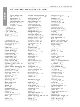
Fitoterapia 82 (2011) 969–975
Contents lists available at ScienceDirect
Fitoterapia
j o u r n a l h o m e p a g e : w w w. e l s ev i e r. c o m / l o c a t e / f i t o t e
New cassane diterpenes from Caesalpinia echinata
Betania Barros Cota a,⁎, Djalma Menezes de Oliveira b, Ezequias Pessoa de Siqueira a,
Elaine Maria Souza-Fagundes c, Adriano M.C. Pimenta d, Daniel M. Santos d,
Ana Rabello e, Carlos Leomar Zani a
a
Laboratório de Química de Produtos Naturais, Centro de Pesquisas René Rachou, Fundação Oswaldo Cruz, Av. Augusto de Lima, 1715, Belo Horizonte, MG,
30190-002, Brazil
b
Universidade Estadual do Sudoeste de Bahia (UESB), Campus Jequié, Avenida José Moreira Sobrinho s/n, Jequié, Bahia, 45206-191, Brazil
c
Departamento de Fisiologia e Biofísica, Universidade Federal de Minas Gerais, Av. Carlos, 6627, Belo Horizonte, MG, 31270-901, Brazil
d
Departamento de Bioquímica e Imunologia, Av. Antônio Carlos, 6.627, Belo Horizonte, MG, 31270-901, Brazil
e
Laboratório de Pesquisas Clínicas, Centro de Pesquisas René Rachou, Fundação Oswaldo Cruz, Av. Augusto de Lima, 1715, Belo Horizonte, MG, 30190-002, Brazil
a r t i c l e
i n f o
Article history:
Received 9 March 2011
Accepted in revised form 17 May 2011
Available online 27 May 2011
Keywords:
Caesalpinia echinata
Leguminosae
Diterpene
Cassane, Leishmania
a b s t r a c t
An investigation of the ethanolic extract from stems of Caesalpinia echinata Lam (LeguminosaeCaesalpinioideae) led to the isolation of five new cassane diterpenes along with known
lambertianic acid. Their structures were determined based on spectroscopic methods. A
preliminary study on leishmanicidal activity demonstrated that compounds 1, 2 and 6 were
found to inhibit the growth of amastigote-like forms of Leishmania amazonensis without
affecting mononuclear cells obtained from human peripheral blood.
© 2011 Elsevier B.V. Open access under the Elsevier OA license.
1. Introduction
Caesalpinia echinata Lam. belongs to the LeguminosaeCaesalpinioideae family. It is a tropical tree that may reach up
to 30 m in height and blooms yellow flowers from September
until December. The species originally had a wide distribution
in the Atlantic rainforest, ranging from Amazonas to São
Paulo, especially in the coasts of Pernambuco and Rio de
Janeiro [1]. Nowadays it is an endangered species [2] and it is
found only in 7% of the original Atlantic Forest [3].
The kernel has astringent and tonic properties [1], but there
are no pharmacological studies about C. echinata. Rezende et al.
[4] analyzed the volatile constituents of C. echinata by GC-MS.
E-β-ocimene was the major constituent of extracts obtained
from flowers by a static cryogenic headspace while (E)-3hexen-1-ol was the major constituent of extracts obtained from
leaves by hydrodistillation in a Clevenger apparatus.
⁎ Corresponding author. Tel.: + 55 31 33497845; fax: + 55 31 32953115.
E-mail address: betania@cpqrr.fiocruz.br (B.B. Cota).
0367-326X © 2011 Elsevier B.V. Open access under the Elsevier OA license.
doi:10.1016/j.fitote.2011.05.014
Previous investigations on species of the genus have
displayed interesting biological activities such as antidiabetic
[5], antiiflammatory [6], antiviral [7], antimalarial [8], antimicrobial [9] and antiplasmodal [10]. As part of an investigation of the chemical constituents of Caesalpinia echinata we
report here the isolation and structure elucidation of five new
cassane diterpenes 1–5, and their preliminary evaluation of
leishmanicidal activities.
2. Experimental procedures
2.1. General experimental procedures
TLC analyses were conducted on pre-coated silica gel G60/F254 (0.25 mm, Merck) eluted with mixtures of CHCl3/
MeOH (65:50) and Hex/EtOAc (9:1). Spots were visualized
after spraying the plates with vanillin–H2SO4. Preparative
high speed co-current chromatography was carried out with
a CCC-1000 model (High Speed Countercurrent Chromatograph, Pharma-Tech Research Corp) equipped by two pumps
970
B.B. Cota et al. / Fitoterapia 82 (2011) 969–975
P-500 model (Pharmacia). Preparative circular chromatography (CCP) was carried out by Chromatotron 7924T model
(Harrison Research). Semi-preparative HPLC purifications
were carried out by a Shimadzu chromatograph, equipped
with a LC6AD pump and a dual wavelength detector (SPD10A),
using a Shim-pack® C18 column (5 μm, 250 × 20 mm i.d.) eluted
with mixtures of MeCN/H2O at a flow rate of 10 ml/min
and detection at 210 nm and 254 nm. Electron impact mass
spectrometry (EI-MS, 70 eV) was measured by Shimadzu
QP5050A spectrometer, equipped with a direct insertion
probe. Electrospray ionization mass spectrometry (ESI-MS)
was recorded on a Thermo Finnigan LCQ-Advantage spectrometer. ESI-Q/ToFMS analyses were performed using a Q-ToF
MicroTM instrument (Micromass, Manchester, UK). Samples
were diluted in methanol/Milli-Q1 water and introduced using
a syringe pump with flow rates of 5–10 ml/min (electrospray).
UV spectra (200–400 nm) were obtained by Shimadzu SPD M10A VP Diode Array Detector. Infrared spectra were obtained by
Shimadzu FTIR-8400 spectrometer, with the samples in KBr
pellets. 1H (400 MHz), 13CNMR (100 MHz), DEPT-135, COSY,
HMBC and NOESY experiments were carried out in a Bruker
DRX 400 spectrometer. Chemical shifts were recorded in δ
(ppm) using CDCl3 as solvent. Complete assignments of the 1H
and 13C chemical shifts of isolated compounds were solved by
interaction using the Perch NMR Software (University of
Kuopio, Finland). Conformational analyses were carried out
by Perch's molecular modeling system (MMS). 3D molecular
models were built with energetic optimization by Merck
Molecular Force Field (MMFF94) based on superior force fields.
2.2. Plant material
Stems of C. echinata were collected at Fundação ZooBotânica, Belo Horizonte, Minas Gerais, Brazil, in March 2007.
A voucher specimen (BHZB 6458) in complete form with
flowers was deposited at the Herbarium of Fundação ZooBotânica of Belo Horizonte.
2.3. Extraction and isolation
Air-dried stems (55 g) of C. echinata were extracted with
EtOH under ultrasonication at room temperature. The solvent
was evaporated under reduced pressure to yield 1.6 g of
crude EtOH extract. The crude extract (1.5 g) was purified by
preparative high-speed co-current chromatography. The apparatus was equipped with three polytetrafluoroethylene preparative coils (total volume, 300 ml) and a 10 ml sample loop.
With the rotor stopped, the coils were filled with the lower
phase composition of a biphasic liquid system H2O/MeOH/
CH2Cl2 (4:6:5) at flow rate of 6 ml/min. The coils were then
rotated at 1000 rpm and the upper phase was pumped in tailto-head direction, at a flow rate of 4.5 ml/min. The extract
(1.5 g) was dissolved in 10 ml of the biphasic solvent mixture
and injected into the column. Lower phase was pumped at
3.0 ml/min and upper phase was pumped at 1.5 ml/min to give
22 fractions after TLC analysis. Fractions 17 and 18 (44 mg)
were subjected to semi-preparative reversed-phase HPLC with
MeCN:H2O 40:60→ 60:40 in 40 min, 60:40 in 10 min, at a flow
rate of 10 ml/min, to yield 1 (2 mg), 2 (2.5 mg) and 3 (6.5 mg).
Fraction 19 (45 mg) was purified at the same conditions used
before and yielded the compounds 4 (2.5 mg) and 5 (2 mg).
Fraction 20 (300 mg) was subjected to semi-preparative
circular chromatography on 1 mm plates of silica gel at a flow
rate of 2–4 ml/min with hexane and ethyl acetate mixtures
to furnish 23 subfractions. Compound 6 (4 mg) was obtained
of subfractions 13–15 (18 mg) after column chromatography
(silica gel) with Hex/EtOAc mixtures. Compound (6, Fig. 1):
lambertianic acid, white powder; 1H NMR (CDCl3, 400 MHz): δ
0.61 (s, 3 H, CH3-20), 1.00–1.06 (m, 1 H, H-1a), 1.07–1.09, (m,
1 H, H-3a), 1.24 (br s, 3 H, CH3-19), 1.31 (dd, J = 10.0 Hz, 5.0 Hz,
1 H, H-5), 1.47–1.56 (m, 1 H, H-2a), 1.54–1.62 (m, 1 H, H-9),
1.64–1.66 (m, 1 H, H-11a), 1.75 (td, J = 9.0, 9.0, 4.0 Hz, 1 H, H11b), 1.79–1.86 (m, 1 H, H-1b),1.86–1.93 (m, 3 H, H-2b, H-6a
and H-7a), 1.97 (dd, J = 11.0 Hz, 5.0 Hz, 1 H, H-6b), 2.15 (dd,
J = 9.0, 4.0 Hz, 1 H, H-3b), 2.26 (dd, J = 14.0, 9.0 Hz, 1 H, H-12a),
2.43 (dd, J = 8.5, 3.0 Hz,1 H, H-7b), 2.56 (ddd, J = 14.0, 9.0,
4.0 Hz, 1 H, H-12b), 4.58 (m, 1 H, H-17a), 4.89 (m, 1 H, J = 1.9,
1.4 Hz, H-17b), 6.25 (m, 1 H, H-14), 7.19 (m, 1 H, H-16), 7.34
(m, 1 H, H-15), 10.57 (br s, 1 H, COO-H); 13C NMR (CDCl3,
100 MHz): δ 12.9 (C-20), 19.91 (C-2), 23.60 (C-12), 24.29 (C11), 26.09 (C-6), 28.99 (C-19), 38.03 (C-3), 38.71 (C-7), 39.07
(C-1), 40.39 (C-10), 44.13 (C-4), 55.27 (C-9), 56.24 (C-5),
106.53 (C-17), 110.97 (C-14), 125.47 (C-13), 138.75 (C-15)
142.69 (C-16) 147.84 (C-8), 182.08 (C-18); EI-MS m/z 316
([M]+), 279 (3), 223 (1.5), 167 (2.9), 149 (16), 139 (0.8), 104
(1), 88 (70), 70 (100), 61 (73).
2.4. Assay with Leishmania (Leishmania) amazonensis
Leishmanicidal activity was determined against amastigotelike forms that were obtained as previously described
by Callahan et al. [11] and cell viability was determined using
the methyl thiazolyl tetrazolium (MTT) assay described by
Teixeira et al. [12].
2.5. Lymphocyte proliferation assay
Peripheral blood mononuclear cells (PBMCs) were prepared using the protocol previously described by Gazzinelli
et al. [13] and the cell proliferation was determined by the
MTT assay described by Jiang and Xu [14].
3. Results and discussion
As a part of an ongoing program devised for drug discovery from natural products, the EtOH extract from stems of
C. echinata killed 94% of amastigotes-like forms of Leishmania
(Leishmania) amazonensis and presented moderate inhibitory
activity against human PBMC cells, stimulated with PHA, at
20 μg/ml (Table 3). The crude extract was fractionated by cocurrent HSCCC, preparative reversed phase HPLC and silica
gel chromatography to give compounds 1–6 (Fig. 1).
Compound 1 was isolated as white amorphous powder
and gave a molecular ion peak at m/z 495 [M − H] − in the
negative ESI-MS spectrum and m/z 497.5985 [M + H] +in the
positive ESI-Q-TOF spectrum (calcd for C29H3707, 497.5999).
The UV spectrum exhibited absorption bands at 210 nm
and 279 nm, whereas the FT-IR (KBr) υmax spectrum showed
absorption bands at 3588, 2936, 2869, 1732, 1675, 1450,
1391, 1191 1144 and 950 cm − 1. The 1H NMR spectrum of 1
(Table 1) showed resolved signals for one olefinic proton at δ
5.72 (1 H, br s) that was assigned for H-15 of α, β-unsaturated
B.B. Cota et al. / Fitoterapia 82 (2011) 969–975
971
O
O
HO
12
11
20
H
9
14
H
1
17
3
18
19
20
14
1
17
3
OR
18
H
19
18
5'
R2 =
3: R1 = COOH
3'
1'
OH
3'
7'
9'
O
Hb
19
7'
6
OH
O
5'
1'
17
O
9'
2: R =
8
HOOC
OR2
5'
1'
10
5
7
R1
O
1: R =
H a 15
9
H
5
7
H
HOOC
O
11
15
H
9
1
5
13
11
20
3
16
16
O
16
5'
4: R1 = CH2OH
3'
9'
R2 =
1'
7'
9'
5: R1 = COOH
OH
3'
7'
OH
R2 = H
Fig. 1. Structure of compounds isolated from Caesalpinia echinata.
γ-lactone moiety, two tertiary methyl groups at δ 1.32 (s)
and δ 1.08 (s), one secondary methyl group at δ 1.13 (d,
J = 7.5 Hz) and two α and β-carbonilic methylene protons at
δ 2.96 and δ 2.64, respectively. One monosubstituted benzene
ring was assigned by resonances at δ 7.18–7.26. Most of the
methylene signals appeared as complex and overlapped
multiplet and were assigned by HSQC, HMBC and COSY
correlations (Table 1). The 13C NMR and DEPT-135 spectra
presented 29 carbon signals, eight quaternary including three
carbonyl (δ 181.60, 172.26, 170.16), two olefinic carbons
(δ 172.17 and δ 140.15), one carbon of a hemiketal (δ 105.24)
and two others (C-4, δ 47.29 and C-10, δ 37.09). Also, these
spectra showed the presence of seven methylene carbons,
eleven methine carbons, comprising five aromatic carbons
(δ 128.50 (2x), 128.23 (2x), 126.39), one olefinic carbon
(δ 113.68), and five methine carbons (δ 72.09, 49.25, 45.09,
35.87, 35.81), besides two tertiary methyl groups (δ 18.35,
17.63) and one secondary methyl group (δ 12.94). The 13CNMR spectrum revealed all signals consistent with the
structure of an α,β-butenolide hemiketal ring and hydrocinnamoyl ring moiety. The deshielded nature of the
carbon at δ 72.09 and the proton attached to it at δ 5.13 (br
d, J = 3.0 Hz) suggested that the hydrocinnamoyl moiety is
attached to C-6. Small vicinal coupling constants values
between H-6 and H-5 ( 3J6eq,5ax = 3.0 Hz) suggested an αequatorial hydrogen attached to C-6. Besides, H-6 methine
showed COSY and NOESY correlations to both vicinal proton
signals at C-7. Other NOESY correlations observed were
consistent with the configuration proposed for compound 1.
NOESY correlations between H-6 and H-5 at δ 1.96 (br d,
J = 3.0 Hz) and H-9 at δ 1.60 (m) suggested that protons were
on the same face. A double doublet at δ 2.76 (J = 7.5 and
3.0 Hz) was assigned to H-14 coupled with both CH3-17
(δ 1.13 d, J = 7.5 Hz) and an axial H-8 at δ 1.59 (m). H-14
proton was depicted as β-equatorial based on NOESY correlations and its small scalar coupling constant (3.0 Hz) with
H-8. From these data, compound 1 was identified as 6β-O2',3'-dihydrocinnamoyl-12-hydroxy-(13)15-en-16,12-olide18-cassaneoic acid.
Compound 2 was obtained as white amorphous powder
and it showed a molecular ion [M-H] - at m/z 493 in the negative
ESI-MS and m/z 495.5800 [M+ H]+in the positive ESI-Q-TOF
(calcd for C29H3507, 495.5841). The UV spectrum exhibited
absorption bands at 218 nm and 279 nm, whereas the F-TIR
(KBr) υmax spectrum showed absorption bands at 3459, 2939,
2869, 1690, 1440, 1400, 1209 and 1139 cm− 1. The 1H and 13C
(Table 1) NMR spectroscopic data were closely related to those
of 1 except at C-6, where the dihydrocinnamoyl ester was
replaced by a cinnamoyl moiety (δ 6.38, d, J = 16.0 Hz, H-2'
and δ 7.66, d, J = 16.0 Hz, H-3'). The configuration at C-6
was the same as in compound 1, since the coupling constants
of the associated protons were similar, and this was confirmed
by NOESY experiments. Thus, compound 2 was established
as 6β-O-cinnamoyl-12-hydroxy-(13)15-en-16,12-olide-18cassaneoic acid.
Compound 3 was isolated as a white powder and the
negative ESI-MS showed a molecular ion [M-H] - at m/z 493
and exhibited fragment ions at m/z 179, suggesting the
presence of the caffeoyl ester moiety. It showed the
quasimolecular ion at m/z 495.5820 [M + H] +in the positive
ESI-Q-TOF (calcd for C29H3507, 495.5841). The UV spectrum
exhibited absorption bands at 220 nm, 309 nm and 327 nm,
whereas the FT-IR (KBr) υmax spectrum showed absorption
bands at 3423, 2939, 2869, 1704, 1514, 1450, 1269 and 1157
cm − 1. A pair of doublets at δ 7.22 (d, J = 1.9 Hz) and at δ 6.17
(d, J = 1.9 Hz) in its 1H NMR spectrum (Table 2) suggested the
presence of a 2,3-disubstituted furan ring. The 1H NMR spectrum revealed the presence of two terciary methyl groups at δ
972
B.B. Cota et al. / Fitoterapia 82 (2011) 969–975
Table 1
NMR spectroscopic data of the compounds 1 and 2 (1H NMR, 400 MHz;
13
C NMR,100 MHz, CDCl3; δ in ppm, multiplicities, J in Hz).
1
2
Position
δH (mult. J, Hz)
1eq(β)
1ax(α)
2ax(β)
2eq(α)
3eq(β)
3ax(α)
4
5ax(α)
6eq(α)
7
1.78 m
1.18 ddd (12.8, 12.8, 5.0)
1.61 m
1.58 m
1.76 dd (12.8, 3.0)
1.69 dd (12.8, 5.0)
8ax(β)
9ax(α)
10
11ax(β)
11eq(α)
12
13
14β
15
16
17α
18
19β
20β
1'
2'
3'
4'
5'
6'
7'
8'
9'
1.59 m
1.60 m
1.96 br d (3.0)
5.13 br d (3.0)
1.61 m (2H)
1.37 d (13.0)
2.39 dd (13.0, 3.0)
2.76 dd (7.5, 3.0)
5.72, br s
1.13. d (7.5, 3H)
1.32 s (3H)
1.08 s (3H)
2.64 td (2× 7.5, 3.0, 2H)
2.96 td (2× 7.5, 3.0, 2H)
7.19
7.27
7.18
7.26
7.19
m
m
m
m
m
δC DEPT
41.03
COSY
HMBC
NOESY
δH (mult. J, Hz)
1α, 2α
1β
3, 9
1α, 11α
1β
19β
1.84 m
1.25 m
1.66 m (2H)
41.09
1β
1.79 m
1.70 m
39.05
6α, 3α
5α, 7α, 9α, 19β
6α
14β
6α
2.10 br d (1.8)
5.30 td (3.0, 1.8, 1.8)
1.86 m
1.73 m
1.96 ddd (10.0, 6.0, 3.0)
1.75 m
8β, 11β
1β, 11α
1.49 d (13.0)
2.47 dd (13.0, 3.0)
17.97
39.00
47.29
49.25
72.09
34.95
35.81
45.09
37.09
37.57
105.24
172.17
35.87
113.68
170.16
12.94
181.60
18.35
17.63
172.26
36.30
30.85
140.15
128.23
128.50
126.39
128.50
128.23
5, 10
1β
2α
2β
6α
5α, 7α
6α
9α, 14β
8β, 11α
11α
11β
8β, 17α
14β
5α
5α
5´, 9´
3´, 7´
8´
5´, 9´
6´
5´, 7´
5, 20
1
5, 19
2, 4, 10, 18, 20
8, 17
17
5
5
20
15
17
17
12, 16
15
8, 13, 14, 16
19
3, 5, 18
1, 5, 9, 10
5',
3',
7',
4',
5',
4',
5',
9'
6', 8'
9'
8'
9'
6'
7'
1.47 and 1.37 and one secondary methyl group at δ 0.97 (d,
J = 7.0 Hz), five aliphatic methylenes (δ 1.79 and 1.23, 1.69
and 1.60, 1.86 and 1.73, 1.96 and 1.73, 2.65 and 2.52) and five
aliphatic methines (δ 5.40, 2.57, 2.21, 2.09, 1.72). A caffeoyl
ester ring moiety was confirmed by signals of aromatic
protons found at δ 6.88–7.11, and two coupled doublets at δ
7.55 (J = 15.8 Hz) and at δ 6.17 (J = 15.8 Hz) assigned for
olefinic protons. Moreover, the 13C NMR and DEPT-135 of 3
(Table 2) showed a total of 29 carbon signals, nine quaternary
represented by two carbonyl (δ 181.06, 166.66) and five
olefinic carbons that includes two furan carbons (δ 149.44
and 122.01) and three aromatic carbons (δ 148.34, 145.80
and 126.33). These spectra showed the presence of five
methylene carbons (δ 41.39, 18.22, 39.00, 35.87, 21.44),
twelve methine carbons, comprising three aromatic carbons
(δ 121.34, 115.12 and 113.99), two furan olefinic carbons (δ
140.16 and 109.26), two olefinic carbons in caffeoyl ester
moiety (δ 144.80 and 115.29) and five others (C-5, δ 49.72, C6, δ 72.14, C-8, δ 31.19, C-9, δ 45.58, C-14, δ 30.87). In addition,
13
C-NMR spectrum of 3 revealed the signals of two tertiary
methyl groups (δ 17.99, 17.72) and one secondary methyl
group (δ 17.44). The configuration of compound 3 was
determined by analysis of coupling constants and NOESY
data. The small coupling constant between H-5 at δ 2.21 (br
d, J = 2.0 Hz) and H-6 at δ 5.40 (br d, J = 2.0 Hz) indicated
that H-6 is at α-equatorial position. In the NOESY spectrum
17α
14β
2.94 dd (7.0, 5.0)
5.72 br s
14β
1.19 d (7.0, 3H)
20
19
1.39 s (3H)
1.26 s (3H)
3'
2'
6.38 d (16.0, 2H)
7.66 d (16.0, 2H)
6'
5', 7'
6', 8'
9', 7'
8'
7.52
7.39
7.40
7.39
7.52
(m)
(m)
(m)
(m)
(m)
δC DEPT
17.96
47.41
49.41
72.12
35.17
36.15
45.18
37.25
37.68
105.31
172.31
35.96
113.72
170.25
12.96
182.20
18.38
18.02
166.27
118.30
145.28
134.18
128.15
128.94
130.53
128.94
128.15
COSY
HMBC
1α, 2α
1β
1α, 1β, 3α, 3β
3
5, 10
19
2β
1β, 3β
19
1, 4, 5
5, 19
2, 4, 10
6α, 7α
5α, 7α, 7β
6α, 7α, 9α
6α, 7β
14β, 7α, 9α
8β,11β, 11α
9α, 11α
9α, 11β
9α, 17
14β
3β
11β
3'
2'
6'
5'
5',9'
7'
8'
8
17
20
5, 20
20
8, 20
15
17
17
12, 16
15
8, 13, 14
19
3, 4, 10, 18
1, 5, 9, 10
3'
4'
1', 5', 9'
2'
3', 8'
5', 9'
5', 6', 8', 9'
5', 9'
3', 6'
H-6 had cross-peaks with H-5 and H-5 with H-9, confirmed
that H-5 was axial and that the C-6 was equatorial. These
observations allowed for 3 to be proposed as being 6β-O-6',7'
dihydroxycinnamoyl-18-vouacapaneoic acid.
Compound 4 showed the molecular ion [M-H]− at m/z 480 in
negative ESI-MS spectrum and m/z 479.5805 [M−H]− in the
negative ESI-Q-TOF (calcd for C29H3706, 479.5845). The UV
spectrum exhibited absorption bands at 218 nm, 309 nm and
327 nm, whereas the FT-IR (KBr) υmax spectrum showed
absorption bands at 3441, 2929, 2855, 1695, 1518, 1446, 1382,
1273 and 1177 cm− 1. The 1H and 13C NMR spectral data
(Table 2) of 4 revealed the same cassane-type skeleton as 3
which contains a furan and a 6',7'-dihydroxy-trans-cinnamoyl
moieties. The major difference was the replacement of the acid
carboxilic group by a hydroxymethylene group at C-18. The 1HNMR spectrum presented two coupled doublets at δ 3.64
( J =11.0 Hz) and at δ 3.18 ( J=11.0 Hz), and the 13C-NMR
spectrum showed an oxygenated carbon signal at δ 71.6
corresponding to hydroxymethylene group. The hydroxymethyl
proton H-18A at δ 3.18 showed HMBC correlation with carbon
signal of methyl group (C-19), and H-19 and H-20 showed HMBC
correlations with C-18. Thus, the structure of compound 4 was
determined as 6β-O-cinnamoyl-18-vouacapaneol.
Compound 5 exhibited molecular peak [M+H] + at m/z 333
by positive ESI-MS and m/z 331.4198 [M− H]− in the negative
ESI-Q-TOF (calcd for C20H2706, 331.4259). The UV spectrum
Table 2
NMR spectroscopic data of the compounds 3–5 (1H NMR, 400 MHz;
13
C NMR,100 MHz, CDCl3; δ in ppm, multiplicities, J in Hz).
3
4
δ (mult. J, z)
1eq(β)
1ax(α)
1.79 d (12.5)
1.23 dd (12.5, 4.0)
2ax(β)
2eq(α)
3eq(β)
3ax(α)
4
5ax(α)
6eq(α)
7(β)
1.69
1.60
1.86
1.73
7(α)
8ax(β)
9ax(α)
10
11ax(β)
11eq(α)
12
13
14β
15
16
17α
18B
18A
19β
20β
1'
2'
3'
4'
5'
6'
7'
8'
9'
m
dd (13.0, 3.0)
dd (13.0, 4.0)
m
2.21 br d (2.0)
5.40 br d (2.0)
1.96 td (14.0,
3.0, 3.0)
1.73 m
2.09 m
1.72 m
2.52 dd (17.0, 10.0)
2.65 dd (17.0, 6.5)
2.57
6.17
7.22
0.97
dd (7.0, 5.0)
d (1.9)
d (1.9)
d (7.0)
1.47 s (3H)
1.37 s (3H)
6.17 d (15.8)
7.55 d (15.8)
7.11 d (1.9)
6.88 d (8.0)
6.92 d (8.0, 1.9)
δC DEPT
5
COSY
HMBC
NOESY
δ (mult. J, z)
41.39
1α
2β
3, 10, 20
1α
1β
18.22
3β, 5α
3β
3α
3β
5, 19
11β
2β
19
5α
1.76 br d (13.0)
1.11 dt (13.0,
13.0, 3.0)
1.72 m
1.55 dd (14.0, 4.0)
1.69 m
1.22 m
7α, 9α
5α, 7α, 7β
14
1.75 m
5.60 ddd (4.0, 3.0, 3.0)
2.34 m
6α
11β, 14β, 20
6α, 11α, 17
1.88 ddd (14.0, 3.0, 3.0)
2.10 m
1.64 m
1β
9α, 1α
2.52 m
2.65 ddd (15.0,
15.0, 7.0)
39.00
47.23
49.72
72.14
35.87
31.19
45.58
37.26
21.44
149.44
122.01
30.87
109.26
140.16
17.44
181.06
17.99
17.72
166.66
115.29
144.80
126.33
113.99
145.80
148.34
115.12
121.34
7β, 3α
5α, 7α, 7β
7β, 8β
7β, 8β
7α, 7β
11α, 11β
9α
9α
17
16
15
14β, 8β, 9α
3β, 20
19
3'
2'
9'
9'
8'
1, 5, 19
5, 19
2, 4, 9, 10, 18, 19
8, 10
5, 8
6, 11
5, 11, 20
1, 5, 6, 11, 20
8, 9, 10, 12, 13, 20
11, 15, 16
11, 15, 16, 17
12, 13, 17
12, 13, 16
15
12, 13, 14
3, 4, 5, 19
1, 3, 4, 5, 18
1, 5, 9, 10
2', 3', 5', 9'
3', 5', 9'
5', 7', 9'
2', 8'
3', 9'
5', 8'
5', 9'
4', 6'
3', 5'
7β, 8β, 17
16
15
9α, 14β, 15,
3β, 6α
1β, 2β, 8β, 2'
2.57
6.18
7.23
0.92
3.64
3.18
0.96
1.34
m
d (1.8)
d (1.8)
d (7.0, 3H)
d (11.0)
d (11.0)
s (3H)
s (3H)
5', 9', 20
5', 9', 20
6.22 d (16.0)
7.54 d (16.0)
2', 3'
7.07 d (2.0)
9'
2', 3'
6.86 d (8.5)
6.99 dd (8.5, 2.0)
δC DEPT
COSY
HMBC
δ (mult. J, z)
41.72
3β
2β, 3a, 20
3, 20
2
18.17
1β, 2α
1β, 2β
2α, 3α
2β, 3β
1
1.72 m
1.18 ddd (12.5,
12.5, 4.0)
1.59 m
1.69 m
1.69 m
37.08
38.31
48.40
69.73
36.46
31.12
45.67
37.78
21.80
6α
5α, 7α, 9α
9α
8β
7α, 9α
6β, 7β, 11β, 14β
5, 19
5, 19
4, 9, 20
5, 8
7, 11
20
5, 20
8
7α, 9α, 11β
149.45
122.55
31.14
109.49
140.44
17.60
71.60
19.56
18.07
167.18
116.62
145.24
127.70
114.30
146.19
148.77
115.52
121.64
8β, 17
16
15
14
18A
18B
1α
3'
2'
8', 9'
5'
5'
11
11
12, 17
16
15
8, 13, 14
19
3, 4, 5, 18
1, 5, 9, 10, 18
3'
4'
1',5'
2', 8'
3', 7', 9'
4, 8'
5'
4', 6'
4', 5'
1.85 br s
4.15 m
1.80 ddd (13.0,
13.0, 4.0)
1.69 m
2.18 m
1.62 m
2.48 dd (16.0, 10.0)
2.61 dd (16.0, 7.0)
2.61
6.19
7.23
0.98
m
d (2.0)
d (2.0)
d (7.0, 3H)
1.62 s (3H)
1.27 s (3H)
δC DEPT
COSY
HMBC
41.44
5α, 20β
1β, 2β
9, 10
18.06
2α
2β
40.40
47.98
50.73
70.18
38.74
30.70
46.22
37.25
21.71
149.35
122.27
31.26
109.52
140.45
18.06
183.59
18.87
17.84
19
6α
5α, 7β, 7α
5, 7
1
6α, 7α
7β
7α, 7β, 9α
8β, 11β
11β
9α, 11α
17α
16
15
14β
1, 10, 20
5, 20
12
11
15
13
15
13
8
19
B.B. Cota et al. / Fitoterapia 82 (2011) 969–975
Position
3, 5, 18
1, 5, 9, 10
973
974
B.B. Cota et al. / Fitoterapia 82 (2011) 969–975
exhibited absorption bands at 220 nm and 279 nm, whereas
the FT-IR (KBr) υmax spectrum showed absorption bands
at 3441, 2930, 2871, 1695, 1459, 1395, 1199 and 1172 cm− 1.
The 1H NMR spectrum (Table 2) exhibited signals of two
tertiary methyl groups at δ 1.62 (s) and δ 1.27 (s), and a
secondary methyl group at δ 0.98 (d, J = 7.0 Hz). In the low field
region of the spectrum, two protons of a 1,2-disubstituted furan
resonated at δ 7.23 (d, J = 2.0 Hz, H-16) and at δ 6.19 (d,
J = 2.0 Hz, H-15). Analysis of the 13C NMR and DEPT-135
spectra of 5 revealed signals of one carbonyl at δ 183.59, four
carbons of the furan ring (δ 149.35, 140.45, 122.27, 109.52) and
one oxygenated carbon at δ 70.18. One oxymethine proton at δ
4.15 (m) was assigned for H-6 that showed correlations with H5 and H-7 in the COSY experiment. The configurations of
compound 5 were the same as in compound 3 and 4 and it was
identified as 6β-hydroxi-18-vouacapaneoic acid.
Compound 6 exhibited molecular peak at m/z 316 ([M] +)
by EI-MS suggesting the molecular formula C20H28O3. All the
spectroscopic data observed for compound 6 were identical
to those described for lambertianic acid [15].
Compounds 1 and 2 can be considered as derivatives from
vinhaticoic acid, a furanocassane-type diterpene isolated from
Dipteryx lacunifera Ducke (Leguminosae-Papilionoideae), a
species that occurs in Brazil [16]. Previous studies on species
of this genus reported the same skeleton with a methine on C-5
[17–21]. Compounds1–5 are cassane diterpenes with transcinnamoyl or trans-hydrocinnamoyl groups as side chain at C-6.
The relative configuration of compounds 1–5 was defined by
NOESY results and built with energy-minimized conformation
(Fig. 2). All structures adopt trans-junction of the three
hexagonal rings and the trans-cinnamoyl ester groups were
placed on opposite face of COOH groups.
1
2
Table 3
Results of the biological assays of the isolated compounds.
Compounds*
% Leishmanicidal
activitya
%Inhibition of PBMC
proliferationb
1
2
3
4
5
6
EtOH extract
AMB**
DMSO***
56 ± 13
69 ± 8
26 ± 19
43 ± 21
27 ± 0
62 ± 9
94 ± 6
70 ± 10
6±2
114 ± 8
112 ± 37
124 ± 13
–
122 ± 12
120 ± 28
60 ± 5
–
100
*Extract and compounds were tested at 20 μg/ml. **Amphotericin B at 0.2 μg/ml
was used as positive drug control. ***DMSO was tested at 0.1% v/v. (–) not
tested.
a
Amastigote-like forms of Leishmania (Leishmania) amazonensis.
b
Human peripheral blood mononuclear cells, stimulated with PHA.
Compounds 1, 2 and 6 were tested against Leishmania
(Leishmania) amazonensis (Table 3) and neither of them is
more active than the crude extract, but they are not toxic to
human peripheral blood mononuclear cells in vitro at a
concentration of 20 μg/ml. In this paper, we reported for the
first time, the isolation and elucidation of furan and hemiketal
cassane-type diterpenes and also the evaluation of their
leishmanicidal activity.
Acknowledgements
We are grateful to the Fundação Oswaldo Cruz and FAPEMIG
for financial support. We are also grateful to Fundação ZooBotânica de Belo Horizonte for supplying the plant material and
Daniela Nabak Bueno Maia for technical assistance.
3
5
4
6
Fig. 2. Conformational analyses of compounds 1–6 by PERCH NMR software.
B.B. Cota et al. / Fitoterapia 82 (2011) 969–975
References
[13]
[1] Corrêa MP. Dicionário das plantas úteis do Brasil e das exóticas
cultivadas. Rio de Janeiro: Ministério da Agricultura; 1984.
[2] Lima HC, Lewis GP, Bueno E. Pau-Brasil. In: Bueno E, editor. Pau-brasil:
uma biografia. São Paulo: Axis Mundi; 2002. p. 39–76.
[3] Morellato L, Haddad CFB. The Brazilian Atlantic forest. Biotropica
2000;32:786–92.
[4] Rezende CM, Corrêa VFS, Costa AVM, Castro BCS. Constituintes
químicosvoláteis das flores e folhas do pau-Brasil (Caesalpinia echinata,
Lam.). Quim Nova 2004;27:414–6.
[5] Sharma SR, Dwivedi SK, Swarup D. Hypoglycaemic, antihyperglycaemic
and hypolipidemic activities of Caesalpinia boncucella seeds in rats.
J Ethnopharmacol 1997;58:39–44.
[6] Hikino H, Taguchi T, Fujimura H, Hiramatsu Y. Antiinflammatory
principles of Caesalpinia sappan wood and of Haematoxylon compechianum wood. Planta Med 1977;31:214–20.
[7] Jiang RW, But PP, Ma SC, Ye WC, Chana SP, Maka TCW. Structure and
antiviral properties of macrocaesalmin, a novel cassane furanoditerpenoid lactone from the seeds of Caesalpinia minax Hance. Tetrahedron
Lett 2002;43:2415–8.
[8] Kuria KAM, De Coster S, Muriuki G, Masengo W, Kibwage I, Hoogmartens J,
et al. Antimalarial activity of Ajuga remota Benth (Labiatae) and Caesalpinia
volkensii Harms (Caesalpiniaceae): in vitro confirmation of ethnopharmacological use. J Ethnopharmacol 2001;74:141–8.
[9] Ragasa CY, Hofilen JG, Rideout JA. New furanoid diterpenes from
Caesalpinia pulcherrima. J Nat Prod 2002;65:1107–10.
[10] Pudhom K, Sommit D, Suwankitti N, Petsom A. Cassane furanoditerpenoids from the seed kernels of Caesalpinia bonduc from Thailand. J Nat
Prod 2007;70:1542–4.
[11] Callahan HL, Portal AC, Devereaux R, Grogl M. An axenic amastigote
system for drug screening HL. Antimicrob Agents Chemother 1997;41:
818–22.
[12] Teixeira MC, de Jesus Santos R, Sampaio RB, Pontes-de-Carvalho L, dos
Santos WL. A simple and reproducible method to obtain large numbers
[14]
[15]
[16]
[17]
[18]
[19]
[20]
[21]
975
of axenic amastigotes of different Leishmania species. Parasitol Res
2002;88:963–8.
Gazzinelli G, Katz N, Rocha RS, Colley DG. Immune response during
human schistosomiasis mansoni X. Production and standartization of
an antigen-induced mitogenic activity by peripheral blood mononuclear cells from treated but not active cases of schistosomiasis. J
Immunol 1983;130:2891–5.
Jiang J, Xu Q. Immunomodulatory activity of the aqueous extract from
rhizome of Smilax glabra in the later phase of adjuvant-induced arthritis
in rats. J Ethnopharmacol 2003;85:53–9.
Tolstikova TG, Sorokina IV, Dolgikh MP, Kharitonov YV, Chernov SV,
Shul'ts ÉÉ, et al. Neurotropic activity of lambertianic acid adducts with
n-substituted maleinimides. Pharm Chem J 2004;38:532–4.
Vieira Júnior GM, Silva HRE, Bittencourt TC, Chaves MH, Simone CA.
Terpenos e ácidos graxos de Dipteryx lacunifera Ducke. Quim Nova
2007;30:1658–62.
Kinoshita T, Haga Y, Narimatsu S, Shimada M, Goda Y. The isolation and
structure elucidation of new cassane diterpene-acids from Caesalpinia
crista L. (Fabaceae), and review on the nomenclature of some
Caesalpinia species. Chem. Pharm Bull 2005;53:717–20.
Yodsaoue O, Cheenpracha S, Karalai C, Ponglimanont C, Chantrapromma S,
Fun HK, et al. Diterpenoids from the seeds of Caesalpinia sappan Linn.
Phytochemistry 2008;69:1242–9.
Yadav PP, Arora A, Bid HK, Konwar RR, Kanojiyad S. New cassane
butenolide hemiketal diterpenes from the marine creeper Caesalpinia
bonduc and their antiproliferative activityI. Tetrahedron Lett 2007;48:
7194–8.
Dickson RA, Houghton PJ, Hylands PJ. Antibacterial and antioxidant
cassane diterpenoids from Caesalpinia benthamiana. Phytochemistry
2007;68:1436–41.
Cheenpracha S, Srisuwan R, Karalai C, Ponglimanont C, Chantrapromma
S, Chantrapromma K, et al. New diterpenoids from stems and roots of
Caesalpinia crista. Tetrahedron 2005;61:8656–62.
Download