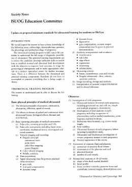
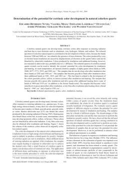
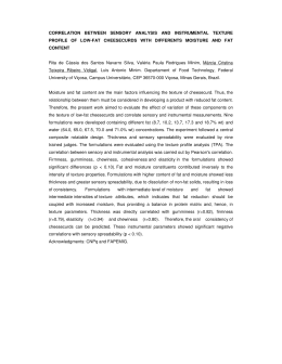
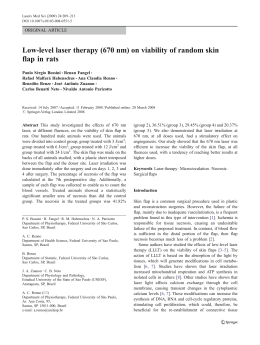
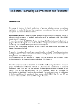
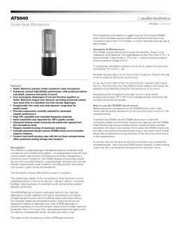
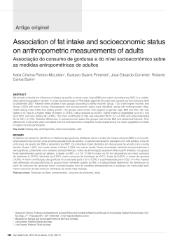
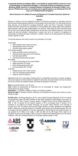
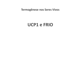
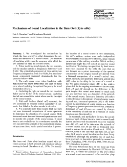
Pathophysiology 9 (2002) 13 /19
www.elsevier.com/locate/pathophys
Effect of ultrasound application on fat mobilization
Hirohide Miwa a, Masato Kino a, Li-Kun Han b, Kunihiro Takaoka c,
Takahiro Tsujita c, Hiroshi Furuhata d, Masahiro Sugiyama e, Hiromasa Seno f,
Yusuke Morita f, Yoshiyuki Kimura g, Hiromichi Okuda b,*
a
Miwa Science Laboratory Inc., 6-7-10 Miyazaki, Miyamae-ku, Kawasaki, Kanagawa 216-0033, Japan
Faculty of Environmental and Symbiotic Sciences, Prefectural University of Kumamoto,Tsukide 3-1-100, Kumamoto 862 /8502, Japan
c
Central Research Laboratory, School of Medicine, Ehime University, Shigenobu-cho, Onsen-gun, Ehime 791-0295, Japan
d
Medical Electronics Laboratory, Jikei Medical College, Minato-ku, Nishishinbashi, Tokyo 105-8461, Japan
e
Laboratory of Exercise Physiology, Faculty of Education, Ehime University, Bunkyou-cho, Matsuyama, Ehime 790-0855, Japan
f
Department of Physiology, School of Medicine, Tokushima University, Kuramoto-cho, Tokushima 770-8503, Japan
g
Second Department of Medical Biochemistry, School of Medicine, Ehime University, Shigenobu-cho, Onsen-gun, Ehime 791-0295, Japan
b
Received 25 December 2000; received in revised form 22 February 2001; accepted 8 March 2001
Abstract
The aim of this experimental trial was to study the effect of ultrasound application on the lipolysis in adipose tissue. Rats were
administered to pentobarbital (Nembutal) anesthesia and their abdomens were shaved. Rat abdomen was subjected to 24 kHz /1
MHz ultrasound for 10 min to investigate frequency and power-intensity dependency for fat mobilization. Blood was taken from the
tail vein to estimate plasma free fatty acids (FFA). For frequency dependency two regions around 100 kHz and 300 /500 kHz were
effective for fat mobilization. For power-intensity dependency, effective regions were found to be from 24 to 1090 kHz. In the
effective regions on frequency and power-intensity, application of ultrasound caused increases in plasma FFA and norepinephrine
concentration of extra-cellular fluid of perirenal adipose tissue. These results suggest that ultrasound application stimulates fat
mobilization through a local increase in norepinephrine secretion under the conditions of effective frequency and intensity. # 2002
Elsevier Science Ireland Ltd. All rights reserved.
Keywords: Ultrasound frequency; Power-intensity; Fat mobilization
1. Introduction
It is well known that obesity predisposes to the
development of chronic illness such as non-insulin
dependent diabetes mellitus (NIDDM), hypertension,
hyperlipidemia, coronary heart diseases and arteriosclerosis. Tokunaga et al. [1] classified obesity into two
type, a visceral type and a subcutaneous type, using
computed tomography (CT). They measured areas of
visceral fat tissue and subcutaneous fat tissue on an axial
CT section at the level of the umbilicus and calculated
the ratio of visceral fat area to subcutaneous fat area.
Recently, Tadokoro et al. [2] reported that the amount
* Corresponding author. Tel.: /81-96-383-2929; fax: /81-96-3846765
E-mail
addresses:
[email protected];
[email protected]. (H. Okuda).
of visceral and subcutaneous fatty tissues of obese
subjects was measured using ultrasonography, and
that preperitoneal and subcutaneous fat thickness had
positive correlations with serum total cholesterol and
LDL-cholesterol and a negative correlation with serum
HDL-cholesterol. Total body multi-slices magnetic
resonance imaging (MRI) also gives valid estimates of
the amount and distribution of body fat [3]. Thus, CT,
MRI and ultrasonography are used as the non-invasive
diagnosis of obesity. There are a number of reports that
the development of obesity is prevented by the inhibition
of dietary fat absorption from small intestine, the
enhancement of lipolysis in adipose tissue and exercise
etc. [4 /7]. We previously reported that chitin-chitosan
[6], caffeine and saponins of oolong tea [7] and saponins
of Platycodi radix [8] prevented the high-fat diet-induced
obesity mice through the inhibition of dietary fat
absorption from small intestine and the enhancement
0928-4680/02/$ - see front matter # 2002 Elsevier Science Ireland Ltd. All rights reserved.
PII: S 0 9 2 8 - 4 6 8 0 ( 0 2 ) 0 0 0 1 7 - 2
14
H. Miwa et al. / Pathophysiology 9 (2002) 13 /19
of norepinephrine-induced lipolysis in fat cells. The
application of ultrasound machine is mainly used as
the diagnostic machine, but it has not been used as
therapeutic machine for obesity. It is well known that
catecholamines such as epinephrine and norepinephrine
stimulate lipolysis in fat cells. Thus, hydrolysis of
triglyceride in fat cells and the successive fat mobilization stimulated by catecholamines are essential for
reduction of stored triglyceride in adipose tissues, and
consequently obesity should be improved. Therefore, we
speculate that application of ultrasound stimulates
lipolysis in adipose tissue through increase in secretion
of norepinephrine from sympathetic nerve and the part
irradiated by ultrasound frequency may be caused
lipolysis, and consequently the irradiated part of body
may be thinned. To clarify whether or not irradiation of
ultrasound to rat abdominal area caused fat mobilization, we examine the changes of plasma Free Fatty
Acids (FFA), epinephrine and norepinephrine, and the
secretions of norepinephrine from sympathetic nerve in
peripheral adipose tissues after ultrasound irradiation
under the various conditions (bathing at 36 8C water of
rat and irradiation of various ultrasound frequency).
2. Materials and methods
2.1. Animals
Young male Crj: Wistar rats, weighing between 175
and 260 g, were given a standard laboratory diet
(Oriental Yeast, Tokyo, Japan) and water ad libitum.
They were cared for in the Laboratory Animal Center at
Ehime University. In the experiments on ultrasound
application (likewise throughout), rats were administered general anesthesia with nembutal and their
abdominal hair was removed. NEFA C test kits
purchased from Wako Pure Chemical Industries.
(Osaka, Japan). Other chemicals were of reagent grade.
Injections of pentobarbital sodium (Nembutal) was
obtained from Dainippon Pharmacy Co. Ltd. (Osaka,
Japan).
2.2. Conditions of ultrasound irradiation
Specifications of the transducers are listed in Table 1.
Sound field of a disc transducer extends a cylinder-like
near-field and adjacent cone-like far-field. In the near
field, the sound field is very locally irregular, and shows
a speckle-like pattern of intensity distribution. In the far
field, the sound field is uniform. Around the transition
area from the near field to far field, sound intensity
reaches maximum and is called as ‘Last maximum’. Its
distance ‘R’ from the transducer (TD) is given by the
following equation, and listed in Table 1.
Rlast
max: D2
4l
D : a diameter of the transducer;
l : a wave length
All ultrasound application should be made in this far
field, and the effective area of application should be
greater than the abdominal area. The actual irradiation
distance from the TD are also listed in Table 1.
Ultrasound application was performed in a water bath
measuring 750 mm (width) /450 mm (depth) /450 mm
(height), and the inner wall side was covered with nonreflecting sheet (Fig. 1). Its reflecting coefficient at a 458
incident angle was 6/29% in 100 kHz /1 MHz and /
70% in 24/36 kHz. Furthermore, an oblique sound path
arrangement to the wall and free water surface as shown
in Fig. 1 was employed to eliminate error from interference with the reflected waves. Sound intensity was
measured by a lead zircotitanate sound pressure sensor
(0.5 /10 mm). Its output (in mv) is proportional to the
square root of the power density or intensity: W/cm2.
The proportional constant was determined by the total
watt meter, the revised UPM-DT-1 (Ohmic Instruments
Co, USA) of which assures the accuracy in frequency
ranges over 100 kHz is guaranteed. A pressure distribution was measured at a distance in far field, and its
readings are squared and integrated over the whole
beam cross sectional area at the distance and compared
with the measured total watt. Thus, the proportional
constant was determined. Finally, a calibration curve
between sensor reading (mv) and mW/cm2 was obtained
in wide frequency range of 24 kHz-1 MHz and used
throughout this experiment. However, the calibration in
the range of 24 and 36 kHz might be ambiguous,
because the accuracy of the Watt meter is not assured
in these frequency ranges, and wall reflection still
remains in the bath.
2.3. Determination of rat plasma FFA, epinephrine and
norepinephrine after irradiation of various ultrasound
frequency to rats
Rats were washed with a detergent to allow for
maximum contact of water and fur, and to remove air
bubbles. Then the rat was inserted into a rat holder and
immersed in 36 8C water which was previously boiled to
expel air. Various ultrasound frequency shown in Table
1 was irradiated for 10 min. A 0.2 ml of blood were
taken from the rat tail vein for estimation of plasma
FFA before ultrasound application and 10 min after the
end of the application. Plasma FFA was measured using
NEFA C test kits. Moreover, whole blood was taken to
estimate plasma epinephrine and norepinephrine from
venus puncture of rats after ultrasound irradiation
under various conditions, and then blood was chilled
in ice-cold tube containing 200 mM EDTA solution (50
H. Miwa et al. / Pathophysiology 9 (2002) 13 /19
15
Table 1
Transducers (TD) and their specificatiosns
TD frequency (kHz)
Wave length
TD type
TD diameter (mm) (f )
Window diameter (mm)
Meterial Thickness (mm)
Last max (mm)
Ultrasound application dist (mm)
24.1
62.5
L
45
60
s.s. 3
14
54
37.0
41.7
L
45
60
s.s. 3
23
64
91.1
14.5
L
15
60
s.s. 0.5
62
66
162.0
9.5
C
45
60
s.s. 3
95
104
311.1
4.8
C
50
60
C
130
160
411.5
3.6
C
50
60
C
174
200
517.0
2.9
C
50
60
C
216
250
616.5
2.4
C
50
60
C
260
275
723.0
2.1
C
50
60
C
298
300
820.0
1.8
C
50
60
C
347
350
1090.0
1.4
C
20
20
Ta.
71
256
L, langevin; C, ceramic; s.s. stainless steel, Ta., Tantalum.
ml) and centrifuged at 4 8C to give plasma. The plasma
(1 ml) was treated by adding 2.5% perchloric acid
(PCA), and then the PCA-soluble fraction was analyzed
using catecholamine analyzer (HLC-825CA, Toyosoza
Industry Co. Ltd., Tokyo, Japan) and the contents of
plasma epinephrine and norepinephrine were measured.
collected dialysate was applied to the pre and analytical
columns to determine the norepinephrine concentration.
Microdialysate norepinephrine concentration was corrected for its dilution. Ultrasound was applied to the
abdomen of rat for 10 min under the conditions of 517
kHz and 100 mW/cm2.
2.4. Determination of epinephrine and norepinephrine
secreted from sympathetic nerve in peripheral adipose
tissue using microdialysis procedure
2.5. Preliminary clinical study
Microdialysis and norepinephrine determination in
the dialysate were carried out with DAA 300 microdialysis-liquid chromatography with electrochemical
detector (EICOM Ltd., Kyoto, Japan). For microdialysis, OP-100-01 probe was used. The polycarbonate
dialysis membrane of this probe has a length of 10 min
and a diameter of 0.2 mm. The microdialysis probe was
inserted into left perirenal adipose tissue of rats under
general anesthesia with nembutal through a guide
cannula and continuously perfused with Ringer’s solution at a flow rate of 2 ml/min using ESP32 microinjection pump. Dialysate volume of 30 ml (sampling time 15
min) were collected in microvials. Ten microliter of the
Thirteen adult men were divided into three groups
and performed under the conditions as follows; A group
(five men): 10 min irradiation of 1 MHz frequency and
10 min walking, B group (five men): 10 min irradiation
of 500 kHz frequency and 10 min walking, and C group
(three men): 10 min walking without irradiation of
ultrasound frequency. Walking speed with a treadmill
was determined to be 100 m/min. The irradiation and
walking were performed once a day for 10 days. The site
of ultrasound irradiation was the inner thigh of the right
leg with the left side being used as the control side. After
the period of walking and ultrasound irradiation, the
subcutaneous fat thickness of right and left thighs were
measured using ultrasonography (SSD 500B, Aloka
Co., Tokyo, Japan) (Fig. 2).
Fig. 1. Water bath for ultrasound application.
16
H. Miwa et al. / Pathophysiology 9 (2002) 13 /19
Fig. 2. Measurement of subcutaneous fat thickness of thigh in human.
2.6. Statistical analysis of data
Values are expressed as mean9/standard deviation
(S.D.). Student’s t-test was used to determine the
significance of differences. A P value of B/0.05 was
considered statistically significant.
3. Results
3.1. Effects of bathing in 36 8C water on the changes of
plasma FFA level with or without ultrasound irradiation
under pentobarbital-treated anesthetized rats
Anesthetized rats with pentobarbital (Nembutal)
(control groups) were bathed in 36 8C water for 10
min without ultrasound application. Plasma FFA levels
of rats were slightly increased by bathing for 10 min
(Fig. 3). It seems likely that the elevation of plasma FFA
rats may be caused by stress during bathing at 36 8C for
10 min. However, irradiation of ultrasound frequency
(500 kHz and 100 mW/cm2) in bathing rats was
increased plasma FFA more than those of bathing rats
(Fig. 3).
3.2. Effects of various frequency or intensity of
ultrasound on the changes of plasma FFA level under
pentobarbital-treated anesthetized rats
As shown in Figs. 4 and 5, the increases of ultrasound
frequency (kHz) and intensity (mW/cm2) significantly
caused the elevations of plasma FFA in ultrasound
irradiated-rats, ultrasound frequent- or intensive-dependently. Maximum level for ultrasound irradiation used
in this study was confined under 1 W/cm2, which is the
Fig. 3. Effects of bathing in 36 8C water on the changes of plasma
FFA level with or without ultrasound irradiation under Nembutaltreated anesthetized rats. k, Control group (bathing). m, Ultrasound
irradiation (526 kHz 110 mW/cm2). *, P B/0.05, vs. control group.
maximum level for ultrasound diagnosis recommended
by WFUMB and FDA of the USA, even though a
higher level than 1 W/cm2 can be used for treatment of
ill patients under strict administration by a medical
doctor. From these results, the irradiation of 300/500
kHz of ultrasound frequency and 100 /500 mW/cm2 of
ultrasound intensity was selected and used. Next, we
examined the effects of ultrasound irradiation with the
above conditions on the changes of rat plasma catecholamine levels after ultrasound irradiation for 10 min.
The levels of plasma epinephrine and norepinephrine
had no effect after ultrasound irradiation to rats (data
not shown). Ultrasound frequency irradiation at 500
KHz and 100 mW/cm2 was reached at 320 mm deep and
permeated through rat body. However, liver and kidney
injuries and hemolysis did not cause under this condition from the results of biochemical analysis of rat blood
(data not shown). In addition, when adipose and muscle
tissues isolated from rat were directly irradiated by the
ultrasound frequency at 500 KHz and 100 mW/cm2, the
release of lactate dehydrogenase (LDH) was not caused,
(data not shown). These results suggest that the irradiation of ultrasound frequency at 500 KHz and 100 mW/
cm2 did not cause the destruction of adipose and muscle
tissues.
H. Miwa et al. / Pathophysiology 9 (2002) 13 /19
Fig. 4. Effects of various frequency or intensity of ultrasound
irradiation on the changes of plasma FFA level under Nembutaltreated anesthetized rats. 1, 160 kHz 130 mW/cm2; 2, 311.1 kHz 225
mW/cm2; 3, 411.5kHz 110 mW/cm2; 4, 526 kHz 110 mW/cm2; 5, 616.5
kHz 100 mW/cm2; 6, 836 kHz 200 mW/cm2; 7, 1090 kHz 125 mW/cm2;
*, P B/0.05, vs. control group.
3.3. Effects of ultrasound irradiation on catecholamine
secretion from sympathetic neuroterminal around white
adipose tissues of rats by microdialysis
As shown in Fig. 6, ultrasound irradiation in each
condition significantly caused about two fold increase in
the norepinephrine concentration in the dialysate, which
corresponded to the extracellular fluid of the perirenal
adipose tissue. Significant increase in the norepinephrine
was found after ultrasound irradiation as compared
with the norepinephrine concentration before irradiation. Epinephrine concentration of the dialysate did not
change before and after ultrasound irradiation. (data
not shown).
3.4. Effects of the combination of ultrasound irradiation
and exercise on fat thickness of leg in humans
After exercise for 10 days, the fat thickness of nonirradiated left leg was not changed as compared with
that of before exercise. On the other hand, the fat
17
Fig. 5. Effects of intensity of ultrasound irradiation at 500 kHz on the
chages of plasma FFA level under Nembutal-treated anesthetized rats.
*, P B/0.05, vs. control group.
thickness of ultrasound irradiated right leg was reduced
by the combination of exercise for 10 days and ultrasound irradiation for 10 min with 500 kHz or 1 MHz at
500 mW/cm2, as compared with control leg (nonirradiation right leg) (Table 2).
The changes of body weight, plasma triglyceride,
cholesterol, FFA, catecholamine, GOT and GPT before
and after exercise and ultrasound irradiation, were not
found (data not shown).
4. Discussion
Recently, It has been reported that the amount of
visceral and subcutaneous fat tissues of obese subjects
was measured using ultrasonography [2]. However, the
application of ultrasound machine were not developed
yet as therapeutic machine for obesity. It is well known
that catecholamines such as epinephrine and norepinephrine stimulate lipolysis in fat cells. Therefore, it
seems likely that the irradiation of ultrasound may cause
fat mobilization, and consequently the fat tissue may be
reduced. In the present study, firstly we examined the
effects of various conditions of ultrasound frequency
and intensity on the changes of plasma FFA in
18
H. Miwa et al. / Pathophysiology 9 (2002) 13 /19
Based on these results, next, we examined the effects
of the combination of ultrasound irradiation and
exercise on the subcutaneous fat thickness of thighs in
human. Adult men were divided into three groups;
[group A] 10 min irradiation of 1 MHz frequency and 10
min walking, [group B] 10 min irradiation of 500 kHz
frequency and 10 min walking, and [group C] 10 min
walking without ultrasound irradiation. The irradiation
and walking were carried out once a day for 10 days.
The site of ultrasound irradiation was the inner thigh of
the right leg with the left side being used as control side.
We found that the subcutaneous fat thickness of the
irradiated (right) thighs significantly decreased compared with that of the non-irradiated (left) thighs after
irradiation (Table 2). Body weight between before and
after exercise and ultrasound irradiation was not
changed (data not shown).
Experiments are now in progress to increase the
clinical experimental data and to clarify the mechanism
of ultrasound irradiation-induced secretion of norepinephrine from the sympathetic nerves.
Fig. 6. Effects of ultrasonic irradiation on norepinephrine concentration in extracellular fluid of rat perirenal adipose tissue. Ultrasound
irradiated at 517 kHz and 100 mW/mc2. *, P B/0.05, vs. before
irradiation.
Table 2
Comparision of the difference of subcutaneous fat thickness in
ultasound irradiated legs with that of non-irradiated legs
Ultrasound irradiation
Mean difference (mm)a
Control (non-irradiated left leg)
500 kHz 500 mW/cm2
1 M kHz 500 mW/cm2
0.3690.51
2.0290.51b
2.7990.85b
a
The mean difference of fat thickness of legs was determined by the
differences before and after the exericse with or without ultrasound
irradiation. Values are expressed as mean9S.D.
b
P B 0.05, vs. control group.
Acknowledgements
Authors sincerely thanks to Sigemasa Hosoki, President and Sumi Ikemoto, Adviser of Nido International
Co. Ltd. for the financial support in early stage and
another corporation for the financial support in later
stage. Authors also appreciate Toshiaki Miyamoto,
executive director, and Hideo Kohsaka, Honda Electronics Co. Ltd. for developing specific ultrasound equipment. Thanks are also given to Dr Atsuko Sasaki,
Department of Preventive Medicine, Jikei University,
School of Medicine, and Dr Tetsuo Shuu, President of
Tokyo Obesity Research Lab. for valuable suggestions
and discussions.
References
ultrasound-irradiated rats. After ultrasound was applied
to rat abdomen, the plasma FFA was increased by
intensity below 1 W/cm2 and frequency of 100 and/or
300 /500 kHz (Figs. 3/5). When, in vitro, ultrasound
was applied to the epididymal adipose tissue or fat cells
from rat, no fat mobilization was observed at any
intensity (data not shown). Moreover, ultrasound irradiation was proved to increase significantly norepinephrine concentration of the dialysate which
corresponded to extracellular fluid of the perirenal
adipose tissue (Fig. 6). These results suggest that
ultrasound irradiation induces fat mobilization through
increase in norepinephrine secretion from sympathetic
nerves in the white adipose tissue.
[1] K. Tokunaga, Y. Matsuzawa, K. Ishikawa, S. Tarui, Anovel
technique for the determination of fat by computed tomography,
Int. J. Obes. 7 (1983) 437 /445.
[2] N. Tadokora, S. Murano, T. Nishide, R. Suzuki, S. Watanabe, H.
Murayama, N. Morisaki, Y. Saito, Preperitoneal fat thickness
determined by ultrasonography is correlated with coronary stenosis
and lipid disorders in non-obese male subjects, Int. J. Obes. 224
(2000) 502 /507.
[3] L. Busetto, A. Tregnaghi, M. Bussolotto, G. Sergi, P. Benicà, A.
Ceccon, V. Giantin, D. Fiore, G. Enzi, Visceral fat loss evaluated
by total body magnetic resonance imaging in obese women
operated with laparascopic adjustable silicone gastric banding,
Int. J. Obes. 24 (2000) 60 /69.
[4] T. Abe, T. Sakurai, J. Kurata, Y. Kawakami, T. Fukunaga,
Subcutaneous and visceral fat distribution and daily physical
activity: comparison between young and middle aged women, Br.
J. Sprts. Med. 30 (1996) 297 /300.
H. Miwa et al. / Pathophysiology 9 (2002) 13 /19
[5] M.K. Wendy, M.T. Kohrt, G.P. Malley, P.D. Gail, O.H. Jophn,
Body composition of healthy sedentary and trained, young and
older men and women, Med. Sci. Sports Exerc. 24 (1992) 832 /837.
[6] L.-K. Han, Y. Kimura, H. Okuda, Reduction in fat storage during
chitin-chitosan treatment in mice fed a high-fat diet, Int. J. Obes.
23 (1999) 174 /179.
19
[7] L.-K. Han, T. Takaku, J. Li, Y. Kimura, H. Okuda, Anti-obesity
action of oolong tea, Int. J. Obes. 23 (1999) 98 /105.
[8] L.-K. Han, B.-J. Xu, Y. Kimura, Y.-N. Zheng, H.
Okuda, Platycodi radix affects lipid metabolism in
mice with high fat diet-induced obesity, J. Nutr. 130 (2000)
2760 /2764.
Download