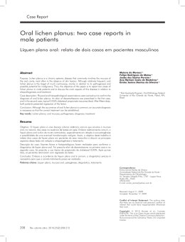
Revista SPDV 73(2) 2015; Alexandre Sisnando, Giuseppe de Figueiredo, Alef Maia, Elaine Melo, Fabio Francesconi;
Qual o seu diagnóstico?/ Which is your diagnosis?
Qual o Seu Diagnóstico?
DERMATOSE COMUM NO PASSADO, MAS QUE AINDA DESAFIA
OS DERMATOLOGISTAS - QUAL O SEU DIAGNÓSTICO?
Alexandre Sabino Sisnando1, Giuseppe Lemos Pertoti de Figueiredo2, Alef Alioscha Andrade Maia2, Elaine Dias Melo2,
Fabio Francesconi3
1
Dermatology resident, Fundação de Medicina Tropical Heitor Vieira Dourado (FMT-HVD)
2
Medical student, Faculdade de Medicina da Universidade Federal do Amazonas (FM-UFAM)
3
Dermatologist, Master of tropical diseases, Preceptor of residency in dermatology (FMT-HVD) and Professor of
dermatology (FM-UFAM)
* Manuscrito apresentado como poster eletrônico/ Presented as a electronic poster, 69º Congresso da Sociedade
Brasileira de Dermatologia, Recife, Brasil, nos dias 27 a 30 de setembro de 2014.
RESUMO – Os autores apresentam um caso de prurigo de Hebra, com o quadro típico numa criança com o mesmo
perfil socioeconômico e de fatores predisponentes descritos por Hebra em Viena no século XIX. Essa doença se manifesta por múltiplas pápulas pequenas e extremamente pruriginosas. Devido à escoriação, ocorrem erosões e liquenificação e infecções secundárias, evoluindo para cicatrizes hiper ou hipopigmentadas.. As crianças afetadas geralmente
são desnutridas e têm adenomegalias inguinal, axilar ou cervical. Normalmente, são crianças atópicas com hipersensibilidade cutânea a picada de insetos, num contexto de higiene inadequada e má nutrição.
PALAVRAS-CHAVE – Prurigo/diagnóstico, Doenças da pele.
Dermatology Quiz
A VERY COMMON DISEASE IN THE PAST THAT STILL
CHALLENGES DERMATOLOGISTS - WHICH IS YOUR DIAGNOSIS?
ABSTRACT – This paper presents a case of Hebra’s prurigo, with the typical picture in a child under similar socioeconomic conditions and predisposing factors described by Hebra in Vienna, in the nineteenth century. This disease is an
eruption of multiple extremely itchy small papules. Because of scratching, there are abrasions or lichenification, frequent
secondary infection and progression to scars with dyschromia. Affected children are usually malnourished and have increased inguinal, axillary or cervical lymph nodes. They are generally atopic children with hypersensitivity to insect bite,
in a context of inadequate hygiene and poor nutrition.
KEY-WORDS – Prurigo/diagnosis; Skin diseases.
Conflitos de interesse: Os autores declaram não possuir conflitos de interesse.
No conflicts of interest.
Suporte financeiro: O presente trabalho não foi suportado por nenhum subsídio ou bolsa.
No sponsorship or scholarship granted.
Direito à privacidade e consentimento escrito / Privacy policy and informed consent: Os autores declaram
que pediram consentimento ao doente para usar as imagens no artigo. The authors declare that the patient gave
written informed consent for the use of its photos in this article.
Recebido/Received - Fevereiro/February 2015; Aceite/Accepted – Março/March 2015
309
Revista SPDV 73(2) 2015; Alexandre Sisnando, Giuseppe de Figueiredo, Alef Maia, Elaine Melo, Fabio Francesconi;
Qual o seu diagnóstico?/ Which is your diagnosis?
Qual o Seu Diagnóstico?
Correspondência:
Dr. Alexandre Sabino Sisnando
Fundação de Medicina Tropical Heitor Vieira Dourado (FMT-HVD)
Av. Pedro Teixeira, 25 – Dom Pedro, Manaus – AM
CEP: 69.040-000, Brazil
Tel.: +55(92)982224746.
Email: [email protected]
CASE REPORT
A 6-year-old boy, from the countryside of Amazonas - Brazil, with a history of allergic reactions to
insect bites since he was 6 months old, had little improvement on topical corticosteroids and oral antihistamines. The child has marasmus and disseminated
highly pruritic rash composed of small erythematous
papules with excoriation in recent lesions and lichenification with dyschromia in the older ones. Lesions
predominated in extensor surface of limbs and face.
He had painful lymphadenopathy in the axillary and
inguinal region, and loss of weight (Fig. 1).
WHICH IS YOUR DIAGNOSIS?
A. Persistent scabious nodules
B. Hyde’s prurigo (Nodular prurigo)
C. Hebra's prurigo
D. Atopic dermatitis
DIAGNOSIS - C. Hebra's prurigo
This is a case of Hebra’s prurigo, presenting with the
typical picture in a child living under similar socioeconomic conditions as those described by Hebra in Vienna in
the 19th century. This rare disease consists of an eruption
of grouped dried extremely itchy papules of 2-3mm, localized in the whole body, mainly in the extensor surface
of upper limbs and face, Because of scratching, there
are abrasions or lichenification, leaving scars with dyschromia. Secondary infection of the lesions may occur.
The child affected is usually malnourished and has increased inguinal, axillary or cervical lymph nodes. They
are generally atopic children with hypersensitivity to insect bite (experimentally induced) and positive skin tests
to insect extracts.1
Scabious nodules are persistent lesions in patients
with evidence of current or previous scabies, and are not
recurrent lesions after treatment of the underlying disease, although they may persist for months after treatment.2
Histologically, the lesions of Hyde’s prurigo (nodular
prurigo) and Hebra’s prurigo may be identical. But the
310
Fig 1 - Hebra’s prurigo. Multiple papules associated
with signs of excoriation, lichenification and scaling,
some with brownish or yellowish crusts, especially on the
extensor surface of limbs and face.
key clinical features of Hyde’s prurigo are large papules
and more-or-less symmetrical nodules, 1-3 cm in diameter, with a raised, warty surface. Cases occur at all
ages, but mainly from 20 to 60 years. Both sexes are
equally affected and patients have intense, often distressing pruritus. The lesions may arise as a secondary event
in a wide range of cutaneous and systemic diseases, but
patient may have an idiopathic nodular prurigo.2
Atopic dermatitis presents as eczema (acute, subacute or chronic), usually without papules or nodules.2
MICROSCOPIC FINDINGS
Histopathological examination revealed interruption
of the epidermis, replaced by fibrinopurulent exudate.
The dermis showed perivascular inflammatory infiltrate
consisting of eosinophils, lymphocytes and histiocytes,
compatible with prurigo (Fig. 2).
During hospitalization, the patient was treated with
systemic antihistamines, moisturizers for recovery of skin
barrier function, topical antibiotic for secondary infection
Revista SPDV 73(2) 2015; Alexandre Sisnando, Giuseppe de Figueiredo, Alef Maia, Elaine Melo, Fabio Francesconi;
Qual o seu diagnóstico?/ Which is your diagnosis?
Qual o Seu Diagnóstico?
Fig 2 - Fibrinopurulent exudate and perivascular inflammatory infiltrate consisting of eosinophils, lymphocytes and histiocytes, compatible with prurigo. (H&E, 100x).
of the lesions and clinical support. There was improvement of pruritus and he was discharged with a recovery
nutrition plan to be followed. With a prescription as outpatient which included moisturizers and repellent, there
was significant improvement of the clinical picture.
DISCUSSION
Ferdinand von Hebra described the term prurigo referring to papules arising as a result of scratching.3 This
definition could include conditions such as papular urticaria, dermographism, among others. However, most
of the original cases of prurigo described by Hebra referred to wheals arising probably after scratching in atopic children living in precarious social conditions and
exposed to insect bites, especially fleas.3,4 Such lesions
were initiated as tiny normochromic papules, more
palpable than visible, which soon were modified by
excoriation and eczema, becoming larger and erythematous.5 For this dermatosis, in 1892, Besnier gave the
name "Hebra’s prurigo".4
Usually, the clinical picture begins early in life with
pruritic papules associated with erosion by excoriation.
With chronicity, lichenification occurs and there may be
a secondary infection with lymphadenopathy. Lesions
predominate in the extensor areas of extremities, face,
buttocks and abdomen, symmetrically. It is believed to
have a multifactorial predisposition.6
Hebra’s prurigo is considered a chronic and recurrent inflammatory skin disease, with cutaneous hyperreactivity to environmental stimuli (insect bites) possibly
enhanced by skin barrier dysfunction and is often associated with atopy. Skin hyperreactivity has intrinsic
(genetic) and extrinsic multifactorial basis and is closely
associated with inadequate hygiene and poor nutrition.6
A perivascular infiltrate in the central region of the
wheal consisting of lymphocytes and eosinophils is observed both in new and old lesions but differences between new and old lesions are noted in histopathology.
Recent lesions show hypergranulosis and normal stratum corneum, vasodilation with oedema and vascular
endothelial cells protrusion in the lumen as well as increased numbers of eosinophils in the infiltrate, while
the old lesions show with thickened collagen and decreased fibrocytes in the dermis. In old lesions there is
hypogranulosis, acanthosis, papillomatosis and irregular hyperpigmentation of basal layer.7
Regarding immunology, Almeida4 showed that the
serum of patients with Hebra’s prurigo had increased
immunoglobulins, especially IgE fraction (81% of patients), but also IgG (77% of them), which strengthens
the possibility that Hebra’s prurigo could be an atopic
manifestation.
Therefore, based on the literature, this is a typical
case of Hebra’s prurigo, with clinical and histopathological features and within the profile of the socioeconomic and environmental predisposing events. Currently,
there are only few reports in the literature, which is
questionable in view of the worldwide prevalence of
predisposing factors. So, knowledge of Hebra’s prurigo
is important for diagnosis and for the proper approach
of cases.
REFERÊNCIAS
1. Sampaio SA, Rivitti EA. Dermatologia. 3.ed. São
Paulo: Artes Médicas, 2007. p. 280-1.
2. Burns T, Breathnach S, Cox N, Griffiths C. Rook’s
Textbook of Dermatology. 8th. ed. Oxford: Blackwell
Publishing Ltd, 2010.
3. Hebra FV. On Diseases of the Skin. Vol. 2. London:
New Sydenham Society translation; 1868.p.257.
4. Almeida FA. Contribuição ao estudo de aspectos
imunológicos no prurigo de Hebra. An Bras Dermatol. 1988; 63:1-5.
5. Liveing R. Remarks on two cases of Hebra’s prurigo, lately treated at the Middlesex Hospital. Lancet.
1879:5.
6. Boediardja SA, Ramelan W, Cornain S. Genetic
inheritance pattern in prurigo Hebra. Paediatr Indones. 2001; 41:76-81.
7. Boediardja SA, Tjarta A, Comain S, Budimulja U,
Roostini A, Hartati M. The immunohistopathological features of Hebra. Med J Indones 2001; 10:115.
311
Baixar