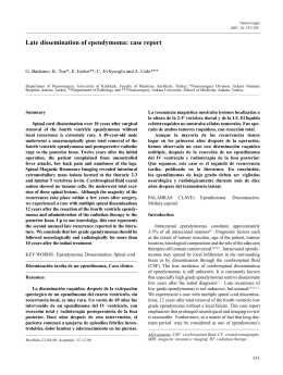
190
Relato de caso
Spontaneous Subarachnoid Hemorrhage Induced by
Warfarin Use - Case Report and Literature Review
Hemorragia subaracnoidea espontânea induzida por Warfarina
- Relato de Caso e Revisão da Literatura
Ricardo Rezende Campos, MD1
Alexandre Martins Cunha, MD, MSc2
Marcelo Eduardo Sette dos Santos, MD1
Guilherme Brasileiro de Aguiar, MD1,2
RESUMO
ABSTRACT
Introdução: A causa mais frequente de hemorragia
subaracnoidea (HSA) espontânea é a ruptura de aneurisma
cerebral. Em aproximadamente 5% dos casos de HSA
espontânea não se observam alterações à angiografia ou
ressonância magnética do sistema nervoso central que explique
o sangramento. O objetivo deste trabalho é apresentar um
caso de HSA espontânea induzida por warfarina, onde não
foi evidenciado nenhuma malformação vascular no sistema
nervoso central. Relato de Caso: Paciente do sexo masculino,
com 59 anos, apresentou quadro súbito de cefaléia durante o
banho. O paciente tinha história de valvulopatia mitral com
troca de vávula há 23 anos e hipertensão arterial sistêmica
acompanhado por cardiologista, em uso de warfarina
sódica ( 5mg diários ). A tomografia computadorizada de
crânio demonstrou HSA Fisher III. Os exames laboratoriais
mostraram uma razão normalizada internacional (RNI)
de 2.2 (normal 1 a 1,2). Foram realizadas ressonância
magnética do neuroeixo e angiografia cerebral que não
demonstraram aneurisma ou qualquer outra malformação
vascular. Conclusão: A ocorrência de hemorragia cerebral
associada ao uso de warfarina é um evento bem documentado
na literatura. Entretanto a ocorrência de HSA isolada nesses
pacientes rara. Para atribuir a hemorragia subaracnoidea ao
uso de warfarina é necessário excluir a presença de aneurisma
ou outras malformações vasculares pelos exames de imagem.
Introduction: The most frequent cause of spontaneous
subarachnoid hemorrhage (SAH) is the rupture of a brain
aneurysm. Approximately 5% of cases of spontaneous SAH
did not show changes on magnetic resonance imaging (MRI)
or angiography that explain such bleeding. The objective of
this study was to report a case of spontaneous SAH induced
by warfarin, where there was no evidence of any vascular
malformation. Case Report: A 59 year-old male patient
developed a sudden onset severe headache while in the
shower. The patient had a history of mitral valve disease
and valve replacement 23 years before and hypertension
monitored by a cardiologist with 5 mg warfarin sodium daily.
Computed tomography (CT) scan revealed a SAH. Laboratory
tests showed an International Normalized Ratio (INR) of 2.22
(normal: 1 to 1.2). Cerebral angiography and neuraxis MRI
were performed and showed no aneurysms or other vascular
malformations.Conclusion: The occurrence of hemorrhage
associated with warfarin treatment is a well-documented
event in the literature. However, the isolated occurrence of
SAH in these patients is a rare event. To prove that SAH is
indeed attributed to anticoagulation, it is necessary to exclude
the presence of aneurysms and other vascular diseases by
imaging techniques.
Palavras-chave: hemorragia subaracnoidea, hemorragia
cerebral, warfarina, angiografia cerebral.
Key words: cerebral hemorrhage, subarachnoid hemorrhage,
warfarin, cerebral angiography.
1 - Division of Neurosurgery, Santa Marcelina de Itaquaquecetuba Hospital - São Paulo / SP / Brazil.
2 - Division of Neurosurgery, Department of Surgical Specialties, State University of Rio de Janeiro - Rio de Janeiro / RJ / Brazil.
Recebido em 10 de julho de 2011, aceito em 16 de setembro de 2011
Campos RR, Cunha AM, Santos MES, Aguiar GB - Spontaneous Subarachnoid Hemorrhage Induced by Warfarin Use - Case Report and Literature Review
J Bras Neurocirurg 22 (4): 190-193, 2011
191
Relato de caso
Introduction
A
B
C
Spontaneous subarachnoid hemorrhage (SAH) accounts for
approximately 5% of all strokes1. The estimated incidence
varies from 8.1 to 23 cases per 100,000 inhabitants1,13, with
high morbidity and mortality rates that can reach up to 45%1. In
85% of SAH cases, a ruptured cerebral aneurysm is evident on
computed tomography (CT) scans1,13. Ruptured arteriovenous
malformations, dural fistula or sinus thrombosis, among others,
represent other causes of spontaneous SAH13.
D
E
F
Of patients with spontaneous SAH, 15% to 20% have no
aneurysm detectable by cerebral angiography10,11. Rinkel et
al.10 listed perimesencephalic idiopathic hemorrhage, cocaine
use and coagulation disorders as possible causes of SAH in the
absence of aneurysms and vascular malformations by imaging
studies10.
The objective of this article was to report a case of spontaneous
SAH induced by warfarin, in which there was no evidence of
an underlying vascular malformation. We also performed a
brief review of the literature on this subject.
Figure 1. Axial head CT without contrast: A,B,C: Fisher III subarachnoid hemorrhage filling the basal cisterns and sylvian fissures; D,E,F: no signs of intracranial
hemorrhage.
A
B
C
D
E
F
Case Report
A 59-year-old male had a sudden onset of severe headache while
he was in the shower. Neurological examination revealed that
the patient was alert and lucid, without any focal neurological
deficits, with a mild degree of neck stiffness (graded as a HuntHess I SAH). The patient had a mitral valve disease with a valve
replacement 23 years previously and hypertension that was
under follow-up by a cardiologist. The patient was under antihypertensive medication, and on 5 mg warfarin sodium daily.
A CT scan demonstrated a SAH (Fisher III) (Fig. 1A, 1B, 1C).
Laboratory tests showed an International Normalized Ratio
(INR) of 2.22 (normal range: 1 - 1.2) and Prothrombin Time
Activity (PTA) by 21.8” and 42% (normal ranges: 10.2 to 14.2”
/ 70-130%). The patient remained in the intensive care unit of
our hospital as part of the initial standard treatment for SAH, in
addition to the discontinuation of his treatment with warfarin.
Cerebral angiography was performed (Fig. 2) and showed no
vascular malformation. Neuraxis MRI was performed, with no
alterations.
With the discontinuation of warfarin, the patient’s coagulation
function normalized after 6 days. The patient left the
hospital after 14 days asymptomatic, and continued to attend
cardiological and neurological follow-up appointments. The
most recent CT scan prior to his discharge from hospital was
normal (Fig. 1D,1E,1F).
Figure 2. Cerebral angiography. Right carotid artery injection, frontal (A) and lateral
(B) view. Left carotid artery injection frontal (C) and lateral (D) view; Left vertebral artery injection, frontal (E) and lateral (F) views. No evidence of vascular malformation.
Campos RR, Cunha AM, Santos MES, Aguiar GB - Spontaneous Subarachnoid Hemorrhage Induced by Warfarin Use - Case Report and Literature Review
J Bras Neurocirurg 22 (4): 190-193, 2011
192
Relato de caso
Discussion
factors, such as the level of consciousness of the patient on
admission, volume of the hemorrhage and site of bleeding, as
bleeding induced by use of such medications can range from
asymptomatic microbleedings to massive bleeding3,9,12,14.
The most common cause of SAH is trauma13. If only spontaneous
SAH is considered, its main cause is rupture of an intracranial
aneurysm1,13. Among the cases of spontaneous SAH, between 15
and 20% present with normal cerebral angiography10,11, which
requires further investigation to diagnose other possible causes.
Intraparenchymal hemorrhage associated with warfarin is an
event that is well-documented in the literature. However, the
occurrence of isolated SAH in these patients is a rare event.
For SAHs in anticoagulated patients to indeed be attributed to
anticoagulation, it is necessary that the presence of aneurysms
and other intracranial vascular diseases are excluded by imaging
techniques, such as cerebral angiography and MRI of the
neuraxis. Once confirmed, any SAH induced by warfarin should
be managed with normalization of the INR to decrease the risk
of further rebleeding, and the imposition of a standard treatment
protocol for SAH.
Intraparenchymal hemorrhage in patients with coagulation
disorders, either through primary causes or in the presence
of anticoagulation therapy, is well-reported in the medical
literature3,10,11,13,14. Oral anticoagulation is a cause of intracerebral
hemorrhage3,14, and has been reported as the most serious
complication with the long-term use of this therapy in patients4.
The location of anticoagulation-induced intracranial hemorrhage
occurs, in decreasing order of frequency, deep in the brain , in
the cerebral lobes, cerebellum and brainstem3.
Olsen et al.8 in a population study, showed that there is no
increase in the risk of SAH associated with the use of oral
anticoagulation8. SAH-induced anticoagulation alone is a rare
event, with only a few reported cases5,3,7. Mattle et al.6 reported
76 cases of extracerebral intracranial bleeding associated with
use of oral anticoagulation. Of these, seven had hemorrhage in
the subarachnoid space, and only four had no history of trauma.
Of the four patients with spontaneous SAH, two were diagnosed
with a cerebral aneurysm and the other two were considered
to have SAHs that were directly related to the use of vitamin
K antagonists. There was no information on any additional
investigation with cerebral angiography in these two patients, or
elsewhere6.
Zubrov et al.14 reported a retrospective study of patients presenting
with intracranial hemorrhage in use of oral anticoagulants
who were treated with drug withdrawal, the administration of
antihypertensive drugs to maintain a mean arterial pressure below
130 mmHg, the correction of the INR with vitamin K associated
with fresh frozen plasma infusion, besides the administration of
recombinant factor VII for cases whose response to vitamin K and
fresh frozen plasma were unsuccessful14. In contrast, Flaherty et
al.3 reported during the same year that there was no consensus
with regards to indications for the suspension or manteinance
of oral anticoagulation in cases of intracranial hemorrhages3.
Claassen et al.2 published a prospective study in 2008, which
reported that there was no difference in relation to rebleeding in
cases where anticoagulation was suspended completely or early
resumed (between 2 weeks and 2 months post-SAH), or late (>2
months of bleeding)2.
As regards to prognosis, the severity of hemorrhagic stroke in
the presence of oral anticoagulation therapy depends on several
Disclosure
The authors have no conflict of interest involved.
References
1. Bederson JB, Connolly Jr ES, Batjer HH, Dacey RG, Dion
JE, Diringer MN, et al. Guidelines for the management of
aneurysmal subarachnoid hemorrhage: a statement for healthcare
professionals from a special writing group of the Stroke Council,
American Heart Association. Stroke. 2009; 40(3): 994-1025.
2.
Claassen DO, Kazemi N, Zubkov AY, Wijdicks EF, Rabinstein
AA. Restarting anticoagulation therapy after warfarin-associated
intracerebral hemorrhage. Arch Neurol. 2008; 65(10): 1313-8.
3. Flaherty ML, Tao H, Haverbusch M, Sekar P, Kleindorfer
D, Kissela B, et al. Warfarin use leads to larger intracerebral
hematomas. Neurology. 2008; 71(14): 1084-9.
4.
Foerch C, Arai K, Jin G, Park KP, Pallast S, van Leyen K, et
al. Experimental model of warfarin-associated intracerebral
hemorrhage. Stroke. 2008; 39(12): 3397-404.
5.
Mathiesen T, Benediktsdottir K, Johnsson H, Lindqvist M, von
Holst H. Intracranial traumatic and non-traumatic haemorrhagic
complications of warfarin treatment. Acta Neurol Scand. 1995;
91(3): 208-14.
6. Mattle H, Kohler S, Huber P, Rohner M, Steinsiepe KF.
Anticoagulation-related intracranial extracerebral haemorrhage.
J Neurol Neurosurg Psychiaty. 1989; 52:829-37.
Campos RR, Cunha AM, Santos MES, Aguiar GB - Spontaneous Subarachnoid Hemorrhage Induced by Warfarin Use - Case Report and Literature Review
J Bras Neurocirurg 22 (4): 190-193, 2011
193
Relato de caso
7. Nilsson OG, Lindgren A, Stahl N, Brandt L, Säveland H.
Incidence of intracerebral and subarachnoid hemorrhage in
southern Sweden. J Neurol Neurosurg Psychiatry. 2000; 69:
601-7.
8.
Olsen M, Johansen MB, Christensen S, Sorensen HT. Use of
vitamin K antagonists and risk of subarachnoid haemorrhage:
a population-based case-control study. Eur J Intern Med. 2010;
21(4): 297-300.
9.
Orken DN, Kenangil G, Uysal E, Forta H. Cerebral microbleeds
in ischemic stroke patients on warfarin treatment. Stroke. 2009:
40(11): 3638-40.
10. Rinkel GJ, van Gijn J, Wijdicks EF. Subarachnoid hemorrhage
without detectable aneurysm. A review of the causes. Stroke.
1993; 24(9): 1403-9.
11. Sarabia R, Lagares A, Fernández-Alén JA, Arikan F, Vilalta
J, Ibáñez J, et al. Idiopathic subarachnoid hemorrhage: a
multicentre series of 220 patients. Neurocirugia (Astur). 2010;
21(6): 441-51.
12. Ueno H, Naka H, Ohshita T, Kondo K, Nomura E, Ohtsuki T, et
al. Association between cerebral microbleeds on T2*-weighted
MR images and recurrent hemorrhagic stroke in patients treated
with warfarin following ischemic stroke. Am J Neuroradiol.
2008; 29(8): 1483-6.
13. van Gijn J, Rinkel GJ. Subarachnoid haemorrhage: diagnosis,
causes and management. Brain. 2001; 124(2): 249-78.
14. Zubkov AY, Mandrekar JN, Claassen DO, Manno EM, Wijdicks
EF, Rabinstein AA. Predictors of outcome in warfarin-related
intracerebral hemorrhage. Arch Neurol. 2008; 65(10): 1320-5.
Corresponding Author
Guilherme Brasileiro de Aguiar, MD
Rua Abílio Soares, 121 – apto 84 – Paraíso
São Paulo – SP. Brazil
04005-000
Email: [email protected]
Campos RR, Cunha AM, Santos MES, Aguiar GB - Spontaneous Subarachnoid Hemorrhage Induced by Warfarin Use - Case Report and Literature Review
J Bras Neurocirurg 22 (4): 190-193, 2011
Download