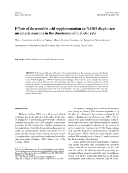
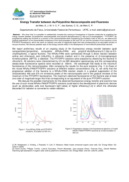
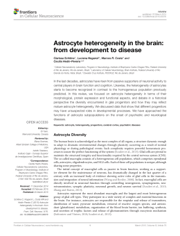
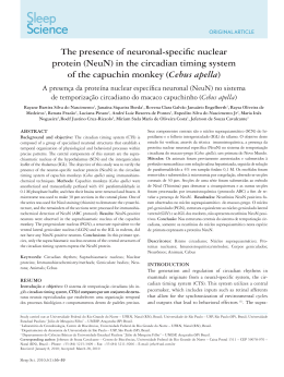
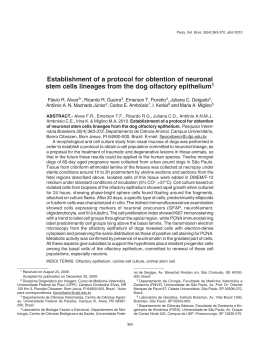
Journal of Cerebral Blood Flow & Metabolism (2009), 1–12
& 2009 ISCBFM All rights reserved 0271-678X/09 $32.00
www.jcbfm.com
On the fate of extracellular hemoglobin and heme
in brain
Flavio A Lara1,2,3, Suzana A Kahn1, Anna CC da Fonseca1, Carlomagno P Bahia4,
João PC Pinho4, Aurélio V Graca-Souza2, Jean C Houzel4, Pedro L de Oliveira2,
Vivaldo Moura-Neto1 and Marcus F Oliveira5,6
1
Laboratório de Morfogênese Celular, Departamento de Anatomia, Instituto de Ciências Biomédicas,
Universidade Federal do Rio de Janeiro, Cidade Universitária, Rio de Janeiro, Brazil; 2Programa de Biologia
Molecular e Biotecnologia, Laboratório de Bioquı´mica de Artrópodes Hematófagos, Instituto de Bioquı´mica
Médica, Universidade Federal do Rio de Janeiro, Cidade Universitária, Rio de Janeiro, Brazil; 3Laboratório
de Microbiologia Celular, Instituto Oswaldo Cruz, Rio de Janeiro, Brazil; 4Laboratório de Fronteiras em
Neurociências, Programa de Neurociências Básico e Clı´nico, Instituto de Ciências Biomédicas, UFRJ;
5
Programa de Biologia Molecular e Biotecnologia, Laboratório de Bioquı´mica Redox, Instituto de Bioquı´mica
Médica, Universidade Federal do Rio de Janeiro, Cidade Universitária, Rio de Janeiro, Brazil; 6Laboratório
Associado de Inflamação e Metabolismo, Instituto Nacional de Biologia Estrutural e Bioimagem,
Universidade Federal do Rio de Janeiro, Cidade Universitária, Rio de Janeiro, Brazil
Intracerebral hemorrhage (ICH) is a major cause of disability in adults worldwide. The
pathophysiology of this syndrome is complex, involving both inflammatory and redox components
triggered by the extravasation of blood into the cerebral parenchyma. Hemoglobin, heme, and iron
released therein seem be important in the brain damage observed in ICH. However, there is a lack of
information concerning hemoglobin traffic and metabolism in brain cells. Here, we investigated the
fate of hemoglobin and heme in cultured neurons and astrocytes, as well as in the cortex of adult
rats. Hemoglobin was made traceable by conjugation to Alexa 488, whereas a fluorescent heme
analogue (tin-protoporphyrin IX) was prepared to allow heme tracking. Using fluorescence
microscopy we observed that neurons were more efficient in uptake hemoglobin and heme than
astrocytes. Exposure of cortical neurons to hemoglobin or heme resulted in an oxidative stress
condition. Viability assays showed that neurons were more susceptible to both hemoglobin and
heme toxicity than astrocytes. Together, these results show that neurons, rather than astrocytes,
preferentially take up hemoglobin-derived products, indicating that these cells are actively involved
in the ICH-associated brain damage.
Journal of Cerebral Blood Flow & Metabolism advance online publication, 1 April 2009; doi:10.1038/jcbfm.2009.34
Keywords: intracerebral hemorrhage; orphyrin; reactive oxygen species; toxicity; trauma
Introduction
Cerebrovascular diseases and their consequences,
including strokes, represent the second most frequent cause of death, after ischemic heart diseases.
Correspondence: Dr FA Lara, Laboratório de Microbiologia Celular,
Sala 27, Pavilhão Hansenı́ase, Fundação Oswaldo Cruz, Instituto
Oswaldo Cruz, Av Brasil, número 4365, CEP 21045-900, Rio de
Janeiro, Brazil.
E-mail: [email protected]
This study was supported by CNPq (through Institutos Nacionais
de Ciência e Tecnologia 2008), FAPERJ (MFO through Jovens
Cientistas do Nosso Estado 2007 and Edital de Apoio a Grupos
Emergentes de Pesquisa do Rio de Janeiro, 2008), ICGEB, HHMI.
Received 26 August 2008; revised 10 March 2009; accepted 13
March 2009
Intracerebral hemorrhage (ICH) has been estimated to
account for 10% to 15% (Sudlow and Warlow, 1997)
of the 5.7 million deaths caused annually by stroke
worldwide (WHO, 2006). More importantly, ICH has
an unfavorable prognosis, with an extremely high
mortality rate in the first month (approximately 45%)
and the majority of survivors show debilitating
neurologic sequels (Fogelholm et al, 1992). The
pathophysiology of ICH comprises a complex chain
of events starting with blood–brain barrier disruption
and infiltration of blood components into the brain
parenchyma, resulting in a progressive edema, which
starts in the first 24 h and remains elevated during
several days (Koeppen et al, 2004). In many cases,
inflammatory cell recruitment and activation contribute to the cerebral parenchyma damage, resulting
Fate of blood products in brain
FA Lara et al
2
in neuronal death, not only inside, but also around
the perifocal reactive zone (Gong et al, 2000; Rogers
et al, 2003).
The destructive action of blood components to the
central nervous system can be analyzed under two
overlapping perspectives: inflammation and oxidative stress. Heme has recently been shown to be an
immunoactive molecule, able to promote neutrophil
and macrophage activation and migration both
in vitro and in vivo (Figueiredo et al, 2007; Porto
et al, 2007; Graça-Souza et al, 2002). The free-radical
hypothesis for ICH is based on the cytotoxicity
triggered by blood components and its degradation
products, such as heme and iron. Heme catabolism
by heme oxygenase (HO) results in the release of
carbon monoxide, biliverdin, and iron. Although HO
is frequently described as an antioxidant enzyme, it
is well known that iron is a potent pro-oxidant atom,
capable to generate highly reactive oxygen species,
such as hydroxyl, alkoxyl, and peroxyl radicals
(Ryter and Tyrrell, 2000). Curiously, derrangements
in iron metabolism have been described in other
neuropathologies, such as Alzheimer’s neurodegenerative disorder (Smith et al, 1997).
Although much progress has been made toward
the functions of HO activity and iron metabolism in
the brain, the cellular mechanisms involved in the
clearance of blood components during an ICH episode
are still not well understood. There is a consensus
that a first wave of microglial recruitment occurs in
areas surrounding the lesion 24 to 48 h after the
hemorrhage, followed by recruitment of blood phagocytes that are essential to blood clot resolution and,
later, by the invasion of neutrophils (Gong et al, 2000).
However, intracortical injections of purified hemoglobin seem to interfere dramatically with brain physiology, as this promotes epileptic seizures (Rosen
and Frumin, 1979). In agreement with this finding,
reductions in plasma levels of haptoglobin are also
associated with epileptic seizures (Panter et al, 1985).
A number of observations converge to the fact that
hemoglobin toxicity is cell specific, being neurons
more sensitive than astrocytes (Rogers et al, 2003;
Chen-Roetling and Regan, 2006; Regan and Panter,
1993). Conversely, HO-1 is involved in astrocyte and
neuron heme tolerance (Regan et al, 2000; ChenRoetling and Regan, 2006), whereas the function of
HO-2 in neuronal protection against heme remains
controversial (Doré et al, 1999; Rogers et al, 2003).
Zinc protoporphyrin IX, a potent HO inhibitor, has
recently shown to be beneficial in a rabbit model of
ICH, indicating that iron released from heme degradation may be involved in the pathogenesis of ICH
(Gong et al, 2006). Expression of HO-1 and ferritin
during ICH suggested that not only microglia, but
also astrocytes, were involved in hemoglobin digestion, heme degradation, and subsequent iron storage
(Koeppen et al, 1995; Turner et al, 1998). These
findings strongly suggest that hemoglobin, heme,
and iron are pivotal in the pathogenesis of ICHinduced brain damage.
Journal of Cerebral Blood Flow & Metabolism (2009), 1–12
To understand the fate of hemoglobin and heme in
the brain after ICH, we monitored the localization of
these molecules in neurons and astrocytes both
in vitro and in vivo, using a fluorescent heme
analogue and/or fluorescent-labeled hemoglobin.
We show that neurons were more efficient in the
uptake of globin, hemoglobin, and a heme analogue,
compared with astrocytes. In addition, neurons
revealed to be more susceptible to hemoglobin and
heme than astrocytes, in a mechanism involving
oxidative stress.
Materials and methods
Primary Astrocyte and Neuronal Cell Cultures
Primary cultures of astrocytes were prepared from newborn
(0- to 1-day-old) Wistar rat cerebral cortex, as previously
described by Gomes et al, 1999. Briefly, dissociated cells
were plated in Dulbecco’s modified Eagle’s medium/F12
with 10% fetal bovine serum and expanded in 25 cm2
tissue culture flasks at 371C in a humidified 5% CO2
atmosphere. After 3 days, weakly bound cells, mostly
neurons, were washed away. After 1 week, when confluence was achieved, the culture was treated with trypsin
and transferred to 15.5 mm diameter poly-L-lysine-treated
glass coverslips in 24-well plates for microscopy, or to
96-well plates for viability assays.
Primary neuronal cultures were prepared from embryonic day 18 or newborn (0- to 1-day-old) Wistar rats cerebral
cortex, as previously described. Briefly, the cerebral
cortices were dissociated in Dulbecco’s modified Eagle’s
medium/F12 medium supplemented with 33 mmol/L
glucose, 2 mmol/L glutamine, and 3 mmol/L sodium
bicarbonate. From this suspension, 5 104 cells were
plated on poly-L-lysine-treated coverslips placed on a
24-well plate for neuronal cultures or plated over normal
astrocytes monolayers for neuroastrocytes coculture experiments. Both cultures and cocultures were maintained
in Dulbecco’s modified Eagle’s medium/F12 medium
without serum to 24 or 48 h. Finally, using cytochemistry
staining by isolectin B4 (Sigma, St Louis, MO, USA) we
observed that microglia contaminants in our neuronal
cultures were approximately 0.5% (data not shown).
Tin-Protoporphyrin IX Fluorescence Analysis
Tin-Protoporphyrin IX (SnPPIX; Frontier Scientific, Logan,
UT, USA) solutions were prepared in dimethyl sulfoxide to
a final concentration of 20 mmol/L. Then a 10 mmol/L
SnPPIX was prepared by diluting this stock solution 1:2 in
0.1 N NaOH. For spectrofluorometric analysis 10 mmol/L
SnPPIX solutions were prepared from the 10 mmol/L
solution by diluting them in methanol, albumin, or water
and data were acquired in a Cary Eclipse spectrofluorimeter (Varian, Palo Alto, CA, USA). To assess SnPPIX
uptake, cells were incubated with a 10 mmol/L SnPP IX
solution diluted in culture medium without serum for 2 h.
Afterwards, cells were washed with phosphate-buffered
saline (PBS), fixed in 4% paraformaldehyde, and observed
Fate of blood products in brain
FA Lara et al
3
in an Axioplan 2 epifluorescence microscope (Zeiss,
Göttingen, Germany). The fluorescence images were
obtained using a filtered 100 W mercury lamp as the
excitation light source with a filter set BP 400 to 410 nm/FT
510 nm/LP 515 nm. Quantification of fluorescent intensities was performed by measuring bitmaps generated by
ImageJ software (available at http://rsb.info.nih.gov/ij/).
Hemoglobin Labeling
Adult rat hemoglobin was purified as follows: heparinized
blood was centrifuged at 100g for 10 min and erythrocytes
were resuspended in 50 mmol/L Tris-HCl (pH 8.5), with
1.7% NaCl and 1 mmol/L EDTA. This step was repeated
twice. After, erythrocytes were lysed in the same buffer
without NaCl and cell debris was pelleted by centrifuging
at 1,000g for 30 mins. Hemoglobin was finally precipitated
by salting out in ammonium chloride at 80% saturation for
12 h at 41C. The insoluble hemoglobin was pelleted by
centrifuging at 1,000g for 30 min and then solubilized by
dialysis against 10 mmol/L phosphate buffer. The final
hemoglobin preparation was quantified in five replicates
by using the Folin method (Lowry et al, 1951). Alexa 488
(Molecular Probes, Eugene, OR, USA) labeling was performed as indicated by the manufacturer. Briefly, 10 mg of
Alexa 488 solubilized in dimethyl sulfoxide was added to
10 mg of hemoglobin in 0.1 mol/L sodium bicarbonate
buffer (pH 8.3), for 1 h at room temperature. The reaction
was stopped by adding 1.5 mol/L hydroxylamine (pH 8.3).
Unbound probe was excluded by gel filtration, using PBS
as mobile phase. Contamination of endotoxins in all
globin, hemoglobin, and heme preparations was assessed
by using the Limulus amebocyte lysate assay (QCL-1000;
BioWhittaker, Walkersville, MD, USA) following the
manufacturer’s procedures.
Globin Preparation
The globin solution was prepared as follows: purified
hemoglobin from rat blood was denatured and heme was
removed in diethylketone (HCl 0.02 mol/L kept in an
ethanol) dry ice bath, as described (Teale, 1959). Precipitated protein was washed once and renaturated by
sequential dialysis against water.
Stereotaxic Injection of Alexa 488-Hemoglobin in Adult
Rat Cerebral Cortex
Wistar male rats (n = 4, 3-month old) were obtained from
the animal facility at the Universidade Federal do Rio de
Janeiro. Experimental procedures followed local rules for
use of laboratory animals, based on the Guide for the Care
and Use of Laboratory Animals (NIH, no. 86-23, revised
1985) and all measures were taken to reduce the number of
animals and avoid their suffering. On the following day,
anesthesia was induced with an intramuscular mixture of
xylazine (4 mg/kg; Rompun, Bayer, Germany) and ketamine
chloride (46 mg/kg, Ketalar; Parke-Davis, Detroit, MI, USA).
Body temperature was maintained at 371C through a
heating pad, respiration and heart rates were monitored
and nociceptive reflexes were checked periodically so that
additional doses of anesthetic could be subsequently
administered if necessary. After head immobilization in
the stereotaxic apparatus, an incision was made through
the scalp and the skull overlying the parietal cortex of both
hemispheres was opened with a dental drill. At standard
stereotaxic coordinates (2.8 mm posterior to bregma and
5.2 mm lateral from the midline) a small incision was
opened in the dura and a glass capillary with 40 to 50 mm
internal tip diameter, connected through a mineral oilfilled tubing to a Hamilton microsyringe, was lowered
1.200 mm below the pial surface. Four microliters of
solutions containing 10 mg of Alexa 488-hemoglobin or
10 mg of unlabeled hemoglobin was slowly injected into the
right and left hemispheres, respectively. The opening was
closed with Gelfoam and bone wax and the incision was
sutured. Animals were allowed to recover with food and
water ad libitum. After 2 days, they were administered a
lethal dose of anesthetic and perfused transcardially with
pH 7.4, 0.1 mol/L PBS followed by 4% paraformaldehyde
in 0.1 mol/L PBS. The brain was dissected, cryoprotected
in 30% buffered sucrose for 12 h, blocked in OCTTissueTek (Sakura, São Paulo, Brazil), and frozen in liquid
nitrogen. Serial, coronal, 15 mm thick sections were cut
using a CM1900 cryostat (Leica Microsystems GmbH,
Wetzlar, Germany).
Immunolocalization of b-Tubulin III and Glial
Fibrillary Acidic Protein
For coronal brain sections, immunohistochemistry for
b-tubulin III was performed as follows: sections were
blocked using 5% bovine serum albumin and 0.05% Triton
X-100 in PBS for 4 h, before overnight incubation at 41C
with mouse anti-b-tubulin III IgG (Promega, Madison, WI,
USA) diluted 1:500 in PBS with 1% bovine serum albumin
and 0.05% Triton X-100). Sections were then washed and
the stained with CY3-conjugated antimouse secondary
antibody (Sigma), diluted 1:5,000 in PBS with 1% bovine
serum albumin. For culture assays, immunocytochemistry
was performed as follows: cultured cells plated on glass
coverslips were fixed with 4% paraformaldehyde for
15 mins and permeabilized with 0.2% Triton X-100 in
PBS for 5 mins at room temperature. After blocking with
5% bovine serum albumin for 30 mins, cells were incubated with primary antibodies: mouse anti-b-tubulin III
IgG (1:1,000) or anti-GFAP IgG (1:500) (Dako, Glostrup,
Denmark) overnight at 41C followed by PBS washes
and incubation with antimouse IgG conjugated with CY3
or antirabbit conjugated with fluorescein isothiocyanate
(FITC) for 1 h at room temperature, to localize b-tubulin III
and glial fibrillary acidic protein (GFAP), respectively.
For all immunostainings, negative control sections or
plates were performed by omitting the primary antibody.
Differential interference contrast images of cultures
were obtained using an Axioplan 2 epifluorescence
microscope (Zeiss). Epifluorescence images of Alexa 488
and FITC were obtained using a 100 W mercury lamp
as the excitation light source with a filter set BP 450 to
Journal of Cerebral Blood Flow & Metabolism (2009), 1–12
Fate of blood products in brain
FA Lara et al
4
490 nm/FT 510 nm/LP 515 nm. 4,6-Diamidino-2-phenyl
indole (DAPI) fluorescence was acquired using G 365 nm/
FT 395 nm/LP 420 nm filter set, CY3 signal was obtained
using BP 545-555 nm/FT 580 nm/LP 590 nm as filter set
and BP 410 nm/FT 510 nm/LP 520 nm to observe SnPPIX
fluorescence. Confocal images were acquired using a Meta
510 laser scanning microscope (Zeiss), with an excitation
laser of 405 nm.
paraformaldehyde and finally stained with DAPI
20 mg/mL for 15 mins. Cells were observed in an Axiobserver
fluorescence microscope (Zeiss). Differential interference
contrast images were used to distinguish neurons from
astrocytes in cocultures. In addition, neurons and astrocytes
can also be distinguished through the pattern of DAPI
staining, as neurons exhibit not only stronger fluorescence
signal, compared with astrocytes, but also smaller nucleus.
Cellular Viability
Statistical Analyses
Viabilities of cell cultures were assessed by using the MTT
(3-[4,5-dimethylthiazol-2-yl]-2,5-diphenyltetrazolium bromide) reduction to formazan assay. Briefly, cells were
adhered to 96-well plates and exposed to different
conditions in quintuplicate for 12 h. Viability was assessed
by adding MTT to the cultures at a final concentration of
500 mmol/L. After 2 h, the supernatant was discarded, cells
were disrupted, and formazan was solubilized by addition
of lysis buffer as follows: 20% SDS, 50% dimethylformamide, 2% acetic acid, and 2.5% HCl 1 mol/L. Formazan
light absorption at 540 nm was determined using a
microplate reader Versa Max (Molecular Devices, Sunnyvale, CA, USA). Viability analysis in cocultures was
assessed after 12 h of different insults in two independent
experiments using four cell cultures for each condition.
Dead cells were removed by washing cultures with PBS
three times for 5 mins and the number of live cells was
determined by counting the number of cells attached to the
coverslips. Afterwards, the cells were fixed with 4%
Comparisons between groups were done by the one-way
analysis of variance and a posteriori Tukey’s test for pairwise comparisons. Differences of P < 0.05 were considered
to be significant. Analysis of variance and Tukey’s test were
performed by GraphPad Prism version 4.00 for Windows
(GraphPad Software, San Diego, CA, USA).
Results
Neurons Uptake SnPPIX more Efficiently than
Astrocytes
Our group has recently showed that palladium
mesoporphyrin IX is a fluorescent heme analogue
that can be used to follow heme traffic in living cells
(Lara et al, 2005). Previous works have shown that
SnPPIX also has potential as a tool for real-time
heme-tracking studies (Simionatto et al, 1984). In
Figure 1A we observe that SnPPIX fluorescence
Figure 1 Cortical neurons are able to take up the fluorescent heme analogue tin-protoporphyrin IX (SnPPIX) in vitro. (A) Spectra of
palladium mesoporphyrin IX (red line) and SnPPIX were recorded in water (black line), methanol (green line) or bound to 5 mmol/L
albumin (blue line). Spectra were acquired by excitation at 400 nm as described in the Materials and methods section. Neurons
derived from 18-day embryonic rat cerebral cortexes were cultured for 24 h and exposed to 10 mmol/L of SnPPIX for 2 h in serum-free
medium. Interference contrast (B and D) or fluorescence images (C, E, and G) were acquired and a merged image (F) was
constructed. (C) Neuronal culture presenting a strong red emission conferred by the porphyrin accumulation along cellular bodies
(asterisk) and neurites (arrow). (E) Higher magnification images showing the SnPPIX signal located inside small vesicles (arrow),
located in a neurite as can be seen in the merged image (F). (G) Neuronal culture exposed to SnPPIX at 41C. Scale bar = 20 mm.
Journal of Cerebral Blood Flow & Metabolism (2009), 1–12
Fate of blood products in brain
FA Lara et al
5
emission spectrum is insensitive to changes in the
environmental dielectric constant, because it shows
the same emission spectra regardless the medium
composition. However, SnPPIX exhibited a much
higher quantum yield when compared with palladium mesoporphyrin IX, because a 0.5 mmol/L solution presents three times more signal at its maximal
emission peak than palladium mesoporphyrin IX
50 mmol/L solution. Taking advantage of these features
and also of higher quantum yield of SnPPIX, we were
able to follow its uptake and traffic as a heme
analogue in rat brain and central nervous system cells.
To compare the uptake of this heme analogue by
neurons and astrocytes, we established cell cultures
containing almost exclusively neurons, confirmed
after 2 days in culture by positive immunocytochemistry against b-tubulin III antigens, and presence of
few cells (less than 5%) immunolabeled with GFAP.
Then, we exposed neurons from embryonic rat
cerebral cortex to 100 mmol/L SnPPIX for 2 h (Figure
1B). The cultured neurons were able to take up
considerable amounts of this probe as shown by the
accumulation of fluorescence signals in the cell
bodies (asterisk in Figure 1C) as well as in the
neurites (arrow in Figure 1C). Porphyrins are
amphipathic molecules and part of their cytotoxicity
is based on their ability to associate with lipid
bilayers, disrupting their permeability, and resulting
in porphyrin internalization and accumulation
(Schmitt et al, 1993). To check whether the observed
uptake was indeed a passive accumulation of the
SnPPIX in the neuronal membranes, higher magnification images were acquired. As shown in Figure 1,
the SnPPIX fluorescence was present in the cell body
as well as within endocytic vesicles (arrows), showing that a simple, passive partition across neuronal
membranes is not the major uptake mechanism for
the porphyrin molecules. SnPPIX uptake at 41C was
undetectable on all five experiments performed
(Figure 1F). In contrast, GFAP-positive astrocytes
from glia-enriched cultures did not display detectable levels of SnPPIX accumulation (Figure 2).
Although astrocytes were less efficient in take up
SnPPIX, it is possible that the staining can be
ascribed to contaminant microglial cells present in
our culture conditions (Figure 2C and 2D , asterisks).
In these experiments, cultures were exposed to
SnPPIX for 2 h in a serum-free medium, to avoid
the conjugation of SnPPIX to serum proteins such as
albumin. Additional experiments performed in serum-supplemented medium produced similar results,
reinforcing the observations that astrocytes were
much less prone to take up free, as well as
albumin-complexed SnPPIX (data not shown).
the hemorrhagic spot to perform endocytosis and
lysosomal digestion of blood constituents (Koeppen
et al, 1995). Thus, we next investigated whether
neurons or astrocytes would be able to take up
hemoglobin in a similar manner, by exposing these
cells to fluorescent-labeled hemoglobin. The concentrations used were chosen based on those where
their cytotoxic effects would be expected to be
minimal (Regan and Panter, 1993). Neuron–astrocyte
cocultures were prepared and exposed to hemoglobin conjugated to Alexa 488 for 12 h. Figure 3
shows the aspect of the coculture, which appears
as a carpet of astrocytes under a layer of neurons.
The small round nuclei of neurons were positive
for DAPI staining and contrast with the large
nuclei of the astrocytes (Figure 3B, asterisks). The
neuronal cells were identified in cocultures
through specific immunolocalization of b-tubulin
III antigens (Figure 3C). Merged images of b-tubulin
III and Alexa 488-hemoglobin stainings reveal that
the uptake of hemoglobin was much more efficient in
neurons than in astrocytes in vitro (Figure 3D).
Control experiments performed at 41C showed no
detectable amounts of hemoglobin uptake (data not
shown).
Hemoglobin is known to be deleterious to the
central nervous system, with both neuroexcitatory
and neurotoxic effects (Rosen and Frumin, 1979;
Sadrzadeh et al, 1987). One obvious mechanism for
hemoglobin neurotoxicity is through reactive oxygen
species formation (Sadrzadeh and Eaton, 1988),
which is related to free heme release from hemoglobin digestion and subsequently of free iron from the
breakdown of heme by HO-2 in neurons and/or HO-1
in neuronal and glial cells. To assess whether
hemoglobin uptake and globin exposure result in
oxidative stress conditions in neuronal cytosol, we
exposed newborn rat cortical neurons to 10 mmol/L
hemoglobin for 4 h and assessed the levels of reactive
species by using the fluorescent probe CMH2-DCFDA
(Figure 4). We observed that hemoglobin exposure
for 4 h is enough to promote a dramatic redox
imbalance and an oxidative stress condition throughout neuronal cell body and neurites (Figure 4A and
4D). Similar to hemoglobin, exposure to heme
(Figure 4B and 4E) was able to increase neuronal
oxidative stress. Interestingly, oxidative stress
mediated by hemoglobin and heme seems not to be
a general process because albumin was not able to
generate a similar response (Figure 4C). In addition,
this phenomenon is not related to an indiscriminate
injury compared with the exposure to 1 mmol/L
staurosporine (Figure 4F).
More Efficient Hemoglobin Uptake in Neurons Triggers
Oxidative Stress
Hemoglobin Uptake and Subcellular Distribution is
Regionally Distinct in the Brain and Involves Neurons
in the Process
It is well known that in the first days after a brain
hemorrhagic event, microglial cells are recruited to
Rat brains were exposed to autologous hemoglobin
in vivo, by injecting hemoglobin-Alexa 488 into
Journal of Cerebral Blood Flow & Metabolism (2009), 1–12
Fate of blood products in brain
FA Lara et al
6
Figure 2 Cortical astrocytes do not uptake the fluorescent heme analogue SnPPIX in vitro. Astrocyte-enriched cultures derived from
newborn rat cerebral cortex dissociation were cultivated for 24 h and incubated with 10 mmol/L SnPPIX for 2 h deprived of serum. (A)
Interference image, (B) FITC fluorescence image, (C) SnPPIX fluorescence image, presenting DAPI and FITC fluorescence leak, and
(D) DAPI fluorescence image merged with image (B). (A) Astrocytes-enriched culture presenting heterogeneous aspect; (B) GFAP
immunocytochemistry showing the position of astrocytes in the field; (C) SnPPIX fluorescence in red (arrows) located in nonastrocytic
cells (asterisk), DAPI and FITC emission leak is observed in green; (D) DAPI emission (blue) appearing in astrocytes and nonastrocyte
nuclei (asterisk) merged with GFAP astrocyte images in green (B). Scale bar = 20 mm.
the parietal cortex. In all four injected animals, we
observed an intense hemoglobin uptake in the
parietal cortex and hippocampus, particularly in
the CA1 region, which displayed the most intense
fluorescence signal (Figure 5A–5F, arrows). A strong
hemoglobin uptake was also observed in perivascular regions, probably associated to endothelial cells
(Figure 5A–5B, arrowheads). Interestingly, the distribution of hemoglobin-rich endocytic vesicles was
different according to the brain region, being rather
diffuse in cortical cells and more punctuated in
hippocampal cells (Figure 5G–5H). To investigate
which cortical cell types were responsible for
hemoglobin uptake in vivo, b-tubulin III-immunolabeled sections were analyzed by confocal microscopy. Figure 5I shows that b-tubulin III staining
surrounds discrete green dots suggesting the presence of hemoglobin-Alexa 488 stain inside endocytic vesicles (arrows).
Hemoglobin Neurotoxic Effects are Mediated by Heme
Because neurons, instead of astrocytes, were more
efficient in the uptake of blood products, we assessed
the viability of both cell types on the exposure of
Journal of Cerebral Blood Flow & Metabolism (2009), 1–12
different compounds (Figure 6). MTT reduction
assays showed that hemoglobin, heme, and reconstituted hemoglobin (globin + heme) all exhibited
cytotoxic effects on neuronal cultures, compared
with control and globin (Figure 6A). All hemoglobin
by-products failed to exert a significant deleterious
effect on astrocytes cultures (Figure 6B), corroborating previous data from Regan and Panter
(1993). In addition, these effects were not related to
the presence of microbial-derived contaminants in
hemoglobin preparations because the experiments
were conducted in the presence of polymyxin B
(5 mg/mL; data not shown). Also, addition of Escherichia coli lipopolysaccharide (50 ng/mL) caused
any neuronal death, as expected by the lower
microglia contaminant in these cultures (data not
shown).
Finally, we performed experiments aiming to
determine hemoglobin toxicity to astrocytes cocultured with newborn cortical neurons. Curiously,
Figure 7 shows that hemoglobin exerted a toxic
effect to astrocytes only when neurons were present
in culture. An astrocyte culture with a small
neuronal contaminant showed no detectable cytotoxic effect when exposed to hemoglobin (Figure 7C).
In contrast, the same hemoglobin concentration
Fate of blood products in brain
FA Lara et al
7
Figure 3 Hippocampal neurons are able to take up hemoglobin in vitro. 24 h coculture of astrocytes derived from newborn rat
cerebral cortexes and neurons derived from 18-day embryonic rat cerebral hippocampuses were exposed to 0.5 mmol/L hemoglobin
conjugated to Alexa 488 for 12 h. (A) Phase contrast; (B) DAPI fluorescence image showing the presence of two astrocytes
(asterisks); (C) Anti b-tubulin III immunofluorescence showing the presence of neurons; (D) Alexa 488-hemoglobin fluorescence
image merged with C, showing the absence of hemoglobin uptake by astrocytes compared with stained neurons (asterisks). Scale
bar = 10 mm. (E) Pixel intensity from hemoglobin median curve fluorescence signal measured by ImageJ software in neurons and
astrocytes. Differences of P < 0.05 were considered to be significant.
Figure 4 Hemoglobin and heme induce oxidative stress to cortical neurons. Oxidative stress of neurons derived from 18-day
embryonic rat cerebral cortexes was evidenced by incubation with 2.5 mmol/L of the reactive probe CMH2-DCFDA, after exposed to
different molecules as follow: (A and D) 10 mmol/L of hemoglobin; (B and E) 40 mmol/L of heme; (C) 10 mmol/L of albumin; (F)
1 mmol/L of staurosporine. (A–C) High CMH2-DCFDA fluorescence in cellular bodies; (D–F) detectable CMH2-DCFDA fluorescence in
neuritis. Scale bar = 10 mm. (G) Pixel intensity from CMH2-DCFDA median curve fluorescence signal measured by ImageJ software
in images generated from conditions: A, B, and C.
(10 mmol/L) induced prominent toxic effects to
astrocytes when neurons were added to the culture
(Figure 7D–7F). Interestingly, a higher cytotoxicity
was observed in astrocytes when neuronal density
was increased (Figure 7D–7F). To exclude the
possibility that intracellular components of senescent neurons would be involved in the astrocytic
death related to hemoglobin in cocultures, astrocytes
were challenged for 12 h with a conditioned medium
of neurons previously exposed to 5 mmol/L staurosporine for 4 h. Viability assays indicated that astrocytes were insensitive to this incubation, suggesting
that the effects of hemoglobin on the neuron-derived
astrocyte death are not related to any component
released by apoptotic neurons (data not shown).
Because hemoglobin is not toxic to purified astrocytes (Figure 6), we postulated that neurons exposed
to holo-hemoglobin might develop astrocytic toxic
properties.
Discussion
Intracerebral hemorrhage is a severe manifestation
of cerebral vascular accident, which is highly
neurotoxic not only because of the hypoxia generated
Journal of Cerebral Blood Flow & Metabolism (2009), 1–12
Fate of blood products in brain
FA Lara et al
8
Figure 5 Cortical and hippocampal cells uptake Alexa 488-hemoglobin in vivo. Coronal thin sections of brain right hemisphere from
adult rats that were stereotaxically injected with 1 mg of hemoglobin-Alexa 488 in the parietal cortex. (A, D, G, and H) Alexa 488hemoglobin signal; (B and E) interference contrast merged with images A and D, respectively; (C and F) DAPI signal merged with
images A and D, respectively; (I) confocal b-tubulin III immunolocalization merged with Alexa 488-hemoglobin fluorescence. (A–C)
Alexa 488-hemoglobin punctuated signal in cortex and hippocampus predominantly located in CA1 hippocampal region (arrows)
and also strongly associated to blood vessels (arrowhead). (D–F) Detail of CA1 hippocampal region (arrows) presenting smooth
signal distribution, contrasting with punctuated signal presented in cortical and hippocampal cells. (G and H) Higher magnification of
cortical (G) or hippocampal (H) parenchyma, evidencing hemoglobin-Alexa 488 uptake differences in these two regions of rat brain.
(I) Parietal cortex presenting b-tubulin III localization stained by CY3, which delineates neurons. Notice the presence of hemoglobinAlexa 488 green signals inside vesicles (arrows) in some neurons. Nuclei were evidenced by DAPI, in blue. This image is
representative of 30 fields from three independent experiments. Scale bar values are indicated in the figure.
by vessel disruption or immunologic response
of the parenchyma to blood components, but also
because of the deleterious effects of hemoglobin,
heme, and iron. Here we show, in vitro and in vivo,
that cortical embryonic neurons are more efficient
in taking up hemoglobin as well as the heme
analogue SnPPIX than astrocytes (Figures 1, 2,
and 3). These results are in agreement with previous
data from the literature showing that hemoglobin
exerted more powerful toxic effects to neurons
than to glial cells (Regan and Panter, 1993). In
addition to the clear difference between neuronal
and astrocyte hemoglobin and heme uptake, our
results open new perspectives on the controversial
role of HO-2 in neuronal susceptibility or resistance
to heme and hemoglobin (Doré et al, 1999; Regan
Journal of Cerebral Blood Flow & Metabolism (2009), 1–12
et al, 2004). Conceivably, experimental features
between different models of in vitro ICH would
influence neuronal heme and/or hemoglobin
uptake, generating the apparent discrepancy of
available data.
Heme breakdown by HO generates three products,
which severely disturb the function of neural tissue:
carbon monoxide, biliverdin, which is immediately
converted to bilirubin by biliverdin reductase, and
free iron (Ryter and Tyrrell, 2000). The fact that brain
HO activity, represented mainly by HO-2, overwhelms that of other organs, including liver and
spleen (Zakhary et al, 1996), may indicate the major
function of their products in cerebral metabolism.
Carbon monoxide seems to act as a neuromodulator
and, thus, is likely to be involved in cognition,
Fate of blood products in brain
FA Lara et al
9
Figure 6 Hemoglobin neurotoxic effects are mediated by heme. Newborn cortical neuron and astrocyte cultures viabilities were
measured by observing the reduction of MTT to formazan after exposure for 12 h to different compounds. (A) Neurons were exposed
to 10 mmol/L globin, 10 mmol/L hemoglobin, 10 mmol/L of hemoglobin reconstituted from globin plus heme, 10 mmol/L heme, or
5 mmol/L staurosporine. Control cultures were performed exposing untreated neurons to MTT. (B) Astrocytes were exposed to
10 mmol/L globin, 10 mmol/L hemoglobin, 10 mmol/L of hemoglobin reconstituted from globin plus heme, or 10 mmol/L heme.
Control cultures were performed exposing untreated astrocytes to MTT. All experiments were conducted in the presence of polymyxin
B (5 mg/mL). Bars represent mean±s.e.m. Statistical analyses between groups were performed by using analysis of variance and a
posteriori Tukey’s test. Differences of P < 0.05 were considered to be significant.
Figure 7 Hemoglobin toxicity is inflicted on astrocytes by neurons. Fluorescence microscopy images of nucleus from neurons
and astrocytes cocultures stained by DAPI. (A) Astrocytes (105) culture control, (B) control astrocyte (105) and neuron
(5 104) coculture, (C) astrocyte (105) exposed to 10 mmol/L hemoglobin for 12 h, (D) astrocyte (105) and neuron (5 104)
coculture exposed to 10 mmol/L hemoglobin for 12 h, (E) astrocyte (105) and neuron (105) coculture exposed to 10 mmol/L
hemoglobin for 12 h. Scale bar = 50 mm. (F) Percentage of viable cells in the conditions described above (letters) estimated by the
number of cells per field. Bars represent mean±s.e.m., white bars represent neurons, and black bars represent astrocytes. Statistical
analyses between groups were performed by using analysis of variance and a posteriori Tukey’s test. Differences of P < 0.05 were
considered to be significant.
Journal of Cerebral Blood Flow & Metabolism (2009), 1–12
Fate of blood products in brain
FA Lara et al
10
behavioral, and sympathetic activities (Johnson and
Johnson, 2000). In addition, carbon monoxide is also
able to interact with a number of hemeproteins,
including mitochondrial cytochrome c oxidase,
affecting cellular respiration (d’Amico et al, 2006).
Interestingly, the distribution of hemoglobin-Alexa
488 uptake in sections of injected brains strongly
correlated with blood vessels (Figure 5A and 5B,
arrowheads), which is in agreement with findings
describing high expression of Toll-like receptor 4
(Zhou et al, 2007) and HO-1 (Wang and Doré, 2007)
in brain endothelial cells during ICH episodes. Tolllike receptor 4 is a surface receptor involved in
innate immune response and its signaling activity
has been shown to be activated by heme (Figueiredo
et al, 2007). Heme detoxification by brain endothelial
cells possibly occurs during the initial phase of ICH
and could be involved in the protective response
against brain edema, based on the vasodilator
activity of carbon monoxide in brain microvessels
(Zakhary et al, 1996). However, some evidence from
the literature point out that neurotoxic effects of
hemoglobin seems to be dependent of HO-2, as
deletion of this isoform not only reduced oxidative
stress but also conferred resistance of neurons to
hemoglobin (Rogers et al, 2003). In addition, HO-2
deletion also protected neurons from heme-induced
toxic effects, in a mechanism mediated by oxidative
stress (Regan et al, 2004). Notwithstanding, astrocyte
resistance to hemoglobin is related to HO activity as
both pharmacological inhibition by SnPPIX or deletion of HO-1 gene caused prompt toxic effects
associated to both hemoglobin and heme (Regan
et al, 2000; Chen-Roetling and Regan, 2006). The data
presented here shed new light on these aspects
because resistance of astrocytes to hemoglobin seems
to be related to triggering of a protective response,
increasing the expression of HO-1 (Regan et al, 2000)
but not involving degradation of hemoglobin-derived
heme as we clearly show here that astrocytes are
much less efficient in the uptake of hemoglobin or
heme than neurons (Figures 2C, 3D, and 5). Thus,
hemoglobin and heme are able to induce HO-1
expression (Regan et al, 2000) by a mechanism
that may not involve detectable uptake of these
molecules.
Under our experimental conditions, injections of
hemoglobin-Alexa 488 into the parietal cortex
showed the strongest uptake in the hippocampal
region (Figures 5A–5F). Comparisons of hemoglobin
uptake between cortical and hippocampal neurons in
vitro also indicated that neurons from hippocampus
were more efficient in hemoglobin uptake (data not
shown). Curiously, the regional difference in hemoglobin uptake ability is compatible with differences
in HO-2 expression, which is abundant in the
hippocampus (Scapagnini et al, 2002; Lein et al,
2007). Such variations may explain why some
regions are more susceptible than others to the
damage induced by ICH and could be related to the
severity of disabilities observed in patients.
Journal of Cerebral Blood Flow & Metabolism (2009), 1–12
Colocalization of hemoglobin uptake and HO
activity led to the conclusion that during intracellular hemoglobin degradation generates free iron, a
neurotoxic and pro-oxidant metal. This may explain
the mechanisms involved in the benefits of using
zinc protoporphyrin IX, a potent HO inhibitor, in
ICH events (Gong et al, 2006). The negative effects of
HO-1 activity in early stages of ICH were already
showed by Wang and Doré (2007), observing a
reduction in leukocyte infiltration, microglia and
macrophage activation, and consequent free radical
production in HO-1 / mice. Thus, controversies on
the neuroprotective or neurotoxic effects of HO-2
must take into account the differential uptake of
blood products between different brain cells (Doré
et al, 1999; Rogers et al, 2003; Ryter and Tyrrell,
2000).
The data shown in Figure 6A indicate that the
well-known neurotoxic effect of hemoglobin (Regan
and Panter, 1993; Sadrzadeh et al, 1987) is related to
the heme moiety but not to the globin polypeptide
chain. However, astrocytes showed no sensitivity to
any of the blood products tested (globin, hemoglobin,
hemoglobin-reconstituted globin, or heme) (Figure
6B). Notwithstanding, a mechanistic explanation for
the neurotoxic effect of hemoglobin indicated an
involvement of the signaling cascade of protein
kinase C and casein kinase 2 activities (ChenRoetling et al, 2008). Further, neurotoxic effects of
hemoglobin are mediated by iron regulatory protein
2 that reduces ferritin expression (Chen-Roetling
et al, 2008).
Death of astrocytes in the perifocal zone and their
posterior recolonization has already been described
(Koeppen et al, 1995). Because astrocytes are extremely resistant to hemoglobin, heme, and hypoxia
(Véga et al, 2006; Figure 6B), it is likely that the
initial gliosis in ICH would be related to the
cytotoxic response of neurons during hemoglobin
digestion, likewise observed in Figure 7.
In conclusion, we observed that neurons were
more efficient in the uptake of globin, hemoglobin,
and a heme analogue, compared with astrocytes,
resulting in an oxidative stress condition and
neurotoxicity. We believe that the involvement of
neurons in the immediate response to hemoglobin
would be responsible for most of the ICH symptoms.
Further work on the mechanisms involved in the
preferential uptake of blood products by neurons
should lead to potential new tools for ICH treatment.
Acknowledgements
We thank Professor Roberto Lent, from Departamento
de Histologia (UFRJ), for the use of stereotactic
injection platform; Professor Rosalia Mendez Otero,
from Instituto de Biofı́sica Carlos Chagas Filho
(UFRJ), for the use of the cryosection apparatus;
Program for Technological Development in Tools for
Health—PDTIS-FIOCRUZ; and Dr Bernardo Miguel
Fate of blood products in brain
FA Lara et al
Pascarelli, from Departamento de Patologia (FioCruz
Foundation), for the use of confocal microscope
platform and Professor Ulysses Lins, from Instituto
de Microbiologia (UFRJ), for the use of differential
interference contrast microscope. We also thank Mrs
Rosenilde Holanda for technical assistance; Dr Jorge
Marcondes, from Hospital Universitário Clementino
Fraga Filho (UFRJ); and Dr João Ricardo Menezes
from Departamento de Anatomia (UFRJ), for the
scientific discussions. We especially thank Dra
Cristina Vidal Pessolani, from Pavilhão Hansenı́ase
(FioCruz Foundation), for all support and comprehension during this project. MFO, AVGS, VMN, and
PLO are research scholars from CNPq.
Disclosure/conflict of interest
The authors have no duality of interest to declare.
References
Chen-Roetling J, Regan RF (2006) Effect of heme oxygenase-1 on the vulnerability of astrocytes and neurons to
hemoglobin. Biochem Biophys Res Commun 350:233–7
Chen-Roetling J, Li Z, Regan RF (2008) Hemoglobin
neurotoxicity is attenuated by inhibitors of the protein
kinase CK2 independent of heme oxygenase activity.
Curr Neurovasc Res 5:193–8
d’Amico G, Lam F, Hagen T, Moncada S (2006) Inhibition
of cellular respiration by endogenously produced
carbon monoxide. J Cell Sci 119:2291–8
Doré S, Takahashi M, Ferris CD, Hester LD, Guastella D,
Snyder SH (1999) Bilirubin, formed by activation of
heme oxygenase-2, protects neurons against oxidative
stress injury. Proc Natl Acad Sci USA 96:2445–50
Figueiredo RT, Fernandez PL, Mourão-As DS, Porto BN,
Dutra FF, Alves LS, Oliveira MF, Oliveira PL,
Graça-Souza AV, Bozza MT (2007) Characterization of
heme as activator of Toll-like receptor 4. J Biol Chem
282:20221–9
Fogelholm R, Nuutila M, Vuorela AL (1992) Primary
intracerebral haemorrhage in the Jyvaskyla region,
central Finland, 1985–89: incidence, case fatality rate,
and functional outcome. J Neurol Neurosurg Psychiat
55:546–52
Gomes FC, Garcia-Abreu J, Galou M, Paulin D, Moura Neto V
(1999) Neurons induce GFAP gene promoter of cultured
astrocytes from transgenic mice. Glia 26:97–108
Gong C, Hoff JT, Keep RF (2000) Acute inflammatory
reaction following experimental intracerebral hemorrhage in rat. Brain Res 871:57–65
Gong Y, Tian H, Xi G, Keep RF, Hoff JT, Hua Y (2006)
Systemic zinc protoporphyrin administration reduces
intracerebral hemorrhage-induced brain injury. Acta
Neurochir Suppl 96:232–6
Graca-Souza AV, Arruda MA, de Freitas MS, Barja-Fidalgo
C, Oliveira PL (2002) Neutrophil activation by heme:
implications for inflammatory processes. Blood
99:4160–5
Johnson RA, Johnson FK (2000) The effects of carbon
monoxide as a neurotransmitter. Curr Opin Neurol
13:709–13
Koeppen AH, Dickson AC, McEvoy JA (1995) The cellular
reactions to experimental intracerebral hemorrhage.
J Neurol Sci 134:102–12
Koeppen AH, Dickson AC, Smith J (2004) Heme oxygenase
in experimental intracerebral hemorrhage: the benefit of
tin-mesoporphyrin. J Neuropathol Exp Neurol 63:587–97
Lara FA, Lins U, Bechara GH, Oliveira PL (2005) Tracing
heme in a living cell: hemoglobin degradation and heme
traffic in digest cells of the cattle tick Boophilus
microplus. J Exp Biol 208:3093–101
Lein ES, Hawrylyc MJ, Ao N et al (2007) Genome-wide
atlas of gene expression in the adult mouse brain.
Nature 445:168–76
Lowry OH, Rosebrough NJ, Farr AL, Randall RJ (1951)
Protein measurement with the Folin phenol reagent.
J Biol Chem 193:265–75
Panter SS, Sadrzadeh SM, Hallaway PE, Haines JL,
Anderson VE, Eaton JW (1985) Hypohaptoglobinemia
associated with familial epilepsy. J Exp Med 161:748–54
Porto BN, Alves LS, Fernandez PL, Dutra TP, Figueiredo
RT, Graca-Souza AV, Bozza MT (2007) Heme induces
neutrophil migration and reactive oxygen species generation through signaling pathways characteristic of
chemotactic receptors. J Biol Chem 282:24430–6
Regan RF, Chen J, Benvenisti-Zarom L (2004) Heme
oxygenase-2 gene deletion attenuates oxidative stress
in neurons exposed to extracellular hemin. BMC
Neurosci 17:5–34
Regan RF, Guo Y, Kumar N (2000) Heme oxygenase-1
induction protects murine cortical astrocytes from
hemoglobin toxicity. Neurosci Lett 282:1–4
Regan RF, Panter SS (1993) Neurotoxicity of hemoglobin in
cortical cell culture. Neurosc Lett 153:219–22
Rogers B, Yakopson V, Teng ZP, Guo Y, Regan RF (2003)
Heme oxygenase-2 knockout neurons are less vulnerable
to hemoglobin toxicity. Free Rad Biol Med 35:872–81
Rosen AD, Frumin NV (1979) Focal epileptogenesis after
intracortical hemoglobin injection. Exp Neurol 66:277–84
Ryter SW, Tyrrell RM (2000) The heme synthesis and
degradation pathways: role in oxidant sensitivity. Heme
oxygenase has both pro- and antioxidant properties.
Free Rad Biol Med 28:289–309
Sadrzadeh SM, Eaton JW (1988) Hemoglobin-mediated
oxidant damage to the central nervous system requires
endogenous ascorbate. J Clin Invest 82:1510–5
Sadrzadeh SM, Anderson DK, Panter SS, Hallaway PE,
Eaton JW (1987) Hemoglobin potentiates central
nervous system damage. J Clin Invest 79:662–4
Scapagnini G, D’Agata V, Calabrese V, Pascale A, Colombrita
C, Alkon D, Cavallaro S (2002) Gene expression profiles
of heme oxygenase isoforms in the rat brain. Brain Res
954:51–9
Schmitt TH, Frezzatti WA, Schreierm S (1993) Hemininduced lipid membrane disorder and increased permeability: a molecular model for the mechanism of cell
lysis. Arch Biochem Biophys 307:96–103
Simionatto CS, Anderson KE, Sassa S, Drummond GS,
Kappas A (1984) Fluorometric measurement of
tin-protoporphyrin in biological samples. Anal Biochem
141:213–9
Smith MA, Harris PLR, Sayre LM, Perry G (1997) Iron
accumulation in Alzheimer disease is a source of redoxgenerated free radicals. Proc Natl Acad Sci USA
94:9866–8
Sudlow CLM, Warlow CP (1997) Comparable studies of the
incidence of stroke and its pathological types. Results
from an international collaboration. Stroke 28:491–9
11
Journal of Cerebral Blood Flow & Metabolism (2009), 1–12
Fate of blood products in brain
FA Lara et al
12
Teale FW (1959) Cleavage of the haem–protein link by acid
methylethylketone. Biochim Biophys Acta 35:543
Turner CP, Bergeron M, Matz P, Zegna A, Noble LJ, Panter
SS, Sharp FR (1998) Heme oxygenase-1 is induced in
glia throughout the brain by subarachnoid hemoglobin.
J Cereb Blood Flow Metab 18:257–73
Véga CR, Sachleben L, Gozal D, Gozal E (2006) Differential
metabolic adaptation to acute and long-term hypoxia in
rat primary cortical astrocytes. J Neurochem 97:872–83
Wang J, Doré S (2007) Heme oxygenase-1 exacerbates
early brain injury after intracerebral haemorrhage. Brain
130:1643–52
Journal of Cerebral Blood Flow & Metabolism (2009), 1–12
World Health Organization (2006) WHO STEPS Stroke
Manual: The WHO STEPwise Approach to Stroke
Surveillance. Geneva: World Health Organization
Zakhary R, Gainett SP, Dinerman JL, Ruat M, Flavahant NA,
Snyder SH (1996) Heme oxygenase 2: endothelial and
neuronal localization and role in endothelium-dependent
relaxation. Proc Natl Acad Sci USA 93:795–8
Zhou ML, Wu W, Ding YS, Zhang FF, Hang CH, Wang HD,
Cheng HL, Yin HX, Shi JX (2007) Expression of Toll-like
receptor 4 in the basilar artery after experimental
subarachnoid hemorrhage in rabbits: a preliminary
study. Brain Res 1173:110–6
Download