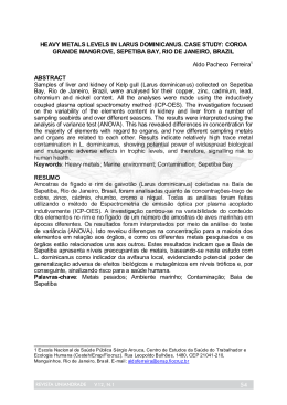
Histology and
Histopathology
Histol Histopathol (2010) 25: 1123-1131
http://www.hh.um.es
Cellular and Molecular Biology
A role for mammalian target of
rapamycin (mTOR) pathway in non
alcoholic steatohepatitis related-cirrhosis
Márcia Saldanha Kubrusly1,3, Maria Lúcia Corrêa-Giannella2, Marta Bellodi-Privato3, Sandra Valéria de Sá2,
Claudia Pinto Marques Souza de Oliveira3, Iberê Cauduro Soares4, Alda Wakamatsu4, Venâncio Avancini
Ferreira Alves4, Daniel Giannella-Neto5, Telesforo Bacchella3, Marcel Cerqueira Cesar Machado3 and Luiz
Augusto Carneiro D’Albuquerque3
1Department
4Department
of Surgery, 2Laboratory for Cellular and Molecular Endocrinology (LIM-25), 3Department of Gastroenterology (LIM-37),
of Pathology (LIM-14) and 5Medical Investigation Laboratories (LIM-07), University of São Paulo School of Medicine,
São Paulo, Brazil
Summary. Non-alcoholic fatty liver disease (NAFLD)
encompasses the whole spectrum of steatosis, nonalcoholic steatohepatitis (NASH), and NASH-related
cirrhosis (NASH/Cir). Although molecular advances
have been made in this field, the pathogenesis of
NAFLD is not completely understood. The gene
expression profiling associated to NASH/Cir was
assessed, in an attempt to better characterize the
pathways involved in its etiopathogenesis. Methods: In
the first step, we used cDNA microarray to evaluate the
gene expression profiles in normal liver (n=3) and
NASH/Cir samples (n=3) by GeneSifter™ analysis to
identify differentially expressed genes and biological
pathways. Second, tissue microarray was used to
determine immunohistochemical expression of
phosphorylated mTOR and 4E-BP1 in 11 normal liver
samples, 10 NASH/Cir samples and in 37 samples of
cirrhosis of other etiologies to further explore the
involvement of the mTOR pathway evidenced by the
gene expression analysis. Results: 138 and 106 genes
were, respectively, up and down regulated in NASH/Cir
in comparison to normal liver. Among the 9 pathways
identified as significantly modulated in NASH/Cir, the
participation of the mTOR pathway was confirmed,
since expression of cytoplasmic and membrane phosphomTOR were higher in NASH/Cir in comparison to
cirrhosis of other etiologies and to normal liver.
Offprint requests to: Dr. Márcia Saldanha Kubrusly, Department of
Gastroenterology, School of Medicine, University of São Paulo, Av. Dr.
Arnaldo 455 3º andar sala 3223, São Paulo, Brazil. e-mail: [email protected]
Conclusions: Recent findings have suggested a role for
the cellular “nutrient sensor” mTOR in NAFLD and the
present study corroborates the participation of this
pathway in NASH/Cir. Phospho-mTOR evaluation
might be of clinical utility as a potential marker for
identification of NASH/Cir in cases mistakenly
considered as cryptogenic cirrhosis owing to paucity of
clinical data.
Key words: Cirrhosis, Non alcoholic steatohepatitis,
Gene expression profile, Microarray analysis, mTOR
pathway
Introduction
Non-alcoholic fatty liver disease (NAFLD)
encompasses the whole spectrum of fatty liver, including
non-alcoholic steatosis, steatohepatitis (NASH), and
NASH-related cirrhosis (NASH/Cir) in patients without
a history of alcohol abuse (McCullough et al., 2002).
NASH associated with cirrhosis can decompensate into
sub acute liver failure, progress to hepatocellular cancer
(HCC) and reoccur post transplantation (Ong et al.,
2001; Bugianesi et al., 2002; Caldwell et al., 2002).
Although the risk factors for NAFLD are well
established and molecular advances have been made in
Abbreviations. NAFLD: Non-alcoholic fatty liver disease; NASH: Nonalcoholic steatohepatitis; NASH/Cir: NASH-related cirrhosis; mTOR:
mammalian target of rapamycin; 4E-BP1: 4E binding protein 1; TMA:
Tissue Microarray
1124
mTOR pathway in NASH-related cirrhosis
knowing the complex pathogenesis of NAFLD
(Sreekumar et al., 2003; Younossi et al., 2005; Chiappini
et al., 2006), the natural history of this condition in
humans is still uncertain.
The study of patterns of intra hepatic gene
expression in normal and diseased liver tissue is a
necessary prerequisite to increase the understanding of
these processes, particularly in human liver disease as
well as seeking to identify molecular markers that will
facilitate early diagnosis and accurate staging of the
disease.
The analysis of differential gene expression has
already been used to identify molecules and pathways
associated to cirrhosis caused by chronic viral hepatitis
(Shackel et al., 2003; Smith et al., 2003),
hemochromatosis, Wilson’s disease, alcoholic liver
disease, autoimmune hepatitis, primary biliary cirrhosis
(Kim et al., 2004) and NASH (Younossi et al., 2005).
The goal of the present study was to assess the role
of mTOR and its downstream effector 4EBP-1 in
NASH-related cirrhosis in an attempt to better
characterize the involvement of this pathway in its
etiopathogenesis.
Materials and methods
Tissue collection and RNA isolation
Hepatic tissue was obtained from three patients (two
male and one female, mean age 64 years) affected by
NASH/Cir during orthotopic liver transplantation at the
Department of Gastroenterology, Liver Transplantation,
Medical School, University of Sao Paulo. Fragments of
liver tissues were fixed in formaldehyde saline (4%) and
processed for hematoxylin-eosin (HE) and Masson
Trichrome stains. The diagnosis of NASH/Cir was made
only after an exhaustive evaluation failed to define other
specific etiologies: absence of serologic markers for
known liver diseases; lack of evidence of chronic
alcohol and potentially hepatotoxic drug ingestion;
negative serologic testing for autoimmune hepatitis
(antibodies to nuclear/mitochondrial/smooth muscle
antigens), for hemochromatosis (ferritin, iron, iron
binding capacity), for Wilson’s disease (ceruloplasmin
concentrations) and for alpha-1 antitrypsin deficiency
(α-1 trypsin levels); negative histopathological findings
for hemochromatosis, for alpha-1 antitrypsin deficiency
and for primary biliary cirrhosis. Histological markers of
NAFLD activity (steatosis 0-3, ballooning 0-2 and
lobular inflammation 0-3) were assessed according to
Histological Scoring System for Nonalcoholic Fatty
Liver Disease (NASH Activity Score or NAS), recently
published by the Pathology Committee of the NASH
Clinical Research Network (Kleiner et al., 2005), which
considers a score 4 consistent with NASH. Two of the
three patients presented a score 4 in the histopathological
evaluation performed on liver sections taken at
transplantation and the third patient presented a score 3,
although a previous hepatic biopsy performed three
years before transplantation presented a score 6,
confirming the histologic diagnostic of NASH.
Normal liver tissue was obtained from donor wedge
biopsies taken at transplantation (two male and one
female donors, mean age 47 years). Biological triplicate
was used to reduce the effect of individual phenotypic
differences for each group. This study was approved by
the Ethical Committee of Hospital das Clinicas of the
University of São Paulo, School of Medicine and in
accordance to The Declaration of Helsinki, with
informed and free consent being required of each subject
or subject’s guardian. All liver tissue samples were
collected in sterile containers and immediately snap
frozen in liquid nitrogen after surgical removal and
stored at -80°C until their use.
Total RNA was isolated from frozen liver samples
after mechanical disruption and extraction by using
TRIzol reagent (Invitrogen, Carlsbad, CA, USA)
according to manufacturer’s guidelines. Further, the
precipitated products were purified with RNeasyTM Mini
Kit (Qiagen, Hilden, Germany).
The quality of total RNA samples was analyzed by
inspection of 18S and 28S rRNA bands following
agarose gel electrophoresis. The concentration of RNA
samples was quantified by measurement of the optical
density using a NanoDrop ND-1000 (NanoDrop
Technologies, Wilmington, DE, USA).
Microarray analysis
We utilized the CodeLink™ Human Whole Genome
Bioarray (GE Healthcare Biosciences, Chalfont St.
Giles, UK) with ~57,000 human transcripts represented
in a single bioarray.
Target preparation, hybridization, and probe array
processing (washing, staining, and scan) were performed
according to the protocols in the manufacturer’s
instructions. Briefly, 5 µg of total RNA was first reverse
transcribed to the single-stranded cDNA and subsequent
cRNA was synthesized using the CodeLink™
Expression Assay Kit (GE Healthcare Biosciences). The
cRNA targets were prepared by in vitro transcription
using a single, labeled nucleotide, biotin-11-UTP at a
concentration of 1.25 mM. Unlabeled UTP was present
at 3.75 mM, while GTP, ATP, and CTP were at 5 mM.
The mixture was incubated at 37°C overnight for 14
hours. The labeled cRNA was then purified using
RNeasy™ mini kit (Qiagen) and subsequently
fragmented in 1x fragmentation buffer (40 mM Trisacetate pH 7.9, 100 mM KOAc, 31.5 mM MgOAc) at
94°C for 20 minutes.
For hybridization, 10 µg of fragmented cRNA in 260
µl of hybridization solution was added to each bioarray
and incubated for 18 hours at 37°C, while shaking at 300
rpm in a shaking incubator. Immediately following
hybridization, the bioarrays were washed and stained
with Cy5™-streptavidin (GE Healthcare Biosciences)
and scanned using a GenePix® 4000B Array Scanner
(Axon Instruments, Union City, CA, USA).
mTOR pathway in NASH-related cirrhosis
Microarray expression analysis
The quality of microarray data was assessed by
monitoring a series of quality control parameters as
suggested by CodeLink™ Expression Array Software
(GE Healthcare Biosciences), including visually
inspecting the array images to confirm scanner
alignment and filtering out genes with missing spots.
The ~57,000 spots’ intensities on the microarray image
were quantified, where the intensity of each spot was
divided by the array’s signal median to provide a scaled
and comparable number across multiple arrays. The
expression values of these genes were normalized
individually across all arrays by taking the intensity
values in increasing order subtracted by 0.5 and divided
by the size of the sample (Q-value).
Expression ratio was calculated by the difference
between the averages of Q-transformed values from 3
NASH/Cir and 3 normal liver arrays, multiplied by 10.
Unpaired Student’s t-test was used for two-group
statistical comparisons. Differentially expressed genes
were selected according to 3 basic criteria: presence of at
least 40% of “G” (good) flag in analyzed arrays,
statistically significant ratio at probability levels of
p<0.05 and differential expression either up- or downregulated by at least 2-fold in the average to normal
livers compared to NASH/Cir data.
Gene ontology analysis using gene ontology
Microarray gene expression data were analyzed by
GeneSifter™ program (http://www.genesifter.net/web/).
Gene ontology routine included in the program was
chosen for further interpretation of the data regarding
molecular function related genes according to Gene
Ontology (GO) Consortium categories (http://www.
geneontology.org/GO.doc.html) (Ashburner et al., 2000).
All the pathways in the GeneSifter™ database were
examined to determine whether a significant number of
altered genes in GO term were affected. We examined zscores that express the frequency of genes fulfilling an
increase ≥2 in each GO term and compared with null
hypothesis expected frequency for that GO term based
on the total number of genes examined on the array. The
z-score was derived by dividing the difference between
the observed number of genes meeting the criterion in a
specific GO term and the expected number of genes
based on the total number of genes in the array meeting
the criterion. This value was then divided by the
standard deviation of the observed number of genes
under a hypergeometric distribution.
Positive z-scores indicate GO terms with a greater
number of genes meeting the criterion than is expected
by chance. Negative z-scores indicate GO terms with
fewer genes meeting the criterion than expected by
chance. A z-score near zero indicates that the number of
genes meeting the criterion approximates the expected
number. A z-score of greater than 2 is considered a
statistically significant association (approximately
1125
equivalent to a p≤ 0.05) between the differentially
regulated genes and their corresponding GO terms
(Ashburner et al., 2000).
Correction for multiple testing was then performed
using the method of Reiner et al., 2003 to derive a false
discovery rate estimate from raw p-values. A false
discovery rate of 5% was also set as a cutoff for
statistical significance.
Tissue microarray (TMA)
From 2003 to 2007, hepatic cirrhosis cases from
different etiologies: NASH/Cir (n=10), HCV-related
cirrhosis (n=9), HBV-related cirrhosis (n=9), autoimmune hepatitis (n=9), primary biliary cirrhosis (n=3),
ethanol cirrhosis (n=1), Wilson’s disease (n=1), α1-antitripsyn deficiency (n=2), primary sclerosing cholangitis
(n=2), and cryptogenic cirrhosis (n=1) were collected
from Anatomic Pathology Division, Hospital das
Clinicas, University of Sao Paulo and spotted on a TMA.
The diagnosis of NASH/Cir was made as described
above. Eleven normal liver samples came from a second
TMA.
In brief, hematoxylin and eosin stained slides from
the donor cases were reviewed to identify viable,
morphologically representative areas of the specimen,
and then the areas were marked (2 spots by cases). After
transferring of the marks to respective donor paraffin
blocks, a TMA block was constructed by punching the
spotted areas (1.0 mm punch) and mounting them onto a
recipient paraffin block at 0.3 mm intervals between the
cores, in a grid system where each core has a coordinate
reference (x-axis, y-axis, in a final grid of 9 lines by 10
columns) using a precision microarray instrument
(Beecher Instruments, USA). The TMA block was cut at
3 µm interval consecutive sections (Leica Instruments,
Germany) (Kononen et al., 1998).
Immunohistochemical protocol
Three µm sections of the TMAs were deparaffinized
in xylene for 10 min followed by hydration in serial
ethanol dilutions and distilled water. Antigen retrieval
was performed by boiling slides in 10 mM sodium
citrate pH 6.0 for 40 min in a steamer (Shi et al., 1991).
Endogenous peroxidase activity was blocked for 30 min
in 6% hydrogen peroxide diluted in methanol.
Subsequent incubation with CASBlock™ (Invitrogen/
Zymed, USA) was carried out for 10 min at 37°C. After
blotting the excess of CASBlock™, slides were
incubated with the primary antibodies (anti-phospho 4EBP1(Thr37/46), rabbit monoclonal, clone 236B4, 1:200,
and/or anti-phospho mTOR (Ser2448), rabbit
monoclonal, clone 49F9, 1:50 (Cell Signaling
Technology Inc, Danvers, USA) for 30 min at 37°C
followed by overnight incubation at 4°C. The next day,
incubation with a short polymer amplification system
(Novolink™, Vision Biosystems, Australia) was applied
for 30 min at 37°C. The peroxidase reactivity was
1126
mTOR pathway in NASH-related cirrhosis
developed by 60 mg 3,3’-diaminobenzidine in PBS
buffer (pH 7.4) for 5 min at 37°C. Between all steps,
slides were washed in phosphate-buffered saline (pH
7.4). Finally, slides were counterstained with Harris’s
haematoxylin for 1 min, dehydrated, and mounted in
Entellan (Merck, USA). Positive control tissue was run
in parallel with another tissue microarray with a varied
group of gastrointestinal carcinomas for both proteins.
Negative control was obtained by omitting the primary
antibody.
Evaluation and scoring
Immunohistochemical staining was quantified
conjointly by 2 observers (ICS and MSK) blinded to
clinical data using a scoring system that incorporates
both staining intensity and percentage of positive
hepatocyte cells. For each TMA spot, the intensity of
each immunostained protein (0, absent; 1, weak; 2,
medium; and 3, strong) was multiplied by the percentage
of positive staining hepatocyte cells (from 0 to 100%,
with classes of 10% increment), resulting in a score
ranging from 0-300 (Herberger et al., 2007). To
represent each case, the mean of the 2 spots of the case
presented on the TMA was calculated. Only spots with
at least 50% of interest tissue were considered in the
computation. Two different cellular compartments were
evaluated: cytoplasm and plasmatic membrane, using the
same scoring system.
Statistical analysis
Statistical analysis was carried out using GraphPad
PRISM software version 2.0 (Kruskal-Wallis and Dunn’s
multiple comparison tests). Non-parametric two-tailed pvalues <0.05 were considered statistically significant.
Results
Genomic expression
Using filtering criteria of at least 2.0-fold-change in
Table 1. The down regulated biological pathways and representative genes in NASH-related cirrhosis.
UNIGENE
GENE SYMBOL
Biosynthesis of steroids
Hs.503134
Hs.632801
Hs.643476
Hs.283652
Hs.130607
Hs.71465
Hs.287749
DHCR7
EBP
FDFT1
IDI1
MVK
SQLE
SC5DL
Pyrimidine metabolism
Hs.473087
Hs.599355
Hs.567352
Hs.458360
Hs.591457
Hs.515122
Hs.643610
CTPS
DTYMK
TXNRD1
UCK2
POLR3C
TK1
POLR3H
Phosphatidylinositol signaling system
Hs.235116
GRK6
Hs.431173
PLCB1
Hs.580527
INPP4A
INPP5A
Hs.523360
HIPK2
Hs.397465
Hs.497487
PIK3C2B
PIK3R2
Hs.371344
EEF2K
Hs.498892
PIP5K1B
Hs.534371
PIK3CG
Hs.32942
CDS2
Hs.472027
mTOR signaling pathway
Hs.411641
EIF4EBP1
Hs.431850
MAPK1
Hs.371344
PIK3R2
Hs.32942
PIK3CG
Hs.78781
VEGFB
PATHWAY
z-score
9.11
3.09
2.68
2.17
GENE NAME
NASH/Cir
Up/Down
7-dehydrocholesterol reductase
Emopamil binding protein (sterol isomerase)
Farnesyl-diphosphate farnesyltransferase 1
Isopentenyl-diphosphate delta isomerase
Mevalonate kinase (mevalonic aciduria)
Squalene epoxidase
Sterol-C5-desaturase (ERG3 delta-5-desaturase homolog, fungal)-like
down
down
down
down
down
down
down
Cytidine 5'-triphosphate synthetase
Deoxythymidylate kinase (thymidylate kinase)
Thioredoxin reductase 1
Uridine-cytidine kinase 2
Polymerase (RNA) III (DNA directed) polypeptide C
Thymidine kinase 1
Polymerase (RNA) III (DNA directed) polypeptide H
down
down
down
down
down
down
down
G protein-coupled receptor kinase 6
Phospholipase C, beta 1 (phosphoinositide-specific)
Inositol polyphosphate-4-phosphatase, type I
Inositol polyphosphate-5-phosphatase
Homeodomain interacting protein kinase 2
Phosphoinositide-3-kinase, class 2, beta polypeptide
Phosphoinositide-3-kinase, regulatory subunit, polypeptide 2 (p85 beta)
Eukaryotic elongation factor-2 kinase
Phosphatidylinositol-4-phosphate 5-kinase, type I, beta
Phosphoinositide-3-kinase, catalytic, gamma polypeptide
CDP-diacylglycerol synthase (phosphatidate cytidylyltransferase) 2
Eukaryotic translation initiation factor 4E binding protein 1
Mitogen-activated protein kinase 1
Phosphoinositide-3-kinase, regulatory subunit, polypeptide 2 (p85 beta)
Phosphoinositide-3-kinase, catalytic, gamma polypeptide
Vascular endothelial growth factor B
Up or down regulation are defined as gene expression in NASH/Cir compared with normal liver tissue.
down
up
down
down
down
down
down
down
down
up
up
down
down
down
up
down
mTOR pathway in NASH-related cirrhosis
Fig. 1. Immunohistochemical analyses of phospho
4E-BP1 (A and B) and of phospho mTOR (D and E)
in NASH/Cir (A and D) and in HCV-related cirrhosis
(B and E) demonstrating cytoplasmic reactivity in
NASH/Cir for both proteins and membrane reactivity
only for phospho mTOR (original magnification, 400x).
Graphics C, F and G elicit, respectively, immunohistochemical scoring for phospho 4E-BP1 (C) and for
cytoplasmic (F) and membrane (G) phospho mTOR in
normal liver, NASH/Cir and cirrhosis of other
etiologies (Others). Data are expressed as mean ±
SEM.
1127
1128
mTOR pathway in NASH-related cirrhosis
expression and a t-test where p<0.05, 244 differentially
expressed genes were identified, 138 of these genes were
up regulated in the NASH/Cir group while 106 genes
were down regulated in comparison to the normal liver
group.
Gene ontology (GO) analysis by GeneSifter TM
Table 2. The up regulated biological pathways and representative genes in NASH-related cirrhosis.
UNIGENE
GENE SYMBOL
T cell receptor signaling pathway
Hs.466907
CBLC
CD28
Hs.591629
Hs.558348
ITK
IL10
Hs.193717
Hs.390616
PAK3
Hs.497487
PIK3C2B
PIK3R2
Hs.371344
Hs.591127
RASGRP1
Hs.32942
PIK3CG
Hs.198998
CHUK
PATHWAY
z-score
2,94
ECM-receptor interaction
Hs.213861
LAMA4
Hs.17441
COL4A1
Hs.508010
FNDC3A
Hs.72550
HMMR
Hs.520525
FNDC1
Hs.591484
LAMC2
2,51
Regulation of actin cytoskeleton
Hs.525572
BDKRB1
Hs.525572
BDKRB2
Hs.479747
BCAR1
Hs.287370
FGF23
Hs.522373
GSN
Hs.199763
SSH1
Hs.410092
F2
Hs.431850
MAPK1
Hs.390616
PAK3
Hs.497487
PIK3C2B
Hs.371344
PIK3R2
Hs.517228
TIAM1
Hs.534371
PIP5K1B
Hs.32942
PIK3CG
Hs.488293
EGFR
2,37
Cytokine-cytokine receptor interaction
Hs.73853
BMP2
Hs.546294
CCL17
Hs.514821
CCL5
Hs.34526
CXCR6
Hs.507590
FLT3
Hs.193717
IL10
Hs.591742
IL7R
Hs.624
IL8
Hs.846
IL8RB
Hs.78781
VEGFB
Hs.488293
EGFR
Complement and coagulation cascades
BDKRB1
Hs.525572
BDKRB2
Hs.525572
F12
Hs.1321
C6
Hs.481992
Hs.93210
C8A
Hs.410092
F2
Hs.516578
TFPI
2,39
2,1
GENE NAME
NASH/Cir
Up/Down
Cas-Br-M (murine) ecotropic retroviral transforming sequence c
CD28 antigen (Tp44)
IL2-inducible T-cell kinase
Interleukin 10
p21 (CDKN1A)-activated kinase 3
Phosphoinositide-3-kinase, class 2, beta polypeptide
Phosphoinositide-3-kinase, regulatory subunit, polypeptide 2 (p85 beta)
RAS guanyl releasing protein 1 (calcium and DAG-regulated)
Phosphoinositide-3-kinase, catalytic, gamma polypeptide
Conserved helix-loop-helix ubiquitous kinase
down
up
up
down
up
down
down
up
up
down
Bone morphogenetic protein 2
Chemokine (C-C motif) ligand 17
Chemokine (C-C motif) ligand 5
Chemokine (C-X-C motif) receptor 6
Fms-related tyrosine kinase 3
Interleukin 10
Interleukin 7 receptor
Interleukin 8
Interleukin 8 receptor, beta
Vascular endothelial growth factor B
Epidermal growth factor receptor
up
up
up
up
up
down
up
up
down
down
up
Bradykinin receptor B1
Bradykinin receptor B2
Coagulation factor XII (Hageman factor)
Complement component 6
Complement component 8, alpha polypeptide
Coagulation factor II (thrombin)
Tissue factor pathway inhibitor (lipoprotein-associated coagulation inhibitor)
up
up
up
down
down
down
up
Laminin, alpha 4
Collagen, type IV, alpha 1
Fibronectin type III domain containing 3
Hyaluronan-mediated motility receptor (RHAMM)
Fibronectin type III domain containing 1
Laminin, gamma 2
Bradykinin receptor B1
Bradykinin receptor B2
Breast cancer anti-estrogen resistance 1
Fibroblast growth factor 23
Gelsolin (amyloidosis, Finnish type)
Slingshot homolog 1 (Drosophila)
Coagulation factor II (thrombin)
Mitogen-activated protein kinase 1
p21 (CDKN1A)-activated kinase 3
Phosphoinositide-3-kinase, class 2, beta polypeptide
Phosphoinositide-3-kinase, regulatory subunit, polypeptide 2 (p85 beta)
T-cell lymphoma invasion and metastasis 1
Phosphatidylinositol-4-phosphate 5-kinase, type I, beta
Phosphoinositide-3-kinase, catalytic, gamma polypeptide
Epidermal growth factor receptor
Up or down regulation are defined as gene expression in NASH/Cir compared with normal liver tissue
up
up
down
down
up
up
up
up
down
up
up
up
down
down
up
down
down
down
down
up
up
mTOR pathway in NASH-related cirrhosis
software identified 9 statistically significant pathways
containing 68 non-redundant genes differentially
expressed. Biosynthesis of steroids, Phosphatidylinositol
signaling system, Pyrimidine metabolism and mTOR
signaling pathway were identified as pathways down
regulated in NASH/Cir in comparison to normal liver,
with z-scores of 9.11, 3.09, 2.68 and 2.17, respectively.
T cell receptor signaling pathway, ECM-receptor
interaction, Cytokine-cytokine receptor interaction,
Regulation of actin cytoskeleton and Complement and
coagulation cascades pathways were identified as
pathways up regulated in NASH/Cir in comparison to
normal liver, with z-scores of 2.94, 2.51, 2.39, 2.37 and
2.1 respectively (Tables 1, 2).
Immunohistochemical analysis
Cytoplasmic immunoreactivity for phospho-mTOR
was demonstrated in 8 out of 10 (80%) NASH/Cir, in 14
out of 37 (38%) cirrhosis of other etiologies and in 4 out
of 11 (36%) normal liver samples, while membrane
immunostaining was found in 8 out of 10 (80%)
NASH/Cir, in 12 out of 37 (32%) cirrhosis of other
etiologies and in 5 out of 11 (45%) normal liver samples.
The evaluation of the phosphorylated form of 4E-BP1, a
downstream effector of mTOR (whose corresponding
gene was identified as down regulated in the microarray
analysis in comparison to normal liver) demonstrated its
expression in 7 out of 10 (70%) NASH/Cir, in 17 out of
37 (46%) of the cirrhosis of other etiologies and in 10
out of 11 (91%) normal liver samples.
Data of semi-quantitative analysis of immunohistochemical stainings are elicited in Figure 1. Expression of
cytoplasmic phospho mTOR was significantly higher in
NASH/Cir compared with cirrhosis of other etiologies
and to normal liver (Graphic F), while membrane
reactivity for phospho mTOR was significantly higher
only in NASH/Cir in comparison to cirrhosis of other
etiologies (Graphic G). Expression of phospho 4E-BP1
was higher in normal liver and in NASH/Cir compared
to cirrhosis of other etiologies, although, statistical
significance was not reached between NASH/Cir and
cirrhosis of other etiologies (Graphic C).
Discussion
The present study identified pathways potentially
involved in the etiopathogenesis of NASH-related
cirrhosis by the use of human whole-genome microarray
hybridization, an approach that allows the obtention of
global transcriptional changes to investigate mechanisms
underlying the development of diseases. The pathway
analysis herein used resulted in the identification of gene
networks such as, Pyrimidine metabolism, Biosynthesis
of steroids, Phosphatidylinositol signaling system and
mTOR signaling pathway, down regulated in NASHrelated cirrhosis in comparison to normal liver, and T
cell receptor signaling pathway, ECM-receptor
1129
interaction, Cytokine-cytokine receptor interaction,
Regulation of actin cytoskeleton and Complement and
coagulation cascades, up regulated in NASH-related
cirrhosis.
Augmented utilization and catabolism of purine and
pyrimidine nucleotides has been described after partial
hepatectomy or hepatic lesion, indicating the
requirement of substrates for salvage nucleotide
synthesis during hepatic regeneration (Hashimoto et al.,
1998). The down regulation of this pathway in NASHrelated cirrhosis is in agreement with previous findings
demonstrating decreased DNA synthesis in cirrhotic
patients and animals submitted to hepatic surgery (Leong
et al., 2006) and probably represent an alteration related
to end-stage liver disease, not necessarily associated
with NASH. On the other hand, down regulation of
several genes belonging to the Steroid biosynthesis
pathway, including four of the seven genes herewith
identified (DHCR, FDFT1, IDI1 and MV) could be
important in the progression of NAFLD. These genes
were previously reported as down regulated in rat
hepatocytes exposed to arginine deprivation and the
authors interpreted this as reflecting an endoplasmatic
reticulum (ER) stress (Ozcan et al., 2004). Additionally,
it has recently been found that chronic ER stress was
present in liver tissue of dietary (high fat diet-induced)
and genetic (ob/ob) models of mouse obesity and was
able to interfere with insulin receptor signaling and
inhibit insulin action in liver cells. Thus, ER stress is
being regarded as a molecular link between obesity,
insulin resistance, and type 2 Diabetes (Marshall, 2006),
which make it plausible to consider its participation in
NAFLD development and progression.
Considering the central role of activated hepatic
stellate cells in extracellular matrix (ECM) production
and hepatic fibrogenesis (Friedman, 2003; Parsons et al.,
2007), the identification of ECM-receptor interaction as
a disturbed pathway, with up regulation of genes which
code for laminin and collagen Type IV in NASH-related
cirrhosis is expected and likely to reflect the end-stage
liver disease, irrespective of etiology. The same is
probably true for the T-Cell receptor signaling pathway,
Cytokine-cytokine receptor interaction, Regulation of
actin cytoskeleton and Complement and coagulation
cascades pathways, whose activation may have a close
relationship with the inflammatory response of injured
liver (Pinzani and Marra, 2001; Boisvert et al., 2003;
Assy et al., 2005).
The mTOR pathway is recognized as an important
mediator of a range of anabolic processes, functioning as
a nutrient-sensing transductional pathway and
interacting with other sensors that detect changes in
nutrient availability, such as insulin and AMPK
signaling pathways (Khamzina et al., 2005; Yang and
Guan, 2007). Recent reports of the involvement of the
mTOR pathway in the development of hepatic steatosis
and insulin resistance following a high fat diet suggested
a role for this pathway in NAFLD (Korsheninnikova et
1130
mTOR pathway in NASH-related cirrhosis
al., 2005; Calvert et al., 2007) and motivated us to
further investigate the gene expression findings,
comparing the protein expression of the active
phosphorylated forms of mTOR and 4E-BP1 between
NASH-related cirrhosis and cirrhosis of other etiologies,
in an attempt to find markers specific for NASH-related
cirrhosis. The expression of phosphorylated mTOR was
significantly higher in NASH-related cirrhosis in
comparison to cirrhosis of other causes, suggesting the
involvement of mTOR pathway until the final stages of
NAFLD. Although these findings might seem
contradictory to the one obtained in the microarray
analysis, which pointed to a down regulation of the
mTOR pathway in NAFLD, it is theoretically possible
that the observed down regulation of the genes
belonging to the mTOR pathway constitutes a
compensatory cellular response to increased pathway
activation.
Given the etiopathogenesis of NAFLD, closely
related to nutrient overload, it seems feasible to propose
that the mTOR pathway may participate in the
pathogenesis of NAFLD until end-stage liver disease
and, going further, that deregulated mTOR signaling is a
more generalized event, as suggested by the findings of
Calvert et al. (2007), who identified members of the
AKT/mTOR pathway as being differentially
phosphorylated in omental adipose tissue from patients
with non-progressive form of NAFLD and those with
NASH. These results deserve further investigation since
characterization of the role of mTOR in NAFLD
progression may open new possibilities of therapeutic
intervention. An interesting aspect that also needs
additional clarification is the absence of differences in
the expression of phospho 4E-BP1, a downstream
effector of mTOR, between NASH/Cir and normal liver
and cirrhosis of other etiologies. One possible
explanation for this dissociation is that a reduction of
cellular ATP levels inhibits mTOR-dependent
phosphorylation of 4E-BP1 (Fingar and Blenis, 2004)
and a condition of energy insufficiency could be present
in human cirrhotic livers, as already demonstrated in
animal models of cirrhosis, where a decreased ratio of
ATP/ADP was detected (Harvey et al., 2000). In favour
of this hypothesis is the finding of a significantly lower
expression of phospho 4E-BP1 when cirrhotic livers of
all etiologies were grouped and compared to normal
livers (data not shown).
Conclusions
In summary, we propose that mTOR signaling may
be specific for the cirrhosis of this etiology, given their
recently described association with impaired insulin
receptor signaling (Khamzina et al., 2005;
Korsheninnikova et al., 2005; Marshall et al., 2006). Our
results corroborate recent findings that mTOR pathway
participates in NAFLD pathogenesis and may provide
potential markers for identification of NASH-related
cirrhosis in cases mistakenly considered as cryptogenic
cirrhosis owing to paucity of clinical data.
References
Ashburner M., Ball C.A., Blake J.A., Botstein D., Butler H., Cherry J.M.,
Davis A.P., Dolinski K., Dwight S.S., Eppig J.T., Harris M.A., Hill
D.P., Issel-Tarver L., Kasarskis A., Lewis S., Matese J.C.,
Richardson J.E., Ringwald M., Rubin G.M. and Sherlock G. (2000).
Gene ontology: tool for the unification of biology. The Gene
Ontology Consortium. Nat. Genet. 25, 25-29.
Assy N., Bekirov I., Mejritsky Y., Solomon L., Szvalb S. and Hussein O.
(2005). Association between thrombotic risk factors and extent of
fibrosis in patients with non-alcoholic fatty liver diseases. World J.
Gastroenterol. 11, 5834-5839.
Boisvert J., Kunkel E.J., Campbell J.J., Keeffe E.B., Butcher E.C. and
Greenberg H.B. (2003). Liver-infiltrating lymphocytes in end-stage
hepatitis C virus: Subsets, activation status, and chemokine receptor
phenotypes. J. Hepatol. 38, 67-75.
Bugianesi E., Leone N., Vanni E., Marchesini G., Brunello F., Carucci
P., Musso A., De Paolis P., Capussotti L., Salizzoni M. and Rizzetto
M. (2002). Expanding the natural history of nonalcoholic
steatohepatitis: from cryptogenic cirrhosis to hepatocellular
carcinoma. Gastroenterology 123, 134-140.
Caldwell S.H. and Hespenheide E.E. (2002). Subacute liver failure in
obese women. Am. J. Gastroenterol. 97, 2058-2062.
Calvert V.S., Collantes R., Elariny H., Afendy A., Baranova A., Mendoza
M., Goodman Z., Liotta L.A., Petricoin E.F. and Younossi Z.M.
(2007). A systems biology approach to the pathogenesis of obesityrelated nonalcoholic fatty liver disease using reverse phase protein
microarrays for multiplexed cell signaling analysis. Hepatology 46,
166-172.
Chiappini F., Barrier A., Saffroy R., Domart M.C., Dagues N., Azoulay
D., Sebagh M., Franc B., Chevalier S., Debuire B., Dudoit S. and
Lemoine A. (2006). Exploration of global gene expression in human
liver steatosis by high-density oligonucleotide microarray. Lab.
Invest. 86, 154-165.
Fingar D.C. and Blenis J. (2004). Target of rapamycin (TOR): an
integrator of nutrient and growth factor signals and coordinator of
cell growth and cell cycle progression. Oncogene 23, 3151–
3171.
Friedman S.L. (2003). Liver fibrosis – from bench to bedside. J. Hepatol.
8, S38-S53.
Harvey P.J., Gready J.E., Yin Z., Le Couteur D.G. and McLean A.J.
(2000). Acute oxygen supplementation restores markers of
hepatocyte energy status and hypoxia in cirrhotic rats. J. Pharm.
Exp. Therap. 293, 641-645.
Hashimoto M., Kothary P.C., Eckhauser F.E. and Raper S.E. (1998).
Treatment of cirrhotic rats with epidermal growth factor and insulin
accelerates liver DNA synthesis after partial hepatectomy. J.
Gastroenterol. Hepatol. 13, 1259-1265.
Herberger B., Puhalla H., Lehnert M., Wrba F., Novak S., Brandstetter
A., Gruenberger B., Gruenberger T., Pirker R. and Filipits M. (2007).
Activated mammalian target of rapamycin is an adverse prognostic
factor in patients with biliary tract adenocarcinoma. Clin. Cancer
Res. 13, 4795-4799.
Khamzina L., Veilleux A., Bergeron S. and Marette A. (2005). Increased
activation of the mammalian target of rapamycin pathway in liver
mTOR pathway in NASH-related cirrhosis
and skeletal muscle of obese rats: Possible involvement in obesitylinked insulin resistance. Endocrinology 146, 1473-1481.
Kim J.W., Ye Q., Forgues M., Chen Y., Budhu A., Sime J., Hofseth L.J.,
Kaul R. and Wang X.W. (2004). Cancer-associated molecular
signature in the tissue samples of patients with cirrhosis. Hepatology
39, 518-527.
Kleiner D.E., Brunt E.M., Van Natta M., Behling C., Contos M.J.,
Cummings O.W., Ferrell L.D., Liu Y.C., Torbenson M.S., UnalpArida A., Yeh M., McCullough A.J. and Sanyal A.J. (2005).
Nonalcoholic Steatohepatitis Clinical Research Network. Design and
validation of a histologic scoring system for NAFLD. Hepatology 41,
1313-1321.
Kononen J., Bubendorf L., Kallioniemi A., Bärlund M., Schraml P.,
Leighton S., Torhorst J., Mihatsch M.J., Sauter G. and Kallioniemi
O.P. (1998). Tissue microarrays for high throughput molecular
profiling of tumor specimens. Nat. Med. 4, 844-847.
Korsheninnikova E, van der Zon G.C., Voshol P.J., Janssen G.M.,
Havekes L.M., Grefhorst A., Kuipers F., Reijngoud D.J., Romijn J.A.,
Ouwens D.M. and Maassen J.A. (2006). Sustained activation of the
mammalian target of rapamycin nutrient sensing pathway is
associated with hepatic insulin resistance, but not with steatosis, in
mice. Diabetologia 49, 3049-3057.
Leong H.X., Simkevich C., Lesieur-Brooks A., Lau B.W., Fugere C.,
Sabo E. and Thompson N.L. (2006). Short-term arginine deprivation
results in large-scale modulation of hepatic gene expression in both
normal and tumor cells: microarray bioinformatic analysis. Nutrition
Metabol. http://www.nutritionandmetabolism.com/content/3/1/37.
Marshall S. (2006). Role of insulin, adipocyte hormones, and nutrientsensing pathways in regulating fuel metabolism and energy
homeostasis: A nutritional perspective of diabetes, obesity, and
cancer. Sci STKE 346, re7.
McCullough A.J. (2002). Update on nonalcoholic fatty liver disease. J.
Clin. Gastroenterol. 34, 255-262.
Ong J., Younossi Z.M., Reddy V., Price L.L., Gramlich T., Mayes J. and
Boparai N. (2001). Cryptogenic cirrhosis and post transplantation
nonalcoholic fatty liver disease. Liver Transpl. 7, 797-801.
1131
Ozcan U., Cao Q., Yilmaz E., Lee A.H., Iwakoshi N.N., Ozdelen E.,
Tuncman G., Görgün C., Glimcher L.H. and Hotamisligil G.S. (2004).
Endoplasmic reticulum stress links obesity, insulin action, and type 2
Diabetes. Science 306, 457-461.
Parsons C.J., Takashima M. and Rippe R.A. (2007). Molecular
mechanisms of hepatic fibrogenesis. J. Gastroenterol. Hepatol. 22,
S79-S84.
Pinzani M. and Marra F. (2001). Cytokine receptors and signaling in
hepatic stellate cells. Sem. Liver Dis. 21, 397-416.
Reiner A., Yekutieli D. and Benjamini Y. (2003). Identifying differentially
expressed genes using false discovery rate controlling procedures.
Bioinformatics 19, 368-375.
Shackel N.A., McGuinness P.H., Abbott C.A., Gorrell M.D. and
McCaughan G.W. (2003). Novel differential gene expression in
human cirrhosis detected by suppression subtractive hybridization.
Hepatology 38, 577-588.
Shi S.R., Key M.E. and Kalra K.L. (1991). Antigen retrieval in formalinfixed, paraffin-embedded tissues: An enhancement method for
immunohistochemical staining based upon microwave oven heating
of tissue sections. J. Histochem. Cytochem. 39, 741-748.
Smith M.W., Yue Z.N., Korth M.J., Do H.A., Boix L., Fausto N., Bruix J.,
Carithers R.L. Jr and Katze M.G. (2003). Hepatitis C virus and liver
disease: Global transcriptional profiling and identification of potential
markers. Hepatology 38, 1458-1467.
Sreekumar R., Rosado B., Rasmussen D. and Charlton M. (2003).
Hepatic gene expression in histologically progressive nonalcoholic
steatohepatitis. Hepatology 38, 244-251.
Yang Q. and Guan K.L. (2007). Expanding mTOR signaling. Cell. Res.
177, 666-681.
Younossi Z.M., Baranova A., Ziegler K., Del Giacco L., Schlauch K.,
Born T.L., Elariny H., Gorreta F., VanMeter A., Younoszai A., Ong
J.P., Goodman Z. and Chandhoke V. (2005). A genomic and
proteomic study of the spectrum of nonalcoholic fatty liver disease.
Hepatology 42, 665-674.
Accepted March 12, 2010
Download