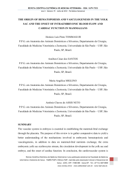
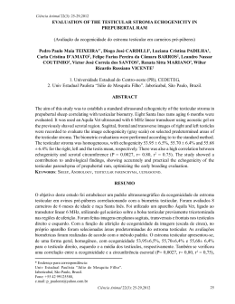
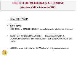
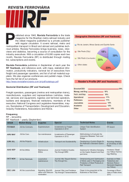
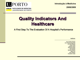
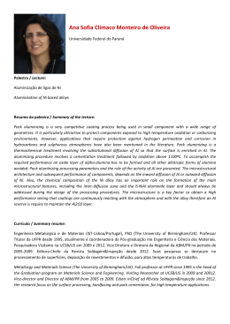
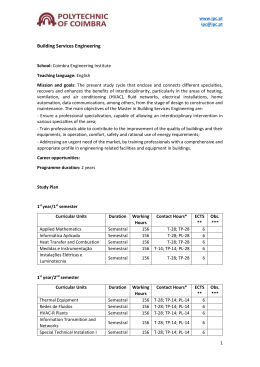
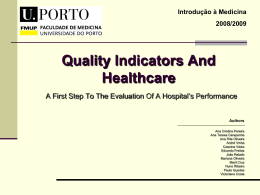
REVISTA CIENTÍFICA ELETRÔNICA DE MEDICINA VETERINÁRIA – ISSN: 1679-7353
Ano X – Número 19 – Julho de 2012 – Periódicos Semestral
TESTICULAR HYPOPLASIA IN BOAR: ASSOCIATION BETWEEN
SEMINAL ANALYSIS AND HISTOPATHOLOGY
Guilherme OBERLENDER
Department of Veterinary Medicine, Federal University of Lavras (UFLA), Lavras,
Minas Gerais, Brazil. E-mail: [email protected]
Luis David Solis MURGAS
Federal University of Lavras (UFLA), Lavras, Minas Gerais, Brazil.
Márcio Gilberto ZANGERONIMO
Federal University of Lavras (UFLA), Lavras, Minas Gerais, Brazil.
Flademir WOUTERS
Federal University of Lavras (UFLA), Lavras, Minas Gerais, Brazil.
Adriana Cristina da SILVA
Federal University of Lavras (UFLA), Lavras, Minas Gerais, Brazil.
Leonardo Pereira MESQUITA
Federal University of Lavras (UFLA), Lavras, Minas Gerais, Brazil.
Pedro Soares Bezerra JÚNIOR
Federal University of Lavras (UFLA), Lavras, Minas Gerais, Brazil.
Luciano José PEREIRA
Federal University of Lavras (UFLA), Lavras, Minas Gerais, Brazil.
Revista Científica Eletrônica de Medicina Veterinária é uma publicação semestral da Faculdade de Medicina
veterinária e Zootecnia de Garça – FAMED/FAEF e Editora FAEF, mantidas pela Associação Cultural e Educacional de
Garça - ACEG. CEP: 17400-000 – Garça/SP – Tel.: (0**14) 3407-8000
www.revista.inf.br – www.editorafaef.com.br – www.faef.edu.br.
REVISTA CIENTÍFICA ELETRÔNICA DE MEDICINA VETERINÁRIA – ISSN: 1679-7353
Ano X – Número 19 – Julho de 2012 – Periódicos Semestral
ABSTRACT
The aim of this study was to describe the occurrence of testicular hypoplasia in boar and
correlate the findings of testicular biometrics and histopathological evaluation with the
seminal parameters. The work was conducted at the Experimental Center of Porcine of
the Federal University of Lavras when four boars were acquired. Since 190±12 days of
age, the boars were subjected to a training protocol for semen collection, and during 130
days, were made andrological evaluations weekly, with testicular weighing, biometrics
and volume. In one animal was observed total absence of sperm in the ejaculated, since
the first semen collect, despite having normal volume of semen and was also found
smaller testicular volume compared to the others. When the boar completed three and
half years old, a surgical castration was performed, being collected samples from both
testis and epididymis for histopathological evaluation. On histological evaluation, there
was a reduced diameter in the seminiferous tubules, which were formed only by Sertoli
cells with rare spermatogenic lineage cells. A marked proportional increase in the
number of Leydig cells in relation to the seminiferous tubules was observed, and the
testis was covered by a thick layer of connective tissue, which formed trabeculae that
radiated into the parenchyma. In the samples from the epididymis were not observed
sperm. Bilateral testicular hypoplasia was present and could be previously detected by
the testicular volume and semen analysis, being confirmed by histopathological
evaluation of the testes and epididymis of the boar, association that is essential for the
definitive diagnosis.
KEY WORDS: Azoospermy; Fertility; Reproduction; Testicles.
INTRODUCTION
Testicular hypoplasia is characterized as a congenital underdevelopment of the
gonads, in wich can be observed, by means of cytology and testicular histology, a very
low number or even absence of germ cells in seminiferous tubules. It can occur in all
farm animals, as in bulls (HUMPHREY; LADDS, 1975), ram (BRUERE ET AL., 1969)
and pigs (HANCOCK; DAKER, 1981; SINGH; PARIHAR, 1998).
Revista Científica Eletrônica de Medicina Veterinária é uma publicação semestral da Faculdade de Medicina
veterinária e Zootecnia de Garça – FAMED/FAEF e Editora FAEF, mantidas pela Associação Cultural e Educacional de
Garça - ACEG. CEP: 17400-000 – Garça/SP – Tel.: (0**14) 3407-8000
www.revista.inf.br – www.editorafaef.com.br – www.faef.edu.br.
REVISTA CIENTÍFICA ELETRÔNICA DE MEDICINA VETERINÁRIA – ISSN: 1679-7353
Ano X – Número 19 – Julho de 2012 – Periódicos Semestral
Considering the swine specie, Singh and Parihar (1998) conducted a study with
88 boars and they found that the frequency of testicular hypoplasia in these animals was
12%. According to Foster and Ladds (2007) this is a hereditary disorder caused by a
pair of recessive genes with incomplete penetrance and variable expressivity. Therefore,
this disorder may occur in total or partial forms, unilateral or bilateral and other
combinations and degrees of involvement.
Regarding to anatomopathology the testicular hypoplasia can be divided into
three types: mild (partial), intermediate and total (severe) (FOSTER; LADDS, 2007). In
severe hypoplasia, all or nearly all of tubules are hypoplasic, presenting a small testis,
consistent with the palpations and resistant to the cut. The animal may present
oligospermia or azoospermia, in this case with high rates of pathological forms of sperm
in the ejaculate. When bilateral, severe hypoplasia makes the animal sterile and the
diagnosis becomes more simple, but when the effect is only partial the animal is
considered sub-fertile or temporarily infertile. In this case, the diagnosis may be more
difficult.
According to Rao et al. (1980) testicular hypoplasia is quite similar to testicular
degeneration, but can be differentiated by its irreversible character. Histologically, there
are no areas of fibrosis or basement membrane thickening such as testicular
degeneration. Also notes total absence of germinal epithelium, only Sertoli cells within
seminiferous tubules, unlike degeneration, in which there are sperm cell lineage, but
with vacuolization of the epithelium at different degrees. Testicular hypoplasia can also
be confused with sexual immaturity, as the prepubertal animal clinically presents
reduced gonads size and a low sperm production typical of hypoplasia; however, the
realization of repeated reproductive examinations allows to observe a progressive
increase in testicular size, quantitative and qualitative evolution of ejaculated, showing
the delayed on the puberty (RAO et al., 1980). Therefore, an appropriate and highly
detailed history is essential for the correct differentiation between testicular hypoplasia,
sexual immaturity and testicular degeneration.
The study aims to report the occurrence of testicular hypoplasia in one boar and
describe the findings in the examinations of testicular biometric, seminal and
Revista Científica Eletrônica de Medicina Veterinária é uma publicação semestral da Faculdade de Medicina
veterinária e Zootecnia de Garça – FAMED/FAEF e Editora FAEF, mantidas pela Associação Cultural e Educacional de
Garça - ACEG. CEP: 17400-000 – Garça/SP – Tel.: (0**14) 3407-8000
www.revista.inf.br – www.editorafaef.com.br – www.faef.edu.br.
REVISTA CIENTÍFICA ELETRÔNICA DE MEDICINA VETERINÁRIA – ISSN: 1679-7353
Ano X – Número 19 – Julho de 2012 – Periódicos Semestral
histopathological evaluation correlating these characteristics with the definitive
diagnosis of this anomaly.
MATERIAL AND METHODS
The study was conducted at the Department of Veterinary Medicine (DVM) and
at the Department of Animal Science (DAS) of the Federal University of Lavras,
Lavras/MG since October 2006 to October 2009. Four high performance boars aged
between five and six months and initial weight of 113.63±3.25 kg were acquired by the
Experimental Center of Porcine (DAS) for replacement of the flock of boars. From 190
days of age, were submitted to a specific training protocol for semen collection.
During 130 days since the beginning of training, when the animals had 190±12
days of age were realized detailed andrological evaluations weekly, a period in which
we determined the weight gain of animals, sexual behavior and monitoring of testicular
development through testicular biometrics parameters. In this latter, we measured the
length and the width of right testis (LRT and WRT) and left (LLT and WLT), following
the model proposed by Schinckel et al. (1984) For the calculating the testicular volume
was used the formula described by Owsianny et al. (1998): T=4/3 x π x a x b2, where:
T=testicular volume (mL) π=3.14, a=1/2 length testicular (cm) and b=1/2 testicular
width (cm).
One of animals presented azoospermia (absence of sperm), despite the normal
semen volume produced and satisfactory sexual behavior. This animal was kept on the
flock just like a ruffian and subjected to the semen collection once a week just to
exhaustion and volume determination, smell and semen appearance. Upon reaching 42
months of age and a total of 152 ejaculations, the animal was subjected to the surgical
castration. Samples from testis and epididymis were collected for histopathological
evaluation and the fragments were fixed in 10% buffered formalin and stained with
hematoxylin and eosin (HE) for subsequent evaluation in an optical microscope.
The histology description of the testis and epididymis of the ruffian associated
with the data of the testicular biometrics, reproductive characteristics and seminal
evaluation were performed.
Revista Científica Eletrônica de Medicina Veterinária é uma publicação semestral da Faculdade de Medicina
veterinária e Zootecnia de Garça – FAMED/FAEF e Editora FAEF, mantidas pela Associação Cultural e Educacional de
Garça - ACEG. CEP: 17400-000 – Garça/SP – Tel.: (0**14) 3407-8000
www.revista.inf.br – www.editorafaef.com.br – www.faef.edu.br.
REVISTA CIENTÍFICA ELETRÔNICA DE MEDICINA VETERINÁRIA – ISSN: 1679-7353
Ano X – Número 19 – Julho de 2012 – Periódicos Semestral
RESULTS
During the evaluation period the four animals evaluated showed similar average
daily weight gain, which was 421 g/day for the hypoplasic animal and 415, 596 and 483
g/day for the others, being 1, 2 and 3, respectively.
During the training period and semen collection, all boars had good libido and
ejaculate. However, in the hypoplasic animal was observed total absence of sperm in all
ejaculates, despite presents a normal semen volume for swine specie, if compared with
the other animals (Table 1). In all collections realized, the ejaculated of this animal
always presented watery aspect, being this feature suggestive of oligospermia or
azoospermia.
Revista Científica Eletrônica de Medicina Veterinária é uma publicação semestral da Faculdade de Medicina
veterinária e Zootecnia de Garça – FAMED/FAEF e Editora FAEF, mantidas pela Associação Cultural e Educacional de
Garça - ACEG. CEP: 17400-000 – Garça/SP – Tel.: (0**14) 3407-8000
www.revista.inf.br – www.editorafaef.com.br – www.faef.edu.br.
REVISTA CIENTÍFICA ELETRÔNICA DE MEDICINA VETERINÁRIA – ISSN: 1679-7353
Ano X – Número 19 – Julho de 2012 – Periódicos Semestral
Table 1. Biometric characteristics and reproductive parameters (mean ± standard deviation) of four boars (Hypoplasic x Normals)
evaluated since 6-7 months of age for 18 weeks.
Characteristics*
*
Animals
Means of
Hypoplasic
Normal 1
Normal 2
Normal 3
Normals
LLT
14.7 ± 1.67
15.7 ± 0.85
15.2 ± 0.87
15.0 ± 1.01
15.3 ± 0.89
WLT
5.7 ± 0.60
8.3 ± 1.09
7.4 ± 0.92
8.5 ± 0.79
8.1 ± 0.88
LRT
14.2 ± 1.39
15.4 ± 1.13
15.1 ± 1.21
15.1 ± 1.16
15.2 ± 1.13
WRT
5.9 ± 0.54
8.2 ± 0.96
7.7 ± 0.74
8.3 ± 0.90
8.1 ± 0.82
VLT
259.9 ± 75.1
585.5 ± 168.2
448.2 ± 122.1
574.7 ± 130.3
533.3 ± 132.9
VRT
268.6 ± 64.1
555.8 ± 142.9
473.0 ± 115.3
560.1 ± 131.7
528.1 ± 125.1
TTV
528.4 ± 136.8
1141.3 ± 302.6
921.2 ± 234.1
1134.8 ± 258.8
1061.4 ± 255.3
ASV
204.2 ± 67.5
250.3 ± 39.8
231.1 ± 60.2
161.9 ± 43.3
214.4 ± 33.1
SNC
0.0 ± 0.0
85.9 ± 26.0
80.3 ± 61.6
93.9 ± 29.8
86.7 ± 29.9
ASC
0.0 ± 0.0
0.429 ± 0.130
0.402 ± 0.308
0.470 ± 0.149
0.434 ± 0.150
rASV/TTV
0.402 ± 0.136
0.233 ± 0.066
0.263 ± 0.095
0.148 ± 0.044
0.211 ± 0.052
rASC/TTV
0.0 ± 0.0
0.178 ± 0.066
0.206 ± 0.217
0.323 ± 0.167
0.213 ± 0.101
LLT, Length of left testicle; WLT, Width of left testicle; LRT, Length of right testicle; WRT, Width of right testicle; VLT, Volume of left
testicle; VRT, Volume of right testicle; TTV, Total testicular volume; ASV, Average seminal volume; SNC, Sperm number counted by
Revista Científica Eletrônica de Medicina Veterinária é uma publicação semestral da Faculdade de Medicina veterinária e Zootecnia de Garça – FAMED/FAEF e Editora FAEF, mantidas pela
Associação Cultural e Educacional de Garça - ACEG. CEP: 17400-000 – Garça/SP – Tel.: (0**14) 3407-8000
www.revista.inf.br – www.editorafaef.com.br – www.faef.edu.br.
REVISTA CIENTÍFICA ELETRÔNICA DE MEDICINA VETERINÁRIA – ISSN: 1679-7353
Ano X – Número 19 – Julho de 2012 – Periódicos Semestral
Neubauer chamber; ASC, Average spermatic concentration; rASV/TTV, Relation between Average seminal volume and Total testicular
volume and rASC/TTV, Relation between Average spermatic concentration and Total testicular volume.
Revista Científica Eletrônica de Medicina Veterinária é uma publicação semestral da Faculdade de Medicina veterinária e Zootecnia de Garça – FAMED/FAEF e Editora FAEF, mantidas pela
Associação Cultural e Educacional de Garça - ACEG. CEP: 17400-000 – Garça/SP – Tel.: (0**14) 3407-8000
www.revista.inf.br – www.editorafaef.com.br – www.faef.edu.br.
REVISTA CIENTÍFICA ELETRÔNICA DE MEDICINA VETERINÁRIA – ISSN: 1679-7353
Ano X – Número 19 – Julho de 2012 – Periódicos Semestral
Regarding the testicular volume, both the left and right, presented itself inferior
in the hypoplasic animal when compared to the other animals evaluated. The biometric
data corroborate with the macroscopic observations of lower testicular volume in the
hypoplasic animal compared to the other, evaluated during its reproductive life in the
flock (Figure 1a and b).
Figure 1. Comparative macroscopic aspect of testes in two animals, being: one
hypoplasic animal (a) showing a testicular volume much smaller than normal animal
(b).
Relating to seminal analysis, the hypoplasic animal had seminal volume similar
to the others and being considered normal for the specie (Figure 2). Considering the
Revista Científica Eletrônica de Medicina Veterinária é uma publicação semestral da Faculdade de Medicina
veterinária e Zootecnia de Garça – FAMED/FAEF e Editora FAEF, mantidas pela Associação Cultural e Educacional de
Garça - ACEG. CEP: 17400-000 – Garça/SP – Tel.: (0**14) 3407-8000
www.revista.inf.br – www.editorafaef.com.br – www.faef.edu.br.
REVISTA CIENTÍFICA ELETRÔNICA DE MEDICINA VETERINÁRIA – ISSN: 1679-7353
Ano X – Número 19 – Julho de 2012 – Periódicos Semestral
normal animals at the flock, we can note that, in the course of the 18 weeks of
evaluation, there was an increase of testicular volume (Figure 3).
Average seminal volume
(mL)
400
350
300
250
Hypoplasic
200
Normal 1
150
Normal 2
100
Normal 3
50
0
1 2 3 4 5 6 7 8 9 10 11 12 13 14 15 16 17 18
Week of evaluation
Figure 2. Average seminal volume of hypoplasic animal and normal animals during
130 days (18 weeks) of evaluation.
Total testicular volume (mL)
1800
1600
1400
1200
1000
Hypoplasic
800
Normal 1
600
Normal 2
Normal 3
400
200
0
1 2 3 4 5 6 7 8 9 10 11 12 13 14 15 16 17 18
Week of evaluation
Figure 3. Total testicular volume of hypoplasic animal and normal animals during 130
days (18 weeks) of testis size evaluation.
Revista Científica Eletrônica de Medicina Veterinária é uma publicação semestral da Faculdade de Medicina
veterinária e Zootecnia de Garça – FAMED/FAEF e Editora FAEF, mantidas pela Associação Cultural e Educacional de
Garça - ACEG. CEP: 17400-000 – Garça/SP – Tel.: (0**14) 3407-8000
www.revista.inf.br – www.editorafaef.com.br – www.faef.edu.br.
REVISTA CIENTÍFICA ELETRÔNICA DE MEDICINA VETERINÁRIA – ISSN: 1679-7353
Ano X – Número 19 – Julho de 2012 – Periódicos Semestral
Regarding the histological evaluation, in compare with a normal anima, the
seminiferous tubules of the hypoplasic boar showed widespread small diameter (Figure
4A and B), being constituted only by Sertoli cells, with rare spermatogenic cells of the
strain which also not showed mitotic figures (Figure 4C and D). There was a marked
proportional increase in the number of Leydig cells in relation to the seminiferous
tubules and the tunica albuginea showed thickened by connective tissue layer, forming
trabeculae that radiating into the parenchyma involving the seminiferous tubules and
Leydig cells. There were no observed sperm within the epididymal ducts of the cauda
epididymis (Figure 4E and F).
Revista Científica Eletrônica de Medicina Veterinária é uma publicação semestral da Faculdade de Medicina
veterinária e Zootecnia de Garça – FAMED/FAEF e Editora FAEF, mantidas pela Associação Cultural e Educacional de
Garça - ACEG. CEP: 17400-000 – Garça/SP – Tel.: (0**14) 3407-8000
www.revista.inf.br – www.editorafaef.com.br – www.faef.edu.br.
REVISTA CIENTÍFICA ELETRÔNICA DE MEDICINA VETERINÁRIA – ISSN: 1679-7353
Ano X – Número 19 – Julho de 2012 – Periódicos Semestral
Figure 4 Comparative microscopic aspect, being (a, c, e and f) animal with testicular
hypoplasia and (b and d) normal animal: (a) seminiferous tubules of small diameter
(arrows); (b) testicular parenchyma showing mainly the larger size of the seminiferous
tubules (arrow) (200x magnification); (c) reduced diameter of tubules, which inside
consists only of Sertoli cells and absence of spermatogenic cells lineage; (d)
seminiferous tubule with large amounts of the spermatogenic cells lineage inside and
evident proliferative activity (400x magnification); (e) cauda epididymis, showing
absence of sperm in epididymal lumen, which demonstrates a lack of sperm storage
inside (200x magnification) and (f) 400x magnification.
Revista Científica Eletrônica de Medicina Veterinária é uma publicação semestral da Faculdade de Medicina
veterinária e Zootecnia de Garça – FAMED/FAEF e Editora FAEF, mantidas pela Associação Cultural e Educacional de
Garça - ACEG. CEP: 17400-000 – Garça/SP – Tel.: (0**14) 3407-8000
www.revista.inf.br – www.editorafaef.com.br – www.faef.edu.br.
REVISTA CIENTÍFICA ELETRÔNICA DE MEDICINA VETERINÁRIA – ISSN: 1679-7353
Ano X – Número 19 – Julho de 2012 – Periódicos Semestral
DISCUSSION
The smaller testis volume of the hypoplasic animal when compared to normal
animals is similar to the findings of Hancock and Daker (1981). In the present study,
this difference was around 50% lower. Evaluating thoroughly, it was found that the left
testis volume was larger than the right testicle in hypoplasic animal and similarly to the
normal animals. These data are similar to those found by Owsianny et al. (1998). This
result suggests that the difference in volume between the right and left testis is not a
decisive factor in the diagnosis of total testicular hypoplasia.
The biometric data and lower testicular volume in hypoplasic animal found in
this study are consistent with Humphrey and Ladds (1975). According to Murgas et al.
(2001) the evaluation of biometrics and testicular volume measurements aid in the
characterization of puberty and sexual maturity in pigs, because it shows the boar
testicular development and suggest its ability to spermatic production. These data are
consistent with the findings of biometric and testicular volume found in this study,
being that the small testicular volume resulted in non sperm production capacity.
The seminal ejaculate volume of boars can vary of 125 to 500 mL and according
to Corrêa et al. (2001) the average volume varies around 200 mL. Therefore, both the
hypoplasic animal as the three normal animals showed normal semen volume for the
specie. An important fact observed in this study was the similar increase of the ejaculate
volume in the hypoplasic animal and sometimes even higher compared to the other
animals during its body development (Figure 2), which accompanied its testicular
growth (Figure 3). Was also observed that seminal volume of animals ranged of very
heterogeneous way during the 18 weeks of evaluation, since the first collection and this
rules out the possibility that the volume can be used as a parameter for the hypoplasia
diagnosis. Moreover, although the hypoplasic animal does not produce sperm, it was
produced a normal semen volume and shows that this reproductive change not affects
the accessory glands of the animals and therefore, there is no impairment in ejaculate
seminal volume.
Since the beginning of the evaluations the hypoplasic animal showed a total
testicular volume (TTV) much lower when compared with the other animals and since
the nine week of analysis, the testicular volume has not grow sharp as in the other boars,
Revista Científica Eletrônica de Medicina Veterinária é uma publicação semestral da Faculdade de Medicina
veterinária e Zootecnia de Garça – FAMED/FAEF e Editora FAEF, mantidas pela Associação Cultural e Educacional de
Garça - ACEG. CEP: 17400-000 – Garça/SP – Tel.: (0**14) 3407-8000
www.revista.inf.br – www.editorafaef.com.br – www.faef.edu.br.
REVISTA CIENTÍFICA ELETRÔNICA DE MEDICINA VETERINÁRIA – ISSN: 1679-7353
Ano X – Número 19 – Julho de 2012 – Periódicos Semestral
remaining almost constant. These data support the hypoplasic testicular occurrence
associated with non-sperm production by the hypoplasic animal.
The increase in testicular volume of normal animals during the 18 weeks of
evaluation agree with Murgas and Zangeronimo (2004) which mentioning that with
increasing age, there are the animals body development, the major testicular
development and, consequently, higher spermatic production ability until the animal
reaches its full sexual maturity. According to these authors, the spermatic production
and the total sperm reserves are directly related to testicular size, which can also be
demonstrated in this study, being these dimensions lower in the azoospermic animal.
According to Valenzuela (1982) the testicular and epididymis dimensions are highly
correlated with age, development and body weight. According to Allrich et al. (1983) an
increase in testicular size is due to the increase in diameter and width of the
seminiferous tubules and the increase in the Leydig cells number.
The testicular development is a parameter widely used in breeding programs due
to be highly correlated with seminal volume and spermatic characteristics of boars.
Thus, the evaluation of biometric and testicular volume may help in the choice of males
to be used in the pig farming.
In young boars, the testicular measures are also positively correlated with the
spermatogenic activity (SCHINCKEL et al., 1983) and with serum levels of testosterone
(SCHINCKEL et al., 1984). There are indications that the testicles size may be
genetically determined (TOELLE et al., 1984; YOUNG et al., 1986). Rathje et al.
(1995) observed increasing of semen production, in a selection of nine generations, in
relation to testicular weight and linear measurements of the testicles.
The higher relation between seminal volume with testicular volume of the
hypoplasic animal during the evaluation period of the animals, can be explained by the
hypoplasic animal presents normal seminal volume despite having a small testicular
volume compared to the others. According to Corrêa et al. (2001) a gradual increase in
the total ejaculate volume and total number of sperm occurs between seven and twelve
months old of the boar, being that maximum levels are reached around the three years of
age. In this study, in the end of the evaluation period, the animals were in the age of
320±12 days. Thus, the variation in the seminal volume can be considered normal due
Revista Científica Eletrônica de Medicina Veterinária é uma publicação semestral da Faculdade de Medicina
veterinária e Zootecnia de Garça – FAMED/FAEF e Editora FAEF, mantidas pela Associação Cultural e Educacional de
Garça - ACEG. CEP: 17400-000 – Garça/SP – Tel.: (0**14) 3407-8000
www.revista.inf.br – www.editorafaef.com.br – www.faef.edu.br.
REVISTA CIENTÍFICA ELETRÔNICA DE MEDICINA VETERINÁRIA – ISSN: 1679-7353
Ano X – Número 19 – Julho de 2012 – Periódicos Semestral
to the animals had not reached full sexual maturity that would result in a more
homogeneous standard.
Relating to spermatic concentration, we observe the development of hypoplasic
animal compared to the others during the 18 weeks of evaluation of reproductive
development. Was observed a total absence of sperm throughout the period, featuring
the total bilateral testicular hypoplasia. Relating to the normal animals, there is mixed
trend of this parameter, which can be explained by the animals age (MURGAS;
ZANGERONIMO, 2004).
All findings on histopathological evaluation are consistent with Foster and
Ladds (2007) who report that, in hypoplasic animals, the most or all of the seminiferous
tubules are small diameter, often surrounded only by Sertoli cells, and perhaps, a layer
basal cells or spermatogonia without mitotic activity to the severe form of testicular
hypoplasia.
The absence of sperm within the epididymal ducts of the cauda epididymis
demonstrates the non-occurrence of spermatogenic activity and sperm storage.
According to Foster and Ladds (2007) the definitive diagnosis of testicular hypoplasia is
confirmed by clinical findings and, especially, the histological examination, which
reveals partial or total absence of spermatogenic lineage cells associated with the
absence of sperm in the epididymis.
CONCLUSION
The bilateral testicular hypoplasia was present and it was previously detected by
testicular volume, semen analysis and confirmed by histopathological examination of
the boar testicles and epididymis, being this association essential for the definitive
diagnosis of total bilateral testicular hypoplasia.
ACKNOWLEDGMENTS
This work was supported in part by FAPEMIG (Research Support Foundation of
the State of Minas Gerais), CNPq (National Council for Scientific and Technological
Revista Científica Eletrônica de Medicina Veterinária é uma publicação semestral da Faculdade de Medicina
veterinária e Zootecnia de Garça – FAMED/FAEF e Editora FAEF, mantidas pela Associação Cultural e Educacional de
Garça - ACEG. CEP: 17400-000 – Garça/SP – Tel.: (0**14) 3407-8000
www.revista.inf.br – www.editorafaef.com.br – www.faef.edu.br.
REVISTA CIENTÍFICA ELETRÔNICA DE MEDICINA VETERINÁRIA – ISSN: 1679-7353
Ano X – Número 19 – Julho de 2012 – Periódicos Semestral
Development), CAPES (Coordination of Improvement of Higher Education Personnel),
Veterinary Medicine and Animal Science Graduate Program and Graduate Directory of
the Federal University of Lavras, Brazil.
REFERENCES
ALLRICH, R. D.; CHRISTENSON, R. K.; FORD, J. J.; ZIMMERMAN, D. R. Pubertal
development of the boar: age-related changes in testicular morphology and in vitro
production of testosterone and estradiol-17 beta. Biology of Reproduction, v. 28, n. 4,
p. 902-909, may. 1983.
BRUERE, A. N.; MARSHALL, R. B.; WARD, D. P. J. Testicular hypoplasia and XXY
sex chromosome complement in two rams: the ovine counterpart of Klinefelter's
syndrome in man. Journal of Reproduction and Fertility, v. 19, n. 1, p. 103-108, jun.
1969.
CORRÊA, M. N.; MEINCKE, W.; LUCIA, Jr., T.; DESCHAMPS, J. C.
Processamento do sêmen. In: CORRÊA, M. N.; MEINCKE, W.; LUCIA, Jr., T.;
DESCHAMPS, J. C. Inseminação Artificial em Suínos. (eds). 1ª Edição. Editora
PRINTPAR, Brasil, p. 85-122, 2001.
FOSTER, R. A.; LADDS, P. W. Male genital system. In: MAXIE, M. G. Jubb,
Kennedy and Palmer’s Pathology of Domestic Animals (ed). 5ª Edição. Saunders
Elsevier, Oxford, England, v. 3, p. 565-619, 2007.
HANCOCK, J. L.; DAKER, M. G. Testicular hypoplasia in a boar with abnormal sex
chromosome constitution (39 XXY). Journal of Reproduction and Fertility, v. 61, n.
2, p. 395-397, mar. 1981.
HUMPHREY, J. D.; LADDS, P. W. Pathology of the bovine testis and epididymis.
Veterinary bulletin, v. 45, n. 11, p. 787-797, nov. 1975.
Revista Científica Eletrônica de Medicina Veterinária é uma publicação semestral da Faculdade de Medicina
veterinária e Zootecnia de Garça – FAMED/FAEF e Editora FAEF, mantidas pela Associação Cultural e Educacional de
Garça - ACEG. CEP: 17400-000 – Garça/SP – Tel.: (0**14) 3407-8000
www.revista.inf.br – www.editorafaef.com.br – www.faef.edu.br.
REVISTA CIENTÍFICA ELETRÔNICA DE MEDICINA VETERINÁRIA – ISSN: 1679-7353
Ano X – Número 19 – Julho de 2012 – Periódicos Semestral
MURGAS, L. D. S.; FIALHO, E. T.; OLIVEIRA, A. I. G.; LIMA, J. A. F. Desempenho
reprodutivo de varrões híbridos alimentados com rações suplementadas com óleo de
soja como fonte de ácidos graxos. Ciência e Agrotecnologia, v. 25, n. 6, p. 1423-1434,
nov./dez. 2001.
MURGAS, L. D. S.; ZANGERONIMO, M. G. Aspectos reprodutivos relacionados
com o varrão. In: MURGAS, L. D. S.; ZANGERONIMO, M. G. Reprodução de
Suínos e Inseminação Artificial. (eds). Editora UFLA, Lavras, Brasil, p. 45-60, 2004.
OWSIANNY, J.; KAWECKA, M.; CZARNECKI, R.; ROZYCKI, M. Relation between
the size of tests and the quantitative parameters of the semen of young boars. Pig News
and Information, v. 19, n. 2, p. 57-60, 1998.
RAO, A. R.; BANE, A.; GUSTAFSSON, B. K. Changes in the morphology of sperm
during their passage through the genital tract in bulls with normal and impaired
spermatogenesis. Theriogenology, v. 14, n. 1, p. 1-12, jul. 1980.
RATHJE, T. A.; JOHNSON, R. K.; LUNSTRA, D. D. Sperm production in boars after
nine generations of selection for increased weight of testis. Journal of Animal Science,
v. 73, n. 8, p. 2177-2185, aug. 1995.
SCHINCKEL, A.; JOHNSON, R. K.; PUMFREY, R. A.; ZIMMERMAN, D. R.
Testicular growth in boars of different genetics lines and its relationships to
reproductive performance. Journal of Animal Science, v. 56, n. 5, p. 1065-1076, may.
1983.
SCHINCKEL, A. P.; JOHNSON, R. K.; KITTOK, R. J. Testicular development and
endocrine characteristics of boars selected for either high or low testis size. Journal of
Animal Science, v. 58, n. 3, p. 675-685, mar. 1984.
Revista Científica Eletrônica de Medicina Veterinária é uma publicação semestral da Faculdade de Medicina
veterinária e Zootecnia de Garça – FAMED/FAEF e Editora FAEF, mantidas pela Associação Cultural e Educacional de
Garça - ACEG. CEP: 17400-000 – Garça/SP – Tel.: (0**14) 3407-8000
www.revista.inf.br – www.editorafaef.com.br – www.faef.edu.br.
REVISTA CIENTÍFICA ELETRÔNICA DE MEDICINA VETERINÁRIA – ISSN: 1679-7353
Ano X – Número 19 – Julho de 2012 – Periódicos Semestral
SINGH, R.; PARIHAR, N. S. Congenital anomalies in swine male genitalia. Indian
Journal of Animal Sciences, v. 68, n. 4, p. 324-327, 1998.
TOELLE, V. D.; JOHNSON, B. H.; ROBISON, O. W. Genetic parameters for tests
traits in swine. Journal of Animal Science, v. 59, n. 4, p. 967-973, oct. 1984.
VALENZUELA, C. A. W. Características físicas e morfológicas do sêmen,
comportamento sexual e biometria testicular de varrões Landrace e Yorkshire de
diferentes regiões do Estado de Minas Gerais. 1982. 78 f. Dissertação (Mestrado em
Medicina Veterinária) – Escola de Veterinária, Universidade Federal de Minas Gerais,
Belo Horizonte, Minas Gerais.
YOUNG, L. D.; LEYMASTER, K. A.; LUNSTRA, D. D. Genetic variation in testicular
development and its relationships to female reproductive traits in swine. Journal of
Animal Science, v. 63, n. 1, p. 17-26, jul. 1986.
Revista Científica Eletrônica de Medicina Veterinária é uma publicação semestral da Faculdade de Medicina
veterinária e Zootecnia de Garça – FAMED/FAEF e Editora FAEF, mantidas pela Associação Cultural e Educacional de
Garça - ACEG. CEP: 17400-000 – Garça/SP – Tel.: (0**14) 3407-8000
www.revista.inf.br – www.editorafaef.com.br – www.faef.edu.br.
Download