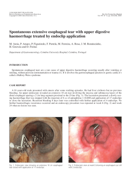
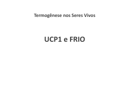
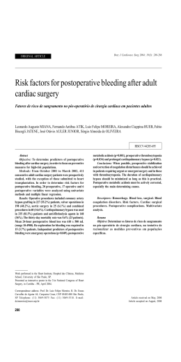
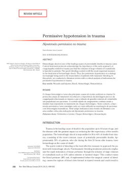
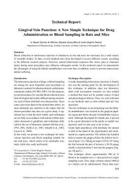
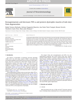
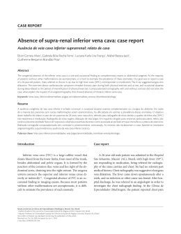
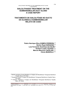
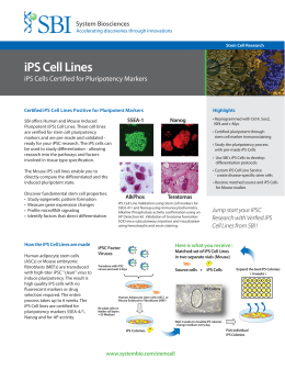
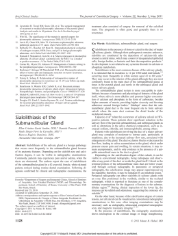
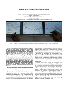
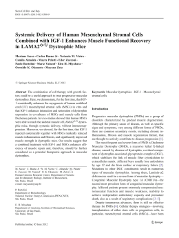
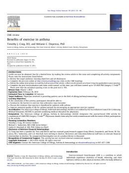
TECHNIQUE
A rapid, simple, and humane method for
submandibular bleeding of mice using a
lancet
William T. Golde, PhD1, Peter Gollobin2 & Luis L. Rodriguez, DVM, PhD1
Methods for obtaining blood samples from mice tend to be difficult, inhumane, or both.
The authors describe an inexpensive, disposable, single-use lancet for submandibular
bleeding of mice that allows investigators to quickly draw 0.2–0.5 ml of blood without
the use of anesthesia.
When using small animals, such as mice, researchers
must be able to acquire samples for testing. Drawing
blood samples from mice is challenging because of the
animal’s small size. Although Institutional Animal Care
and Use Committees will approve protocols including
several blood collection methods, none are particularly
simple or humane.
In the United States, the most common rodent bleeding method is retro-orbital, puncturing the orbital
sinus behind the eye. This method consistently yields a
reasonable blood volume (0.2–0.5 ml) when the investigator is experienced in executing the procedure 1.
Nevertheless, poor technique can blind the animal,
and several countries have banned this method because
officials consider it to be inhumane.
Another common method is cardiac puncture. This
procedure requires anesthesia, which may alter parameters of the experiment. Briefly, one inserts a smallgauge needle into the ventricle and slowly draws out
blood. This procedure requires a skilled investigator
and is most commonly used as a terminal blood collection procedure; however, some investigators are able to
keep animals alive for several blood collections2. This
is not a simple method and is only humane when the
procedure goes very well, leaving minimal damage to
cardiac and pericardial tissues along the needle track.
Missing the heart or passing the needle completely
through the heart could lead to undetected internal
bleeding or other complications. Because the chance
of losing animals is so great, investigators choosing
this method often supplement the number of animals
requested for the research so as to accommodate loss
during an experiment.
A simple method of bleeding mice is the tail clip, in
which a piece of the tail is excised and blood harvested
from the severed tail vein. One can do this repeatedly
for a few sequential bleeds. A major disadvantage is
that to leave enough tail for several future bleeds, the
portion of tail excised must be small, thus yielding a
small blood sample of a few drops (<0.1 ml). Another
problem with this method is that it could lead to cannibalism among cagemates and is not at all humane,
especially for several blood draws. In addition, there
is the tail laceration technique in which the mouse
is restrained and an incision is made in the tail vein
with a scalpel or razor blade. This technique yields as
much as 0.5 ml of blood3; however, it usually requires
anesthesia, and an incision made too deeply can
complicate repair.
Finally, a more humane method that is done without
anesthesia is the saphenous vein puncture4. This is a
complicated and time-consuming method that involves
immobilizing the animals with the rear legs accessible.
The investigator shaves the hair from the caudal surface
of the thigh using a small scalpel or razor. The saphenous
vein of the thigh is visible, and one can puncture it with
a 23- to 25-gauge needle, then collect the blood with a
microvette capillary collection tube. These tubes have a
maximum volume of 0.3 ml. A compress held on the site
will stop the bleeding. This is a more humane method of
blood collection, and with no anesthesia required there
are no side effects to consider. However, this procedure
is slow, requiring extensive time working with each animal, and is not compatible with large trials of pharmaceuticals or biologicals. The time required to do a large
trial (e.g., 50–100 animals) would cause researchers to
1Plum Island Animal Disease Center, ARS, USDA, Greenport, NY 11944. 2MediPoint Inc., Mineola, NY 11501. Correspondence should be
addressed to W.T.G. ([email protected]).
LAB ANIMAL
Volume 34, No. 9 | OCTOBER 2005
39
TECHNIQUE
FIGURE 1 | Bleeding from the mouse cheek targets the retro-orbital and submandibular veins
draining the face of the mouse at the point that they join at the origin of the jugular vein.
design smaller experiments using fewer animals. The
investigators describing this method limit the amount
of blood collected to 0.3 ml, and in practice, the blood
volumes collected are even less. This commonly would
yield ~0.1 ml of serum and limit analysis to a few very
small-volume assays. Hoff has extensively reviewed all
of these techniques5.
SUBMANDIBULAR BLEEDING METHODS FOR MICE
We have been using an alternative method for drawing blood samples from mice that is more rapid and
humane. This is a method that has been used by colleagues for several years but requires a great deal of
practice. A small vascular bundle at the back of the jaw
characterizes the cheek of laboratory mice (Fig. 1). This
is the point where the orbital veins, the submandibular
vein, and other veins draining the facial region join to
form the beginning of the jugular vein. The researcher
holds the animal by the scruff of the neck in the air to
establish the most relaxed situation for the mouse. Using
FIGURE 2 | (a) The Goldenrod animal bleeding lancet removed from the sterile wrapper.
(b) The Goldenrod animal bleeding lancet (5 mm point size) in a sterile wrapper and the
blood collection vessel (Microtainer tube from B/D sold by Fisher Scientific).
40 Volume 34, No. 9 | OCTOBER 2005
a number 11 scalpel, the investigator pokes the cheek
facing him or her with enough force to create a small
stick hole, so that drops of blood exude from the point
of penetration. The blood collection tubes used may or
may not include separating gel for collection of serum
and whole blood, and either heparin or EDTA as an
anticoagulant. The position of the punch is crucial to
obtain a sufficient volume of blood in a very short time,
and with practice the investigator can become proficient
at positioning the scalpel. One can collect as much as 0.7
ml of blood from an adult mouse depending on the frequency of bleeding. Simply applying a sterile gauze pad
or tissue with a little compression for less than a minute
will stop the bleeding. After the mouse is released, it will
groom and within a short time will show no evidence of
the blood draw.
The problem with this method is that control of
the scalpel during the stabbing process requires a
fine touch that comes from many repetitions of the
procedure. Even after much practice, the investigator
often will not penetrate far enough with the scalpel,
or worse, penetrate too deep and cause tissue damage. Investigators who are not yet proficient or who
only occasionally bleed mice have often had frustration with the inconsistency of the procedure. Most
often the application of insufficient pressure yields
little or no blood. A stab that is too strong cuts the
mouse cheek skin, which may require intervention to
arrest the bleeding, and possibly sutures; this complication now elevates the procedure to minor surgery.
In some cases the scalpel pokes through the inside
of the cheek, causing blood to fill the mouth cavity
and putting the animal in danger of death by blood
aspiration and asphyxiation if the bleeding is profuse.
Also, in stabbing high above the jaw, the investigator
may punch into the ear canal causing bleeding from
the ear.
An alternative to the scalpel is to perform the
same procedure using a 19-, 21-, or 23-gauge needle,
depending on the size of the mouse. However, the
disadvantages of needles are similar to those of scalpels; thus they can cause lacerations of the cheek and
complications similar to those cited for the scalpel.
In addition, a significant amount of blood coagulates inside the needle, resulting in poor blood yields.
Some investigators have used this method successfully, but it is difficult to learn because of the need
for consistency.
MOUSE BLEEDING LANCET
We have now developed an inexpensive, disposable,
sterile single-use mouse bleeding lancet for the submandibular bleeding method. We based this device
on the standard lancet used to take blood samples
from infants’ heels or from the fingertips of children
and adults. These lancets cause only momentary pain
www.labanimal.com
TECHNIQUE
FIGURE 3 | (a) Positioning and poking the cheek with the lancet. (b) Collecting blood from the cheek.
at the time of the puncture and leave no significant
tissue damage, as demonstrated by dissection of
the site 3 or 7 days later. For use in the mouse procedure, we determined that the length of the standard fingertip lancet, 3.0 mm, was too short. We
produced and tested 4.0-mm and 5.0-mm lancets
(Fig. 2a). In Figure 2b, we show the sterile, paperwrapped lancet with the blood collection tube, a B/D
microtainer tube (Fisher Scientific, Pittsburgh, PA).
As shown in Figure 3, the method is identical to the
scalpel method, with the advantage that the lancets
control the depth of the skin puncture such that there
is a high degree of consistency in a blood draw with
very little training. As shown in Figure 3a, the optimal
puncture point is at the back of the jaw of the mouse,
FIGURE 4 | The mouse after holding a sterile gauze compress on the bleeding site for
10–30 s.
LAB ANIMAL
very slightly behind the hinge of the jawbones, toward
the ear. One can feel the jawbone with the lancet and
should avoid the bone, instead puncturing the cheek
just behind the point at which the upper and lower
jawbones meet. We have found that holding the animal
by the skin between the shoulder blades rather than
between the ears tends to result in less distortion of the
skin and presumably blood vessels on the face. There
should be enough force in the thrust to quickly insert
the point to the hilt. Once the stick has been made, one
should quickly seize the collection tube, because blood
can exude from the puncture very rapidly and often
the flow slows in a very short time. For more detailed
descriptions and a video example, see the supplier’s
website (http://www.medipoint.com).
The 5.0-mm lancets provided the best results with
very few problems apart from finding the proper location for the lancet puncture. Blood volumes that we
collected were always >0.3 ml, and bleeding could be
stopped once we had collected a sufficient volume.
The 4.0-mm lancets worked very well on young mice,
2- to 6-week-old BALB/c and C57Bl/6, and the 5.0mm lancets worked best for adult mice aged 2 months
and older. For investigators doing long-term studies or
working on aging, we developed a 5.5-mm lancet for
the larger 9-month to year-old mice. After the procedure the animal shows little evidence of the blood draw
(Fig. 4), and after a short period of grooming there is
no evidence of the procedure.
The mouse lancet eliminates the problems associated with the use of a scalpel or needle when drawing
blood from the cheek. It makes the procedure easy,
resulting in consistent quantities of blood of about
between 0.2 and 0.5 ml with little chance of hurting
the mouse. With firm pressure during the stick, we
never made an incision all the way through to the
inside of the mouth. This provides the researcher
with the confidence to push hard enough so that an
Volume 34, No. 9 | OCTOBER 2005
41
TECHNIQUE
and recover the blood volume within 24 h. However,
daily bleeding may have an effect on erythrocytes and
platelets4, which should be monitored.
The mouse lancet also allows for relatively rapid processing of many animals compared with the saphenous
vein bleeding method, in which the researcher must
shave each animal. Dispensing with the need for anesthesia eliminates both the effects on animal physiology and the need to tend to animals as the effects of
the anesthetic subside. In addition, the results of tests
on the same laboratory animal are more accurate than
those obtained using different animals that are identical genetically. Because this lancet device allows the
use of the same animal for several tests, it will improve
reproducibility in animal trials as well as greatly reduce
the number of animals required. Finally, this method
of blood collection leaves the animals unaffected; they
recover instantly and show no signs of distress resulting
from the bleeding.
FIGURE 5 | The 50-lancet clip loaded into the dispenser with the clip drawn out for use.
excessively light touch is no longer a problem. Poking
too high on the face can lead to blood in the ear canal,
but with minimal experience investigators can become
proficient at avoiding this mistake. As with the needle
or scalpel system, the lancets are individually wrapped
and sterile to eliminate cross-contamination between
animals. Unlike scalpels and needles, these lancets are
relatively inexpensive (approximately $0.04/ unit),
and single use is economical.
It is important to note that with this system, one
can draw blood from the same mouse several times
in a day as well as daily; this is not easy to do with any
of the methods reviewed earlier. When doing multiple blood draws, investigators can adjust the volume of blood harvested for the size of the animal,
so that the total daily volumes collected are within
the recommended margin of safety for the animal.
The blood volume of a mouse varies with size but
is typically 6–8% of body weight or 6–8 ml per 100
g total weight4. Taking 1 ml of blood requires fluid
replacement, but mice tolerate draws of 0.5–0.7 ml
LANCET DISPENSER FOR HIGH-VOLUME USERS
The use of the individually wrapped, sterile bleeding lancet is impractical in some circumstances. Most
notably, there are situations in which investigators
are bleeding hundreds of mice and the unwrapping
of each lancet is not practical. In addition, scientists
working in certain containment conditions, such as
glove boxes, require a more readily accessible source
of the lancet. For special-use investigators, we have
developed a system that dispenses a clip of 50 lancets.
One can steam-sterilize both the clip and the dispenser before use. Once the clip has been opened and
mounted on the dispenser, the investigator can easily
extract individual lancets with a single hand (Fig. 5).
Not only is this dispenser usable with a gloved hand,
but it also provides rapid delivery of lancets for the
high-volume user.
The animal bleeding lancet for submandibular
bleeding from the cheek of a mouse is low in cost and
more convenient than devices used in other methods
(Table 1). We are assuming that all methods require
a blood collection tube containing separation gel or
anticoagulant, as required by the procedure. In addition, the absence of anesthesia provides for a more
TABLE 1. A comparison of bleeding techniques for mice.
Method
Anesthesia
Device
Cost/devicea
Retro-orbital
Cardiac puncture
No
Yes
Capillary
Syringe and needle
Saphenous vein
Yes
Submandibular vein
No
aProduct
$.04
$.148 and $.084
Number of
draws possible
4–5
1–3
Other devices
required
No
No
Needle
$.068
>10
Yes
Lancet
$.045
>10
No
Time/
mouse
<1 min
5–10
min
5–10
min
<1 min
prices from Fisher Scientific, Pittsburgh, PA.
42 Volume 34, No. 9 | OCTOBER 2005
www.labanimal.com
TECHNIQUE
rapid processing of mice and no adverse effects on
animal physiology.
For the cost, time requirement, complicating variables
such as the need to monitor animals recovering from
anesthesia, and the number of times an investigator
can bleed the same animal, only retro-orbital bleeding
is as timely and economical as submandibular bleeding. Even in this case, only a few blood draws are possible using retro-orbital bleeding for all but the most
adept investigator.
SUMMARY
The new mouse lancet for submandibular bleeding
is a humane, efficient, and economical method for
bleeding laboratory mice. Similar styles of blood
lancet have been in use for decades to draw blood
from many mammalian species, especially humans,
resulting in very little pain, discomfort, and tissue
damage. We believe that this method will not only
improve scientific design and results in studies using
laboratory mice, but may also have application to
other laboratory animals including rats, hamsters,
and gerbils.
LAB ANIMAL
ACKNOWLEDGMENTS
Photos were provided by E. Clark.
COMPETING INTERESTS STATEMENT
The authors declare competing financial interests. All three authors
are inventors of the patent-pending technology described here,
and developed under a Cooperative Research and Development
Agreement between the US Department of Agriculture, Agricultural
Research Service and MEDIpoint, Inc.
Received 23 June 2005; accepted 12 August 2005.
Published online at http://www.labanimal.com.
1.
2.
3.
4.
5.
Fox, J.G., Anderson, L.C., Lowe, F.M. & Quimby, F.Q. (eds.).
Laboratory Animal Medicine 2nd Edn. (Elsevier Science,
New York, 2002).
Bertani, S., Gantier, J.C., Chabaud, A. & Landau, I. Action
of adrenalin on the circulation of the murine Plasmodium
developing stages, in different blood compartments. Parasite
11(4), 343–350 (2004).
Fields, B.T. & Cunnigham, D.R. A tail artery technique for
collecting one-half milliliter of blood from a mouse. Lab Anim.
Sci. 26(3), 505–506 (1976).
Hem, A., Smith, A.J. & Solberg, P. Saphenous vein puncture
for blood sampling of the mouse, rat, gerbil, guinea pig, ferret
and mink. Lab. Anim. 32(4), 364–368 (1998).
Hoff, J. Methods of blood collection in the mouse. Lab Anim.
(NY) 29(10), 47–53 (2000).
Volume 34, No. 9 | OCTOBER 2005
43
Download