ACTA BIOLOGICA CRACOVIENSIA Series Botanica 51/2: 115–118, 2009
ARE THERE SEED PEDESTALS IN LENTIBULARIACEAE?
BARTOSZ J. PŁACHNO1*, DÉBORA CLIVATI2, VÍTOR F. OLIVEIRA DE MIRANDA2,
AND PIOTR ŚWIĄTEK3
1
Department of Plant Cytology and Embryology, Jagiellonian University,
Grodzka 52, 31–044 Cracow, Poland
2
Plant Systematics Laboratory, Herbarium Mogiense – HUMC,
Universidade de Mogi das Cruzes – UMC, Av. Dr. Cândido Xavier de Almeida Souza,
n.200, CEP 08780-911, Mogi das Cruzes, SP, Brasil
3
Department of Animal Histology and Embryology, University of Silesia,
Bankowa 9, 40–007 Katowice, Poland
Received July 20, 2009; revision accepted October 2, 2009
The term "seed pedestal" was introduced recently to describe a structure of placental origin connecting a seed
with the placenta. Seed pedestals are widespread in Scrophulariaceae and a few adjacent families, but have not
been found in Lentibulariaceae so far. Here their presence is reported for Utricularia reniformis from Brazil,
and their formation during seed development is described. We observed that the formation of this structure was
strictly associated with seed development; seed pedestals were not formed under aborted (unfertilized) ovules.
Key words: Ovule, seed development, placenta, seed pedestal, Utricularia, micromorphology,
carnivorous plants.
INTRODUCTION
Lentibulariaceae, the largest carnivorous plant
family, comprises a highly evolved and specialized
family of the Lamiales (e.g., Fischer et al., 2004;
Müller et al., 2006). They show several morphological modifications ("relaxed morphology") (Brugger
and Rutishauser, 1989; Rutishauser and Isler,
2001; Ellison and Gotelli, 2009), traps for catching
small invertebrates (e.g., Juniper et al., 1989;
Płachno et al., 2007, 2008), and also unusual
embryological characters (e.g., Khan, 1954;
Płachno and Świątek, 2008). As in other
angiosperms, in Lentibulariaceae the seeds provide
important taxonomic information. Taylor (1989)
described the great diversity of seed morphology in
Utricularia. Seed shape and seed testa structure
are important characters in Utricularia taxonomy
on both section and species levels. In Pinguicula
some characters of seed coat structure may be significant at the section level, and other characters at
specific or infraspecific levels (Degtjareva et al.,
2004, 2006). Many authors have studied seed for-
mation in Lentibulariaceae (mainly in Utricularia),
including Merz (1897), Lang (1901), Merl (1915),
Lloyd (1942), Khan (1954) and Farooq (1965), but
less attention has been paid to placental changes
during seed development.
The term "seed pedestal" was recently introduced by Rebernig and Weber (2007) to denote a
structure which is developmentally placental in origin and thus not part of the ovule, and which connects a seed with the placenta. They examined in
detail members of Scrophulariaceae (s.l.) (45
species of 27 genera) and also samples of 11 other
(mainly derived) families of Lamiales, including
Lentibulariaceae (Pinguicula and Utricularia).
However, they did not find evidence for
seed pedestals or comparable structures in
Lentibulariaceae.
This study was intended to determine whether
the seed pedestal occurs in Lentibulariaceae. The
paper is also part of a larger project on the biology
of reproduction in Lentibulariaceae carried out by
B.J.P. (grant N N304 002536 from the Polish
Ministry of Science and Higher Education).
*e-mail: [email protected]
PL ISSN 0001-5296
© Polish Academy of Sciences and Jagiellonian University, Cracow 2009
116
Płachno et al.
MATERIALS AND METHODS
PLANT MATERIAL
Material was taken from two sources: directly from
nature and from greenhouse collections. Buds, flowers and fruits of Utricularia reniformis A.St.-Hil.
(large form) were collected by V.F.O.M. in the Pedra
do Garrafão from a population on a mountain slope
facing the sea, near the town of Biritiba Mirim, São
Paulo State, Brazil (Fig. 1a). Voucher specimens are
deposited in HUMC. Another form, Utricularia reniformis (A.St.-Hil.) "Enfant Terrible" (B.Rice &
M.Studnicka) (Rice and Studnièka, 2004), was provided by the Liberec Botanical Garden (Czech
Republic) and then cultivated successfully in the
Botanical Garden of the Jagiellonian University in
Cracow, Poland. The plants produced flowers in
2008, and after hand-pollination fruits were obtained.
HISTOLOGICAL ANALYSIS
Buds, flowers and fruits were fixed in 2.5% formaldehyde and 2.5% glutaraldehyde in 0.05 M cacodylate
buffer (pH 7.0), rinsed in the same buffer, dehydrated in a graded ethanol series (10%, 30%, 50%, 70%,
96%) for 15 min at each concentration and then kept
1 h in absolute ethanol. Later, samples were infiltrated in mixtures of absolute ethanol and Technovit7100
(2-hydroxyethyl-methacrylate)
(Heraeus
Kulzer) (3:1, 1:1, 1:3 v/v; 1 h in each mixture) and
stored for 12 h in pure Technovit. The resin was
polymerized with the addition of hardener at 37°C.
The material was sectioned 5 μm and 7 μm thick
with a rotary microtome (Microm, Adamas
Instrumenten), stained with toluidine blue or methylene blue and mounted in Entellan (Merck). Part of
the material was embedded in Epon 812 (Fullam,
Latham, NY). Semithin and ultrathin sections were
cut on a Leica Ultracut UCT ultramicrotome and
stained with methylene blue. Microscope sections
were photographed with a Zeiss Axio Cam MRe digital camera with Zeiss Axio Vision 3.0 software and
an Olympus BX60 microscope.
SCANNING ELECTRON AND EPIFLUORESCENCE
MICROSCOPY
The procedures for preparing samples for SEM
were as described earlier (Płachno et al., 2005a,b).
Briefly, flowers and fruits were fixed as for histological analysis. The dried tissues were sputter-coated
with gold and viewed in a HITACHI S-4700 microscope (Scanning Microscopy Laboratory of
Biological and Geological Sciences, Jagiellonian
University). Additionally, whole ovules were examined by epifluorescence microscopy.
RESULTS
The stalked spherical placenta is covered with
numerous sessile, unitegmic ovules (Fig. 1b). The
ovule is ~165 μm long. During ovule development a
small spherical group of cells differentiates near its
base, which forms the placental nutritive tissue
(Fig. 1c; see also Merl, 1915, Fig. 30f). The placental epidermal cells are papilla-shaped and form an
epithelium (Fig. 1c). During seed development the
placental tissues form elaborations (Fig. 1d). Each
seed has one placental elaboration. The seed is
attached at the apex of this elaboration (pedestal)
(Figs. 1d,e). The pedestal is formed by placental
parenchyma, epidermis and nutritive tissue. The
epidermal and parenchyma cells that form the
pedestal change their shape and enlarge. The seed
pedestal is formed only if the ovule is fertilized and
the seed is developing (Fig. 1e). After maturation,
the seeds very easily drop away from the pedestals
(the seed detaches from the site of the endospermal
micropylar haustorium; Fig. 1f).
DISCUSSION
Our results are in contrast to Rebernig and Weber's
(2007) finding that the Lentibulariaceae lack seed
pedestals. However, the focus of that work was on
Scrophulariaceae (s.l.) and the sample size for the
remaining families of the Lamiales was small.
Regarding Lentibulariaceae, Rebernig and Weber
examined Pinguicula (P. agnata, P. esseriana,
P. moranensis) and Utricularia (U. livida,
U. sandersonii). All were found to lack seed
pedestals. The genus Pinguicula is the most basal
genus of the Lentibulariaceae family and a sister
group of Utricularia-Genlisea clade (Jobson et al.,
2003; Müller et al., 2004, 2005). U. livida and
U. sandersonii are classified in sect. Calpidisca,
which is not very evolutionarily advanced according
to Taylor (1989). Utricularia reniformis, on the
other hand belongs to the derived section Iperula
according to the phylogenetic hypotheses of
Utricularia (Jobson et al. 2003; Müller and Borsch,
2005). At this point it is difficult to suggest any taxonomic implications, because our knowledge of
Lentibulariaceae placenta and seed development is
poor. The seed pedestal in Utricularia reniformis is
similar to the Digitalis type of Rebernig and Weber's
(2007) classification.
Our data show that in Utricularia reniformis the
formation of the seed pedestal is strictly connected
with seed development; it did not form under aborted
(unfertilized) ovules. This finding is not in agreement
with Rebernig and Weber (2007), who stated that the
pedestal develops fully even in the case of ovule abor-
Seed pedestals in Lentibulariaceae
117
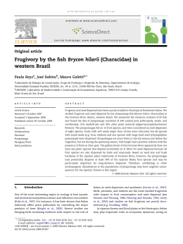
Fig. 1. Utricularia reniformis A.St.-Hil. (a) Flower of large form from the Pedra do Garrafão, Brazil, (b) Ovules on the
placenta, (c) Section through ovule and female gametophyte (FG) and nutritive tissue (star), (d) Section through placenta
(P), seed pedestal (SP) and seed (S), embryo (em), (e) Placenta surface with seed pedestal (SP), seeds (S) and aborted
(unfertilized) ovules (arrow), (f) Apical surface of seed pedestal after seed detachment, haustorium of endosperm (star).
Bar in (a–c) = 36 μm in (d) = 137 μm.
118
Płachno et al.
tion. It may be speculated that seed pedestals facilitate
detachment of seeds from the placenta.
The presence or absence of seed pedestals
needs to be examined in other Utricularia species.
In species exhibiting a very similar pattern of seed
development, the presence of seed pedestals is especially probable.
ACKNOWLEDGMENTS
The first author gratefully acknowledges the support
of an award from the Foundation for Polish Sciences
(Start Programme). This study was funded by grant
N N304 002536 from the Polish Ministry of Science
and Higher Education. Doctoral candidate Débora
Clivati's stay at the Jagiellonian University was made
possible by funding from FAPESP (Fundação de
Amparo à Pesquisa do Estado de São Paulo), grant
2008/52239-9. We thank the Rector of the
Jagiellonian University, Professor Szczepan
Biliński, for kindly supporting our projects, and
Dr. Miroslav Studnièka (Director of Botanical
Garden in Liberec, Czech Republic) for kindly providing U. reniformis "Enfant Terrible" for this study.
REFERENCES
BRUGGER J, and RUTISHAUSER R. 1989. Bau und Entwicklung
landbewohnender Utricularia-Arten. Botanica Helvetica
99: 91–146.
DEGTJAREVA G, CASPER J, HELLWIG F, SOKOLOFF D. 2004. Seed
morphology in the genus Pinguicula (Lentibulariaceae)
and its relation to taxonomy and phylogeny. Botanische
Jahrbücher 125: 431–452.
DEGTJAREVA GV, CASPER SJ, HELLWIG FH, SCHMIDT AR, STEIGER
J, SOKOLOFFS DD. 2006. Morphology and nrITS phylogeny of the genus Pinguicula L. (Lentibulariaceae), with
special attention to embryo evolution. Plant Biology 8:
778–790.
ELLISON AM, and GOTELLI NJ. 2009. Darwin review energetics
and the evolution of carnivorous plants – Darwin's' most
wonderful plants in the world'. Journal of Experimental
Botany 60/1: 19–42.
FAROOQ M. 1965. Studies in the Lentibulariaceae III. The
embryology
of
Utricularia
uliginosa
Vahl.
Phytomorphology 15: 123–131.
FISCHER E, BARTHLOTT W, SEINE R, and THEISEN I. 2004.
Lentibulariaceae. In: Kubitzki K and Kadereit J [eds.],
The Families and Genera of Vascular Plants, vol. VII,
276–282. Springer Verlag. Berlin, Heidelberg, New York.
JOBSON RW, PLAYFORD J, CAMERON KM, and ALBERT VA. 2003.
Molecular phylogenetics of Lentibulariaceae inferred
from plastid rps16 intron and trnL-F DNA sequences:
implications for character evolution and biogeography.
Systematic Botany 28:157–171.
JUNIPER BE, ROBINS RJ, and JOEL JM. 1989. The Carnivorous
Plants. Academic Press, London.
KHAN R. 1954. A contribution to the embryology of Utricularia
flexuosa Vahl. Phytomorphology 4: 80–117.
LANG FX. 1901. Untersuchungen über Morphologie, Anatomie
und Samenentwicklung von Polypompholyx und Byblis
gigantea. Flora 88: 149–206.
LLOYD FE. 1942. The Carnivorous Plants. Chronica Botanica
Company, Waltham, Mass., U.S.A.
MERL EM. 1915. Beiträge zur Kenntnis der Utricularien und
Genlisen. Flora 108:127–200.
MERZ M. 1897. Untersuchungen über die Samenentwicklung
der Utricularien. Flora 84:69–87.
MÜLLER K, BORSCH T, LEGENDRE L, POREMBSKI S, and BARTHLOTT W.
2006. Recent progress in understanding the evolution of
carnivorous Lentibulariaceae (Lamiales). Plant Biology
8: 748–757.
MÜLLER K, BORSCH T, LEGENDRE L, POREMBSKI S, THEISEN I, and
BARTHLOTT W. 2004. Evolution of carnivory in
Lentibulariaceae and the Lamiales. Plant Biology 6:
477–490.
MÜLLER K, and BORSCH T. 2005. Phylogenetics of Utricularia
(Lentibulariaceae) and molecular evolution of the trnK
intron in a lineage with high substitutional rates. Plant
Systematics and Evolution 250: 39–67.
PŁACHNO BJ, ADAMUS K, FABER J, and KOZłOWSKI J. 2005a.
Feeding behaviour of carnivorous Genlisea plants in the
laboratory. Acta Botanica Gallica 152:159–164.
PŁACHNO BJ, FABER J, and JANKUN A. 2005b. Cuticular discontinuities in glandular hairs of Genlisea St.-Hil. in relation to their functions. Acta Botanica Gallica
152:125–130.
PŁACHNO BJ, KOZIERADZKA-KISZKURNO M, and ŚWIĄTEK P. 2007.
Functional ultrastructure of Genlisea (Lentibulariaceae)
digestive hairs. Annals of Botany 100: 195–203.
PŁACHNO BJ, and ŚWIĄTEK P. 2008. Cytoarchitecture of
Utricularia nutritive tissue. Protoplasma 234: 25–32.
PŁACHNO BJ, KOZIERADZKA-KISZKURNO M, ŚWIĄTEK P, and
DARNOWSKI DW. 2008. Prey attraction in carnivorous
Genlisea
(Lentibulariaceae).
Acta
Biologica
Cracoviensia Series Botanica 50/2: 87–94.
RICE B, and STUDNIÈKA M. 2004. Utricularia reniformis 'Enfant
Terrible'. Carnivorous Plant Newsletter 33: 53–55.
REBERNIG CA, and WEBER A. 2007. Diversity, development and
systematic significance of seed pedestals in
Scrophulariaceae (s.l.). Botanische Jahrbücher 127:
133–150.
RUTISHAUSER R, ISLER B. 2001. Developmental genetics and
morphological evolution of flowering plants, especially
Bladderworts (Utricularia): Fuzzy Arberian morphology
complements classical morphology. Annals of Botany
88: 1173–1202.
TAYLOR P. 1989. The genus Utricularia – A taxonomic monograph. Kew Bulletin 14: 1–735
Download