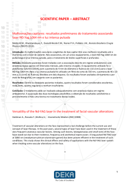
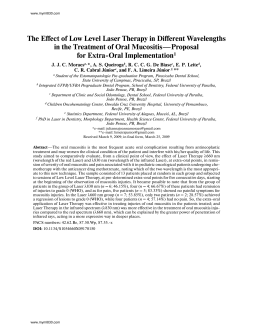
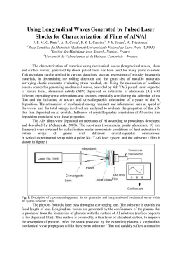
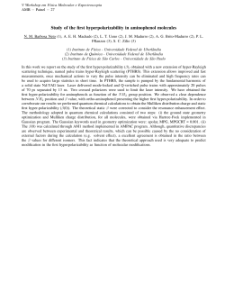
314
Felippe EP, Sales Á, Soglia J, Ribeiro R, Póla C, Mitre J, Rehder JRCL
CASE REPORT
Preretinal hemorrhage
Hemorragia pré-retiniana
Eduardo Felippe1, Álvaro Sales2, Jean Soglia3, Rodrigo Ribeiro4, Cláudio Póla5, Jorge Mitre6, José Ricardo Carvalho
de Lima Rehder7
ABSTRACT
A case of Valsalva hemorrhagic retinopathy treated with Nd:YAG
laser indescribed. The patient presented decreased visual acuity
after coughing, and a preretinal hemorrhage was diagnosed in the
posterior pole; puncturing the posterior hyaloid face was performed
with Nd:Yag laser. Rapid hemorrhage absorption was observed
after the therapy proposed and visual acuity was recovered. Nd:Yag
laser proved to be safe and efficient in the management of preretinal
hemorrhage.
Keywords: Valsalva maneuver; Lasers/therapeutic use; Visual
acuity; Eye hemorrhage
RESUMO
Descreve-se um caso de hemorragia pré-retiniana por Valsalva
tratado com Nd:YAG laser. Paciente apresentou baixa da acuidade
visual após crise de tosse, sendo diagnosticada hemorragia préretiniana no pólo posterior; foi realizado pertuito na hialóide
posterior com Nd:Yag laser. Após tratamento proposto, observouse rápida absorção da hemorragia com o restabelecimento da
acuidade visual. Nd:Yag laser mostrou-se seguro e eficaz no manejo
da hemorragia pré-retiniana.
Descritores: Manobra de Valsalva; Lasers/uso terapêutico;
Acuidade visual; Hemorragia ocular
INTRODUCTION
Preretinal hemorrhage may occur after rupture of
retinal vessels and is associated with physical exercises
and increased venous pressure (Valsalva retinopathy)
or retinal vascular alterations, such as macroaneurismas
and proliferative diabetic retinopathy(1).
Valsalva retinopathy is characterized by a sudden decrease
in visual acuity. It may be treated by vitrectomy, Nd:YAG
laser membranotomy or observation. We described a case of
preretinal hemorrhage, its treatment with Nd:YAG laser
and a favorable progression within a short period of time.
CASE REPORT
Patient I.M.M.L, a white, 40-year old housewife sought
an ophthalmologic service due to decreased visual
acuity for one day and history of dyspnea associated
with coughing and vomiting.
Her blood pressure was borderline and she was on
behavioral and dietetic treatment. She smoked approximately
20 cigarettes/day. Her parents were hypertensive and she
denied any past ophthalmologic history. On ophthalmologic
examination, she presented visual acuity with no correction
of finger counting at 15 centimeters in the right eye (OD)
and 20/20 in the left eye (OL).
On biomicroscopic examination, a slight
conjunctival hyperemia was observed in OD and there
were no alterations in OL. The intraocular pressure
(IOP) was 10 mm Hg in OD and 12 mm Hg in OL, at 4
o’clock. In OD retinal mapping, it was difficult to assess
papillary excavation due to hemorrhage in the papillary
edge from 7 to 1 o’clock. Nevertheless, the visualized
area presented clear edges, preserved neuroretinal rim
with normal color, attached retina with normal color,
slightly increased vascular tortuosity and presence of
preretinal hemorrhage of 10 papillary diameters (PD).
On OL examination, vertically oval papilla with clear
edges, preserved ISNT rule, neuroretinal rim with
normal color and 0.5 vertical/0.4 horizontal excavation,
attached retina with normal color and macula with
preserved foveal reflex. Posterior hyaloid
photodisruption was performed with Nd:YAG laser (21
pulses with 6 mJ-energy, total energy of 142 mJ). In
the first week after Nd:YAG laser, the patient already
Institution: Department of Ophthalmology of the Faculdade de Medicina do ABC.
1
Second-year resident in Ophthalmology in the Faculdade de Medicina do ABC.
2
Second-year resident in Ophthalmology in the Faculdade de Medicina do ABC.
3
Third-year resident in Ophthalmology in the Faculdade de Medicina do ABC.
4
Fifth-year resident in Ophthalmology in the Faculdade de Medicina do ABC.
5
Fourth-year resident in Ophthalmology in the Faculdade de Medicina do ABC.
6
Full professor and Head of the Retina Service of the Faculdade de Medicina do ABC.
7
Full professor and Head of the Department of Ophtalmology of the Faculdade de Medicina do ABC.
Corresponding author: Eduardo P. Felippe - Faculdade de Medicina do ABC - Av. Príncipe de Gales, 821 - Anexo 3 - 1º andar - Departamento de Oftalmologia - CEP 09060-260 - Santo André (SP), Brazil
- Fax (5511) 4993-5408 - e-mail: [email protected]
Received on March 18, 2004 – Accepted on July 15, 2004
einstein. 2004; 2(4):314-5
Preretinal hemorrhage
presented visual acuity of 20/20 in both eyes; however,
cellularity in the anterior chamber and turbid vitreous
++/4+ persisted for 15 days and clinical treatment
with VUEFFE® 3 times/day for 30 days was initiated.
After this period, the patient was asymptomatic and
was submitted to a fluorescein angiography that
demonstrated a hyperfluorescent area uniformly
comprising the macular region in all phases of the exam.
Figure 1a and figure 1b shows the comparison between
the two eyes by retinography.
Figure 1a
Figure 1b
DISCUSSION
Macular preretinal hemorrhage generally causes severe
visual loss. The most common etiologies are
proliferative diabetic retinopathy, macroaneurisma,
Valsalva retinopathy and blood dyscrasias(2).
The frequency of Valsalva retinopathy has not been
estimated due to the reduced number of reported cases.
A sudden increase in intra-abdominal or intrathoracic
pressure against a closed glottis (Valsalva maneuver)
may cause fast rise in venous pressure, leading to
spontaneous rupture of superficial retinal capillaries(1).
According to the literature, the visual acuity of this
patient was within the limits(3-8), as well as hemorrhage
size (3,6). The most common site for hemorrhage in
Valsalva retinopathy is the posterior pole because of a
preexisting anatomical space - premacular bursa. Since
it is difficult to indicate the accurate site of hemorrhage
- between retina and internal limiting membrane, or
internal limiting membrane and hyaloid, we decided
to use the term preretinal hemorrhage.
The presence of a light reflex and the fluid level on
fundoscopy may indicate the involvement of the internal
limiting membrane(1,6). Other therapeutical options are
315
observation, Nd:YAG laser photodisruption and
vitrectomy. Although hemorrhage resulting from Valsalva
retinopathy has spontaneous resolution, it presents some
inconveniences, such as long waiting period for visual
recovery and some complications, such as epiretinal
membrane. In vitrectomy, a more invasive procedure, the
complication rate in both peri- and postoperative periods
is higher and potentially more severe. Faulborn was the
first author to describe photodisruption in the treatment
of premacular hemorrhage(9).
Puncturing the posterior hyaloid of the internal
limiting membrane in the hemorrhagic site is a feasible
alternative to vitrectomy in the management of extensive
premacular hemorrhage(3-8). Laser membranotomy enables
erythrocytes accumulated in the macula to leak to vitreous,
where they are more rapidly absorbed(3-8).
Most studies describing the treatment of premacular
hemorrhage with photodisruption aimed to open a
single passage, the most distant possible from the fovea
and trying to not achieve areas in which there would
be clotted blood. The energy used ranges from 3 to 8
mJ and the number of pulses may increase up to 54(3-8).
In the case mentioned above, we chose treating with
Nd:YAG laser photodisruption since hemorrhage had
more than three papillary diameters, a thick blood
column protecting the retina. We were cautious to use
the lesser possible effective amount of energy(5,10).
CONCLUSION
Nd:Yag laser proved to be an efficient and safe
technique for treating preretinal hemorrhages
considering the safety criteria.
REFERENCES
1. Gas JDM. Stereoscopic atlas of macular diseases: Diagnosis and treatment. 4a
ed. St Louis: CV Mosey; 1997.
2. Gabel VP, Birngruber R, Gunther-Koszka H, Puliafito CA. Nd:YAG laser
photodisruption of hemorrhagic detachment of the internal limiting membrane.
Am J Ophthalmol.1989;107(1):33-7.
3. Stempels N, Tassignon MJ, Worst J. Les hemorragies dans la bourse premaculaire
traitées par le Q-switched Nd:YAG laser. Ophtalmologie. 1990;4(3):314-6.
4. Raymond LA. Neodymium:YAG laser treatment for hemorrhages under the
internal limiting membrane and posterior hyaloid face in the macula.
Ophthalmology. 1995;102(3):406-11.
5. Ulbig MW, Mangouritsas G, Rothbacher HH, Hamilton AM, McHugh JD. Longterm results after drainage of premacular subhyaloid hemorrhage into the
vitreous with a pulsed Nd:YAG laser. Arch Ophthalmol. 1998; 116(11):1465-9.
6. Kaynak S, Eryildirim A, Kaynak T, Durak I, Saatci O, Eryildirim S, et al. Nd:YAG
laser posterior hyaloidotomy in subhyaloid hemorrhage. Ophthalmic Surg.
1994;25(7):474-6.
7. Ezra AND, Dowler JG, Burgess F, Sehmi K, Hamilton PA. Identifying maculopathy
after neodymium: YAG membranotomy for dense diabetic premacular
hemorrhage. Ophthalmology. 1996;103(10):1568-74.
8. Diniz AV. Manifestações do trauma ocular contuso. In: Abujamra S. Retina e
Vítreo: clínica e cirurgia. São Paulo:Roca; 2000. p. 769-77.
9. Faulborn J. Behandlung Einer Diabetischen Praemaculaeren Blutung mit dem
Q-switched Neodym:YAG Laser. Spektrum Augenheilkd. 1998; 18:4-6. German.
10. Dori D, Gelfand Y, Erlik N, Miller B. Nd:YAG laser treatment for premacular
hemorrhage. Ophtalmic Surg Lasers 1998;29(12):998-1000.
einstein. 2004; 2(4):314-5
Download