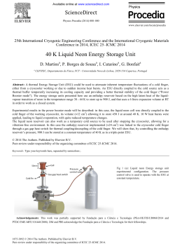
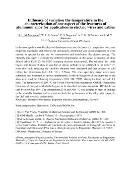
Revista Brasileira de Engenharia Biomédica, v. 23, n. 1, p. 17-23, abril 2007 © SBEB - Sociedade Brasileira de Engenharia Biomédica ISSN 1517-3151 Artigo Original Recebido em 24/10/2005, aceito em 02/03/2007 In site electric field measurements in hospital operating rooms Medição de campos elétricos in situ em centros cirúrgicos Guilherme Nunes Nogueira Neto* Laboratório de Engenharia de Reabilitação - PUC-PR, Parque Tecnológico, Bloco II Rua Imaculada Conceição, 1155 80215-901, Curitiba, PR, Brasil E-mail: [email protected] Marcos Antonio Muniz de Moura Percy Nohama Laboratório de Engenharia de Reabilitação - PUC-PR, Universidade Tecnológica Federal do Paraná (UTFPR) Av. Sete de Setembro, 3165 80230-901, Curitiba, PR, Brasil Sérgio Luiz Bazan de Paula Universidade Tecnológica Federal do Paraná (UTFPR) *Autor para correspondência Abstract Electromagnetic interference can cause malfunctions in elec� tromedical devices (EMD). Among the interfering sources found in the literature, cell phones and electrosurgical units (ESU) have been getting special focus. Commonly, these sources can work simultaneously in small physical spaces, like operating rooms (OR). This study describes the sounding of ambient electric field intensities during surgical interventions inside OR. Employing an isotropic mobile field meter with a calibrated electric field probe, the measurements have been carried out in OR of three hospitals. Seven different types of surgery were monitored. In order to avoid discrepant field magnitudes in the measurements, experiments were made in the available OR that showed similar location of EMD and surgical furniture. The electric field was measured in four test points around the surgical table. Remote communication was established between the meter inside OR and a laptop outside the room by means of an optical cable. Fifteen minutes in each point totalized 1 hour of observation per surgery. When ESU was activated, the meter registered values higher than basal field level. The mean electric field intensities varied between 1.1 ± 0.3 V/m and 3.0 ± 1.2 V/m. Nevertheless, higher electric fields as 9.1 V/m were observed at distances greater than 2.0 m. The electric field measurements result from wideband integration of all fields over the meter operating frequency spectrum. Therefore, the meter cannot determine the electric fields of a specific EMD operating frequency, contrary to spec� trum analyzers. However, it indicated the impact caused by ESU activation on the ambient electromagnetic field. Techni� cians observed the occurrence of electromagnetic interference (EMI) in vital signal and laparoscopy system monitors when ESU was activated. Cell phones were also present inside OR. The mobile meter was suitable for monitoring the OR ambi� ent electromagnetic field during surgeries. Future work will include spectrum analyzer scanning and selective antennas since the mobile meter cannot perform narrowband scanning. Keywords: Electrosurgical device, Electric field, Electromag� netic interference, Measurement, Operating room. 17 E�������� lectric ������ field measurement ������������ ��� in ���������� operating ����� rooms G.N.Nogueira Neto, M.A.M. Moura, P. Nohama, S.L.B. Paula Resumo A interferência eletromagnética pode causar problemas no funcionamento de equipamentos eletromédicos. Dentre as fontes radiantes de interferência encontradas na literatura, os telefones celulares e as unidades eletrocirúrgicas vêm recebendo atenção especial. Rotineiramente, essas fontes operam simultaneamente em pequenos espaços físicos, como as salas cirúrgicas. Este estudo apresenta uma sondagem da intensidade de campo elétrico ambiente em salas cirúrgicas durante a intervenção médica. As medições aconteceram em centros cirúrgicos de três hospitais com o emprego de um medidor isotrópico de campo elétrico com sensor calibrado. Sete diferentes tipos de cirurgia foram monitorados. Para evitar a obtenção de intensidades de campo discrepantes nas medições, os experimentos aconteceram nas salas cirúrgicas disponíveis que apresentaram disposição similar de objetos e móveis cirúrgicos e equipamentos eletromédicos. O medidor de campo adquiriu as medidas em quatro pontos ao redor da mesa cirúrgica. A comunicação remota entre o medidor dentro da sala cirúrgica e um computador portátil fora da sala foi estabelecida por meio de um cabo de fibra óptica. Quinze minutos em cada ponto totalizaram 1 hora de observação por cirurgia. O medidor registrou valores de campo elétrico mais elevados que o nível basal quando do acionamento da unidade eletrocirúrgica. O valor médio de campo elétrico variou entre 1,1 ± 0,3 V/m e 3,0 ± 1,2 V/m. Contudo, intensidades mais elevadas de campo elétrico foram observadas, como 9,1 V/m, para distâncias maiores que 2,0 m. Os campos elétricos registrados resultam da integração de todos os campos existentes no espectro de freqüências que o medidor é capaz de monitorar. Por esse motivo, o medidor não consegue determinar os campos elétricos na freqüência de operação de um equipamento eletromédico específico, diferentemente de um analisador de espectro. Porém, ele apresentou o impacto causado pela ativação da unidade eletrocirúrgica no campo eletromagnético ambiente. Os técnicos observaram ocorrências de interferência eletromagnética em monitores de sinais vitais e de sistemas de videolaparoscopia quando a unidade eletrocirúrgica foi acionada. Telefones celulares também estavam presentes nas salas cirúrgicas. O medidor portátil mostrou-se apropriado para a monitoração do campo eletromagnético ambiente das salas durante a intervenção cirúrgica. Pesquisas futuras incluirão a varredura com analisador de espectro e antenas seletivas, uma vez que o medidor portátil não realiza análise em banda estreita. Palavras-chave: Campo elétrico, Interferência eletromagnética, Medição, Sala cirúrgica, Unidade eletrocirúrgica. 18 Revista Brasileira de Engenharia Biomédica / v. 23 / n. 1 Brazilian Journal of Biomedical Engineering / v. 23 / n. 1 Introduction A complex electrical network supply energy to elec� tromedical devices (EMD) inside hospitals. Although most EMD comply with the electromagnetic compat� ������� ibility��������������������������� regulations, ruled by IEC ������������������� 60601-1-2 standard (ABNT, 1997; IEC, 2001), researchers published a series of electromagnetic interference (EMI) reports due to many electromagnetic sources (AAMI, 1997; Silberberg, 1993; Turcotte & Witters, 1998). Among the interfering sources found in the literature, cell phones (Cabral, 2001; Nogueira-Neto, 2003) and electrosurgical units (ESU) (Nelson & Ji, 1994; Tan & Hinberg, 1993) have been getting special focus. Commonly, these sources can work simultaneously in small physical spaces, like operating rooms (OR), where also work EMD whose immunity to EMI is frequently unknown. There are studies that investigated the presence of RF electromagnetic fields generated by power transmit� ters in and around hospitals (Irnich & Tobisch, 1999; Vlach et al., 1995). Some researchers focused on the determination of fields inside hospital corridors (Davis et al., 1999; 2001) whereas other researchers measured electric fields in and around ambulances (Boivin et al., 1997). The existing electromagnetic fields may also induce electrical currents and cause microshock in surgeries in which patients have part of their bodies connected to EMD. Depending on the path taken by the current through the patient’s chest, it can cause ventricular fibrillation and death. Understanding these fields during surgeries can help improving patient’s and EMD operators’ safety and also lead to recommen� dations concerning EMD acquisition and operation inside OR. During a preliminary inspection of the ambient electric field with a portable meter, the activation of the ESU apparently caused the meter to register higher�������������������������������������������������� electric field values than basal level. It would be interesting to investigate the dynamics of electric field intensities while a patient has surgery. This paper describes the sounding of ambient electric field inten� sities during surgical interventions inside OR. Materials and Methods The experiments took place in three hospitals located in the city of Curitiba, Brazil. Seven different types of surgeries were monitored: radical hysterectomy, posthetomy, Wertheim Wiggs, appendicectomy, in� guinal herniorraphy, gastrointestinal laparotomy and laparoscopy. These surgeries were monitored with the allowance and availability of the hospitals surgical E�������� lectric field ������ measurement ������������ in ��� operating ���������� rooms ����� G.N.Nogueira Neto, M.A.M. Moura, P. Nohama, S.L.B. Paula centres for performing the study. The dimensions of OR and the number of medical staff members present inside the room were taken into account for easing the installation and control of the field meter as well as not interfering with the ongoing surgery. Moreover, once the chosen rooms had the most similar dimensions, it is reasonable to expect that the particular variations in the results were due to the type of surgery taking place. The arrangement and position of EMD, the number of staff members circulating inside the OR, and the type of surgery influence the ambient electromagnetic field. The experiments happened in the available OR that showed similar location of EMD and surgical furniture in order to avoid discrepant field magnitudes in the measurements. The available rooms have the following dimensions: 5.70 ± 0.89 m width and 5.50 ± 1.20 m length. Due to the fact that EMD, ESU, and medical staff stay around the surgical table, the rooms were divided into four regions. The measurements were taken at one single test point per region. Figure 1 illustrates the location of these test points, ESU, and EMD. It also indicates OR mean size and respective standard deviation. The circled numbers 1 to 4 correspond to the positions in which the electric field meter stood during the measurements. The meter stayed approximately at 1.62 m apart from the walls and the probe was 1.20 m above the floor. Table 1 shows the distances from EMD (A), la� paroscopy video system (B), and ESU (C) to the test points. The distance values informed in Table 1 are approximate since positions have often underwent slight changes in order to ease the circulation of mobile surgical furniture and medical staff. The acceptance of such small changes was necessary to ensure patients’ safety and the success of the surgeries. The distance between ESU and laparoscopic video system was 2.26 m, and between ESU and EMD was 2.53 m. The placement of test points around surgical tables aimed to determine the intensity of electric fields pres� ent at the region where the medical staff and EMD usually stay during surgeries. The values registered by the meter are the contribution of all electromag� netic fields that influence the local environment. These fields can come from all possible sources: ESU, EMD, cell phones, illumination systems, surgical lamps, and other external sources. The EMD consisted of���������������������������� anaesthetic���������������� devices, pulse oximeters, vital signal monitors and infusion pumps. The gastrointestinal laparoscopy differed a little be� cause it used a different position for the laparoscopy video system. The estimated duration of the surgeries was 50 ± 15 min. Therefore, for each surgery, the electric fields were measured for 60 min (15 min in each measurement point). The electric fields present in OR during surgeries were continuously monitored for seven hours. The isotropic field intensity meter (Wandel & Golt� ermann EMR-300 with a calibrated electric field probe) has a frequency range from 100 kHz up to 3 GHz. The meter response range extends from 0.6 to 800.0 V/m. Therefore, electric field intensities below 0.67 V/m (0.6 V/m + 10%) were discarded from the statistics. The meter remained at each point for 15 min, begin� ning at point 1 and finishing at point 4. Only the meter stayed inside the OR during the experiments in order Table 1. Distance (m) from existing devices in OR to the test points 1, 2, 3, and 4 (A: EMD; B: laparoscopy video system; C: ESU). Figure 1. Operating room dimensions, EMD (A), laparoscopy video system (B) and ESU (C). EMD consist of anaesthetic device, pulse oximeter, vital signal monitor and infusion pump. Points 1 2 3 4 A 1,05 2,90 3,52 2,20 B 2,72 3,48 2,35 1,00 C 3,25 1,60 1,60 3,25 19 Revista Brasileira de Engenharia Biomédica / v. 23 / n. 1 Brazilian Journal of Biomedical Engineering / v. 23 / n. 1 E�������� lectric ������ field measurement ������������ ��� in ���������� operating ����� rooms G.N.Nogueira Neto, M.A.M. Moura, P. Nohama, S.L.B. Paula to not disturb the ongoing surgery. Remote commu� nication was established between the meter inside OR and a laptop outside the room by means of an optical cable. The laptop acquired remotely 900 electric field measurements at a rate of 1 measurement/s. A techni� cian entered the room and changed the meter position at the end of every 15 min of data acquisition. Results The electric fields measured are illustrated in Figures 2 to 8. In their time axes, four measurement intervals are delimited: the first 15 min, 15 to 30 min, 30 to 45 min, and 45 to 60 min. In Figures 2 to 8, the occurrence of spikes is noti� ceable. These high electric fields are mainly due to the activation of the ESU. The dark regions at the base of the graphics indicate that ambient fields remained at low intensities (usually below 0.5 V/m) while ESU was not in use. Therefore, electric fields with intensities lower than 0.67 V/m were dismissed, favouring the measurements since background electric field noise was severely reduced. Figure 4. Electric field intensities registered in the Wertheim Wiggs surgery at hospital A. Figure 5. Electric field intensities registered in the appendicectomy at hospital B. Figure 2. Electric field intensities registered in the posthetomy at hospital A. 20 Figure 3. Electric field intensities registered in the radical Figure 6. Electric field intensities registered in the gas- hysterectomy at hospital A; cell phone symbol represents trointestinal laparotomy at hospital C; cell phone symbol the moment that phone started ringing. represents the moment that phone started ringing. Revista Brasileira de Engenharia Biomédica / v. 23 / n. 1 Brazilian Journal of Biomedical Engineering / v. 23 / n. 1 E�������� lectric field ������ measurement ������������ in ��� operating ���������� rooms ����� G.N.Nogueira Neto, M.A.M. Moura, P. Nohama, S.L.B. Paula Figure 7. Electric field intensities registered in the gastrointestinal laparoscopy at hospital C; cell phone symbol represents the moment that phone started ringing. Figure 8. Electric field intensities registered in the inguinal herniorraphy at hospital C. In the first moment, when ESU was activated, the meter registered high field intensities (actually visualized in the laptop). Table 2 indicates the mean values, standard deviations and maxima of the ����� meas� ured�������� fields. The results presented low-variation field intensities during the posthetomy (Figure 2) and the gastrointes� tinal laparotomy (Figure 6), as exposed in Table 2. The mean time of ESU operation was 114 ± 11 s. The ESU was only activated 20 min after the beginning of the surgeries. In the Wertheim surgery (Figure 4) and in the appendicectomy (Figure 5), the ESU was in use for 83 s and 608 s, respectively. Fields presented the high� ����� est���������������������������������������������������� standard deviations during the gastrointestinal la� paroscopy (Figure 7) and in the inguinal herniorraphy (Figure 8). The laparoscopy employed the ESU for a short time (41 s) whereas the inguinal herniorraphy employed it for a long time (454 s). The ESU was in use for 1,086 s during the radical hysterectomy (Figure 3) and was activated from the first 15 min on. In general analysis, the mean electric field inten� sities varied between 1.1 ± 0.3 V/m (laparotomy) and 3.0 ± 1.2 V/m (inguinal herniorraphy). Nevertheless, higher electric field intensities were observed, like the maxima registered in the gastrointestinal laparoscopy and the Wertheim Wiggs surgery, respectively 9.1 V/m and 8.8 V/m. Discussion The portable meter used in the study has a wideband operating frequency (100 kHz – 3 GHz). This spec� trum covers the operating frequency of EMD and other radiated RF sources inside and outside OR. The registered field values correspond to the integration of electric field intensities over the whole meter operating frequency. It does not allow finding out the value of ESU operating frequency electric field. For a detailed investigation of the OR ambient electromagnetic field, huge antennas and thick cables should be placed inside the room. A spectrum analyzer and other accessories would also be required. This scenario would probably hamper the study due to antenna dimensions and maneuver, because the OR in this study generally had small dimensions. It would also prevent staff members moving easily (mainly nurses) and cause a negative impact on the ongoing surgery. In addition, there would be the problem of antenna set assepsy. On the other hand, however, the meter has small dimensions and excellent mobility. It provided a good indication of when ESU was activated. Despite informing wideband Table 2. Statistics of measured electric field values (V/m) during the time ESU was in use (s). Type of surgery (Figure #) Posthetomy (2) Radical hysterectomy (3) Wertheim Wiggs (4) Appendicectomy (5) Gastrointestinal laparotomy (6) Gastrointestinal laparoscopy (7) Inguinal herniorraphy (8) Mean 1.2 1.8 2.4 2.4 1.1 2.4 3.0 Standard deviation 0.4 0.5 1.0 0.9 0.3 2.2 1.2 Maximum 2.4 4.5 8.9 6.1 2.1 9.1 6.3 Time (s) 106 1,086 608 83 122 41 454 21 Revista Brasileira de Engenharia Biomédica / v. 23 / n. 1 Brazilian Journal of Biomedical Engineering / v. 23 / n. 1 E�������� lectric ������ field measurement ������������ ��� in ���������� operating ����� rooms G.N.Nogueira Neto, M.A.M. Moura, P. Nohama, S.L.B. Paula 22 electric field, the meter made clear that electric fields reached higher values during ESU activation. The ESU were supposed to be the strongest source of radiated electromagnetic fields inside OR. Furthermore, the meter did not register high electric fields while the ESU was not in use (except fields from cell phones). Never� theless, it would be a mistake to refute the influence of other sources in the obtained results. Figures 2 to 8 show that the surgeon can operate the ESU for a long time depending on the type of surgery. Figures 3 and 4 show an extensive use of the ESU when compared to Figures 2 and 5. Such difference occurs be� cause Figures 3 and 4 have a bigger amount of electric field measurements with ESU in operation. Another issue that deserves analysis is the surgical technique. In some of them, the ESU requires configuration to work in modes that employ more energy to perform incision. Due to this fact, fields measured in one spe� cific surgery can be higher than the ones observed in a different surgery. Actually, this difference can exist in a same surgery. Table 2 shows that practically all surgeries presen� ted mean values lower or equal to 3.0 V/m. In the pos� thetomy, radical hysterectomy, and gastrointestinal la� parotomy the addition of mean and standard deviation resulted in fields lower than 3.0 V/m. However, this did not happen with the other surgeries. On a closer� ������� inspection, the maximum field intensities revealed������ �������������� that only the posthetomy and the gastrointestinal laparo� tomy introduced values lower than 3.0 V/m. The other surgeries presented higher intensities. Analyzing the values of fields registered in certain surgeries, the addition of mean and standard deviation can surpass 3.0 V/m. Despite the fact that electric field wideband integration is a limitation for comparing with the 3.0 V/m of NBR-IEC 60601-1-2/1997, values higher������������������������������������������ than 3.0 V/m can be dangerous to electro� magnetic compatibility between EMD. Such values serve as an alert for clinical engineers to investigate the probability of EMI occurrence in EMD that work inside OR. IEC 60601-1-2/2001 states a limit of 10.0 V/m. Even though the limit has increased, almost all EMD in the OR monitored in this study were manufactured years before 2001. Tan & Hinberg (1993) analyzed the electromagnetic radiation intensities propagated by an ESU and found that the electric field value of 0.8 V/m, measured at the surface of the ESU generator, was enough to cause EMI in other EMD (Tan & Hinberg, 1993). Therefore, when the present study shows the mean values indicated in Revista Brasileira de Engenharia Biomédica / v. 23 / n. 1 Brazilian Journal of Biomedical Engineering / v. 23 / n. 1 Table 2 and the distances given in column A of Table 1, it is possible to assume that the measured fields can also cause EMI in EMD working in OR. And, indeed, this suspicion was confirmed. The electromagnetic field generated by the ESU caused EMI in other EMD, degrading their performance and jeopardizing the vis� ���� ualization������������������������������� of physiological information. Perturbations �������������� in vital signal and laparoscopy video system monitors occurred during the gastrointestinal laparoscopy. In another study, Nelson & Ji (1994) found maxi� mum electric field intensities of 43.5 V/m and 7.7 V/m, respectively, the ESU cut and coagulation modes. The values were measured at the distance of 1.00 m from the source and they are in agreement with the maxima indicated in Table 2. Such high fields can expose EMD to dangerous RF radiation and bring fatal EMI effects, thus, endangering the success of surgeries. Once values such as 8.0 V/m and 9.0 V/m were measured at distan� ces greater than 2.50 m, there is a clear need to discuss the importance of electromagnetic compatibility tests between fields generated by ESU and the other EMD working inside OR. The gastrointestinal laparoscopy employs a tech� nique that allows the surgeon to perform a surgery looking at a monitor screen. Sometimes, however, the mobile support on which stands the laparoscopy video system is metallic and usually has big dimensions. Inside OR, this support represents a reflective object to electromagnetic waves and such property can increase the intensity of electric fields that reflect on it. In addition to ESU, cell phones were also present during all surgeries and, many times, the ambient electromagnetic field suffered from their influence. The contribution of cell phones became more signifi� cant in the moment that these transceivers received a call (indicated in Figures 3, 6 and 7 with a cell phone symbol). In the radical hysterectomy, gastrointestinal laparotomy and laparoscopy, medical staff members answered incoming calls. During a call, the meter registered field intensities of 1.7 V/m with the phone 2.0 m apart. Although cell phones introduced low field intensities, they radiate fields and communicate in a frequency range higher than ESU operating frequency. Thus, EMI from cell phones and ESU may cause diffe� rent effects on EMD. In almost all surgeries, the technicians have ob� ��� served��������������������������������������������������� the use of small metallic trolleys as support for the medical staff members’ cell phones. When there was no such support, the windowsill performed the task. All cell phones were put in line on the window� ������� sill��������������������������������������������������� . Every time a cell phone received a call, a nurse E�������� lectric field ������ measurement ������������ in ��� operating ���������� rooms ����� G.N.Nogueira Neto, M.A.M. Moura, P. Nohama, S.L.B. Paula answered������������������������������������������������� it and, in the case of a call to one of the sur� ���� geons�������������������������������������������������� , the nurse walked towards the surgical table and stood there holding the cell phone at the surgeon’s ear. Then, the surgeon could talk in a ‘hands free’ fashion without interrupting the surgery. Another problem is conducted radiation. The ESU can cause EMI in EMD via 60 Hz electrical network. Not all EMD have effective internal filters in order to reduce conducted EMI. Therefore, later research should also include such investigation. Conclusion This study described a sounding of the electric fields inside OR. Emphasis was given to the fields during surgical intervention. The sounding revealed that ESU can be the strongest source of radiated RF energy. Depending on the surgery, the results showed that ESU contributes to the generation of high intensity electric fields, as high as 9.0 V/m. In the sounding, technicians used a wideband electric field meter that has good mobility e small dimensions being suitable for the occasion. Future work will include spectrum analyzer scanning and selective antennas since the used meter cannot perform narrowband scanning. During ESU activation, some EMD (vital signal and laparoscopy monitors) suffered from EMI effects that jeopardized the visualization of patient information. In a wider context, the study represent an initial step for the identification of EMI problems in OR. After all, every effort to determine if EMD that work in OR are susceptible to EMI is very important. Only after being stressed in EMI tests and showed good performance, EMD can satisfactorily and safely inform trustful physiological signals. Finally, the indiscriminate use of cell phones in OR claims attention not only to the problem of EMI in EMD but also to ethical issues. Acknowledgements Thanks to Dr. Ivo Baptista, Viviane Seki Sassaki, Maria Claudia Hahn, Thaís Ariela Machado, and Graziele Fá� tima Klein for their support in medical terminology. References AAMI – Association for the Advancement of Medical Instrumentation (1997), TIR 18 - Guidance on electromagnetic compatibility of medical devices for clinical/biomedical engineers - Part 1: radiated radio-frequency electromagnetic energy, Arlington. ABNT – Associação Brasileira de Normas Técnicas (1997), NBR-IEC 60601-1-2 - Equipamento eletromédico - Parte 1: prescrições gerais para segurança - Norma colateral - compatibilidade eletromagnética - prescrições e ensaios, São Paulo. Boivin, W.S., Boyd, S.M., Coletta, J.A., Neunaber, L.M. (1997), “Measurement of radiofrequency electromagnetic fields in and around ambulances”, Biomedical Instrumentation & Technology, v. 31, n. 2, p. 145-154. Cabral, S. (2001), Interferência eletromagnética em equipamento eletromédico ocasionada por telefonia móvel celular, Dis� sertação de Mestrado, Departamento de Engenharia Biomédica, FEEC/UNICAMP, Campinas, 141 p. Davis, D., Segal, B., Cinquino, A., Hoege, H., Mastrocola, R., Pavlasek, T. (1999), “Electromagnetic compatibility in hospital corridors”, In: Proceedings of the 1999 IEEE International Symposium on Electromagnetic Compatibility, Seattle, p. 268-272, 2-6 Aug. Davis, D., Segal, B., Martucci, D.M., Pavlasek, T. (2001), “Volumetric 1.9-GHz fields in a hospital corridor: elec� tromagnetic compatibility implications”, In: Proceedings of the 2001 IEEE International Symposium on Electromagnetic Compatibility, Montreal, p. 1131-1134, 13-17 Aug. IEC – International Electrotechnical Commission (2001), Medical electrical equipment - Part 1 - General requirements for safety - 2 Collateral standard - electromagnetic compatibility - IEC 60601-1-2, Geneva. Irnich, W.E., Tobisch, R. (1999), “Mobile phones in hos� pitals”, Biomedical Instrumentation & Technology, v. 33, n. 1, p. 28-34. Nelson, R., Ji, H. (1994), “Electric field strengths created by electrosurgical units”, In: Proceedings of the 1994 IEEE International Symposium on Electromagnetic Compatibility, Chicago, p. 366-370, 22-26 Aug. Nogueira-Neto, G.N. (2003), Metodologia de ensaios ad-hoc e avaliação de interferência eletromagnética em equipamentos eletromédicos, Dissertação de Mestrado, CPGEI/CEFETPR, Curitiba, 126 p. Silberberg, J.L. (1993), “Performance degradation of electronic medical devices due to electromagnetic interference”, Compliance Engineering, v. 10, n. 5, p. 25-29. Tan, K., Hinberg, I. (1993), “Measurement of electric and magnetic fields from an electrosurgical device that interfaces with other medical devices”, In: Proceedings of the 15th International Conference of the IEEE Engineering in Medicine and Biology Society, San Diego, p. 1428-1428, 28-31 Oct. Turcotte, J., Witters, D. (1998), “A practical technique for assessing electromagnetic interference in the clinical setting: ad hoc testing”, Biomedical Instrumentation & Technology, v. 32, n. 3, p. 241-252. Vlach, P., Segal, B., Pavlasek, T. (1995), “The measured and predicted electromagnetic environment at urban hospitals”, In: Proceedings of the 1995 IEEE International Symposium on Electromagnetic Compatibility, Atlanta, p. 4-7, 14-18 Aug. 23 Revista Brasileira de Engenharia Biomédica / v. 23 / n. 1 Brazilian Journal of Biomedical Engineering / v. 23 / n. 1
Download