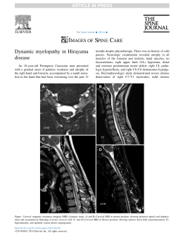
Original
article
Morphometry and spatial correlation of the foramen magnum
and spinal cord through the magnetic resonance in normal
young adults – anatomical and clinical aspects
Damiani, D.1*, Borelli, NS.2, Melo, HJF.3, Lima, RS.3 and Nobeschi, L.4
Biomedical, University of Santo Amaro - UNISA, Neuroscience Professor of Faculdade Integrada Paulista,
Internal Medical Student of City of São Paulo Medical University – UNICID, Bela Cintra Street, suite 09,
CEP 01415-000, São Paulo, Brazil
2
Internal Medical Student of City of Sao Paulo Medical University – UNICID, Bela Cintra Street, suite 09,
CEP 01415-000, São Paulo, Brazil
3
CIMAS Radiology Institute, Federal University of Sao Paulo – UNIFESP,
Rua Borges Lagoa 1341, V. Clementino, Sao Paulo - Brazil
4
Professor of Anatomy, MD and PhD of Federal University of Sao Paulo – UNIFESP, CIMAS Radiology Institute
*E-mail: [email protected]
1
Abstract
Aim: to establish the spatial relationship between the surfaces of foramen magnum (FM) and spinal cord
(SC) identifying how much of the FM surface is occupied by the SC. Material and methods: 40 normal
adults (23 females) with a mean age of 22 yrs (18-25) were evaluated through MRI to evaluate the sagittal
and transversal diameter of foramen magnum and spinal cord. The surface area of FM and spinal cord was
calculated, in the craniovertebral junction. Results: the sagittal diameter was consistently greater than the
transversal diameter in the FM with a mean value of 34.78 mm. On the other hand, the transversal diameter
was bigger in the spinal cord, with the mean value of 12.18 mm. The spinal cord occupies 82.93% of the
FM surface in the craniovertebral junction. There was no difference between sexes. Conclusions: The better
knowledge of craniovertebral junction anatomy is helpful in the neurosurgical planning and in the diagnosis
of diseases that present intracranial hypertension.
Keywords: morphometric, magnum foramen, spinal cord, MRI.
1 Introduction
The foramen magnum (FM) varies in shape in children
and adults (TESTUT, 1947; GARDNER, GRAY,
and O´RAHILLY, 1988; WILLIAMS, WARWICK,
DYSON et al., 1995; PETROIANU, 1999). Lang classified
the shapes into 5 groups, with the following composition:
semicircles; an elongated circle; egg-shaped; rhomboidal
and rounded (ZAIDI and DAVAL, 1988; LANG, 1991).
Brazilian studies classified the FM in 2 groups: egg shaped
and rounded6. Configuration and size of the FM play an
important role in the pathophysiology of various disorders of
the craniovertebral junction. The embryologic development
of the craniocervical junction is a complex process
involving the notochord as an inducer of neuroectodermal
differentiation and the paraxial mesoderm, a precursor
to bone and skeletal muscle in the craniocervical region.
For the formation of the cranial base, a combination of
endochondral ossification and the growth of regional
sutures is necessary. Elongation of the clivus and the anterior
aspect of the foramen magnum results from growth of the
spheno-occipital synchondrosis and sutural growth along the
lateral portion of the base (TUBBS, GRIESSENNAUER,
LOUKAS et al., 2010).
The neural structure located in the region of the FM are the
caudal part of the brainstem, cerebellum and fourth ventricle,
the rostral part of the spinal cord, and the lower cranial and
upper cervical nerves. The accessory nerve is the only cranial
nerve that passes through the FM. The major arteries related
J. Morphol. Sci., 2012, vol. 29, no. 2, p. 87-90
to the FM are the vertebral and posteroinferior cerebellar
arteries (PICA), and meningeal branches of the vertebral, and
external and internal carotid arteries. The venous structures
in the region of the FM are divided into three groups: one
composed of the extradural veins, another formed by the
intradural (neural) veins, and a third constituted by the
dural venous sinuses. The three groups anastomose through
and emissary veins (PETROIANU, 1999; DI DIO, 2002;
MOORE and DALLEY, 2006; RHOTON, 2000a). Other
authors proposed vertebral foramen evaluations using X-ray
methods but very different results have been observed
(MALZAC and BARROS FILHO, 2002; PETROIANU,
1999; MOORE and DALLEY, 2006; AUMÜLLER,
AUST and DOLL, 2009; CATALINA-HERRERA, 1987;
GÜNAYA and ALTINKÖKB, 2000). At this moment
there are no data about spinal cord sectional area and
FM area correlations. Fundamental knowledge of normal
anatomy and basic craniometric measurements for assessing
craniovertebral relations is important to the neurosurgeon
who operates on this anatomy (TUBBS, GRIESSENNAUER,
LOUKAS et al., 2010). The aim of neurosurgical approaches
to this FM region is designed to maximize the restoration
of CSF (cerebrospinal fluid) circulation across the FM
(FURTADO, THAKAR and HEGDE, 2011). Many
surgical approaches have been advocated for the surgery
in the FM region. The surgical approach depends on the
anatomical location of the disease and its extension. Three
87
Damiani, D., Borelli, NS., Melo, HJF. et al.
main routes to FM are anterior, lateral, and posterior. In
general, anteriorly placed lesions may be approached through
a transoral route, which is the most frequently used but is
usually suitable only for extradural lesions. However, the risk
of cerebrospinal fluid leak and infection limits the use of this
approach for most intradural disease. Lateral approaches and
their variants were developed and used to remove lesions
located anteriorly or laterally to the brainstem and FM. The
main advantages of these approaches are wide exposure
and adequate working space. Many variations have been
described for the lateral approach, including: lateral inferior
suboccipital approach, transcondylar approach and far lateral
approach. Posterior midline approach is used for most
lesions. The main advantages of this approach are low risk
of damage to vertebral artery, wide exposure, and extension
of lower exposure by laminectomy of required vertebrae
(TAGHIPOUR, MOIN, ZAMANIZADEH et al., 2004;
RHOTON, 2000a, b; KAWASHIMA, TUNRIOVER and
RHOTON, 2003) (Figure 1). The aim of our study was to
measure the sagittal and transversal diameters of FM and
SC, establishing a spatial relationship between them and
identifying how much of the FM surface is occupied by the
SC in normal adults, through MRI.
2 Material and methods
Forty adults (23 female and 17 male) of both sexes had
their MRIs evaluated and measured. The mean age was
22 years (18 to 25 years-old). Exclusion criteria included
cervical pain, scoliosis, kyphosis, vertebral canal stenosis, and
nervous system disease.
We used MRI acquisition in T1W and T2W sequences
(Siemens 1.5 Tesla) in sagittal and transversal modes.
Bright calibration was performed to make a precise measure
of FM structures. Reference points were determined for
measurements: sagittal diameter of FM was measured by
a virtual line from basal clivus (anterior part) to posterior
border (Figure 2), and for transversal diameter we used
extreme lateral points of FM (Figure 3). For spinal cord we
directly measured the nervous tissue calculating the area.
We used Osirix software to this evaluation; The examiner
evaluated the same image three times. The mean data was
a
used after these three individual values to reduce our error
factor. All data was evaluated using t Student statistical
method (P ≤ 0.05).
3 Results
No difference in the sagittal and transversal diameters of
FM and SC was seen with regard to the sex of the patient,
and all the patients were grouped, independently of the sex,
for our analyses. The FM has a greater sagittal diameter
(mean 34.78 mm) compared with the transversal diameter
(mean of 28.69 mm) (Table 1).
The transversal diameter of the SC was greater (mean of
12.18 mm) than the sagittal (mean of 10.30 mm) (Table 2).
The surface of FM is greater than the surface of the SC.
The mean surface or the FM is 95.25 mm2, and the SC,
79.00 mm2 (Table 3). The SC occupies 82.93% of the FM in
the craniovertebral junction.
4 Discussion
Our results show that the sagittal diameter of FM was
consistently bigger than the transversal diameter, which
is in accordance with studies performed in dried skulls
(HENRÍQUEZ PINO, CRICENTI, and DIDIO, 1995).
In a study of 211 dried skulls, the sagittal diameter of
FM was 36.5 mm (males) and 35.6 mm (females), while the
transversal diameter of FM was 30.6 mm (males) and 29.5 mm
(females), but no statistical significance was obtained between
the sexes (GALDAMES, PEREZ, ZAVANDO et al., 2009).
According to Günaya and Altinkökb (2000) the diameters of
FM were smaller in the females due to the accentuated oval
shape in women (GÜNAYA and ALTINKÖKB, 2000). In
our study, we did not evaluate the shape of the FM.
To our knowledge there is no such data in the medical
literature. The foramen magnum is a fundamental
component in the complex interaction between bony,
ligamentous, and muscular structures composing the
craniovertebral junction. In this complex we can find motor
myelopathy, sensory abnormalities, brainstem and lower
cranial nerve dysfunctions, and signs and symptoms due
to vascular pathology, bone disease, congenital anomalies,
metastatic and degenerative processes, neurological diseases
b
Figure 1. Foramen magnum anatomy (a) and it´s relation with brain structures and possible neurosurgical approaches (b).
88
J. Morphol. Sci., 2012, vol. 29, no. 2, p. 87-90
Morphometry and spatial correlation of the foramen magnum and spinal cord through MRI
Table 1. Foramen magnum sagittal and transversal diameters (mm).
Mean
Minimum
Maximum
SD
Sagittal diameter
34.78
28.44
39.97
2.19
Transversal diameter
28.69
21.34
34.6
2.73
SD – Standard Deviation.
Table 2. Spinal cord sagittal and transversal diameters (mm).
Mean
Minimum
Maximum
SD
Sagittal diameter
10.30
8.12
12.03
2.41
Transversal diameter
12.18
10.47
13.35
2.62
SD – Standard Deviation.
Table 3. Spinal cord and foramen magnum area (mm2).
Figure 2. Sagittal Section. MRI T1W image. A-B Line
(red line): measured diameter of FM. C-D Line (blue line):
measured diameter of spinal cord.
Mean
Minimum
Maximum
SD
FM Area (mm2)
95.25
63.49
125.41
13.86
Spinal cord area (mm2)
79.00
43.82
88.79
12.51
SD – Standard Deviation.
such as meningiomas, neurofibromas, schwannomas,
astr ocytomas, chor domas (clivus), ependymoma,
Arnold‑Chiari type I and II malformations, hydatid cysts,
Beare-Stevenson syndrome and Marchesani syndrome
have been reported (TELLA JUNIOR, PAIVA NETO,
AGUIAR et al., 2006). We believe that our data may be
helpful in the neurosurgical evaluation of craniovertebral
junction, through MRI.
5 Conclusion
Figure 3. Axial section. MRI T1W. E-F Line shows transversal
diameter of FM.
and more rarely inflammatory and infectious diseases
(TELLA JUNIOR, PAIVA NETO, AGUIAR et al.,
2006; TAGHIPOUR, MOIN, ZAMANIZADEH et al.,
2006). The clinical manifestations of FM stenosis include
respiratory failure, vomiting, impotence, cranial nerves
palsy, cerebellar symptoms/ataxia, central cord syndrome,
bulbar symptoms, headache, sensitive and or motor
neurological deficits (OJEMANN, 1992; NANDA,
VINCENT, VANNENREDY et al., 2002; BOULTON
and CUSIMANO, 2003; TUBBS, GRIESSENNAUER,
LOUKAS et al., 2010). Some diseases involving this area
J. Morphol. Sci., 2012, vol. 29, no. 2, p. 87-90
A fundamental knowledge of normal FM neuroanatomy
and its structural correlations are very important for
c l i n i c i a n s ( S I VA R A M A K R I S H N A N , A L P E R I N ,
SURAPANENI et al., 2004). Our results found a transversal
and sagittal FM and spinal cord diameters by MRI and
their respective areas. The spatial correlation between spinal
cord and FM in the craniovertebral junction were analyzed
showing that spinal cord occupied 82.93% of FM total
area without statistical difference between sexes. MRI is an
important diagnostic method to craniovertebral diseases.
Rerefences
AUMÜLLER, G., AUST, G. and DOLL, A. Anatomia. Rio de
Janeiro: Guanabara Koogan, 2009.
BOULTON, MR. and CUSIMANO, MD. Foramen magnum
meningiomas: concepts, classifications and nuances. Neurosurgical
Focus, 2003, vol. 14, n. 6, p. 1-8. http://dx.doi.org/10.3171/
foc.2003.14.6.10
CATALINA-HERRERA, CJ. Study of the anatomic metric values of
the foramen magnum and its relation to sex. Acta Anatomica, 1987,
vol. 130, n. 4, p. 344-7. http://dx.doi.org/10.1159/000146468
89
Damiani, D., Borelli, NS., Melo, HJF. et al.
DI DIO, JAL. Tratado de Anatomia Sistêmica Aplicada. São Paulo:
Atheneu, 2002.
OJEMANN, RG. Foramen magnum meningioma. In TATTER, SB.
Clinical neurosurgery. New York: Saunders, 1992.
FURTADO, SV., THAKAR, S. and HEGDE, AS. Correlation of
Functional Outcome and Natural History with Clinicoradiological
Factors in Surgically Managed Pediatric Chiari I Malformation.
Neurosurgery, 2011, vol. 68, p. 319-328. PMid:21135728. http://
dx.doi.org/10.1227/NEU.0b013e31820206e5
PETROIANU, A. Anatomia Cirúrgica. Rio de Janeiro: Guanabara
Koogan, 1999.
GALDAMES, S., PEREZ, PR., ZAVANDO, DA. and SMITH,
RL. Dimorfismo sexual em lãs dimensiones Del forâmen magno.
International Journal of Morphology, 2009, vol. 27, n. 1, p. 21-3.
RHOTON, AL. The Foramen Magnum. Neurosurgery, 2000,
vol. 47, n. 3, p. S155-S194. PMid:10983308.
GARDNER, E., GRAY, DJ. and O´RAHILLY, R. Anatomia:
estudo regional do corpo humano. 4. ed. Rio de Janeiro: Guanabara
Koogan, 1988.
GÜNAYA, Y. and ALTINKÖKB, M. The value of the size of
foramen magnum in sex determination. Journal of Clinical Forensic
Medicine, 2000, vol. 7, n. 3, p. 147-9. PMid:16083665. http://
dx.doi.org/10.1054/jcfm.2000.0430
HENRÍQUEZ PINO, J., CRICENTI, SV. and DIDIO, LJA.
Morphometry of the foramen magnum and its relantioship to skull
types on brazillian individuals. Revista Chilena de Anatomía, 1995,
vol. 13, n. 2, p. 159-64.
KAWASHIMA, M., TUNRIOVER, N. and RHOTON, AL.
Comparasion of the far lateral and extreme lateral variants of the
atlanto-occipital transarticular approach to anterior-extradural
lesions of the craniovertebral junction. Neurosurgery, 2003, vol. 53,
p. 662-675. PMid:12943582. http://dx.doi.org/10.1227/01.
NEU.0000080070.16099.BB
LANG, J. Clinical Anatomy of the posterior cranial fossa and its
foramina. Stuttgart: Thieme, 1991.
MALZAC, A. and BARROS FILHO, TEP. Morfometria do canal
vertebral no segmento cervical em militares jovens assintomáticos.
Acta Ortopédica Brasileira, 2002, vol. 10, n. 4, p. 40-51. http://
dx.doi.org/10.1590/S1413-78522002000400006
MOORE, K. and DALLEY, A. Clinically Oriented Anatomy. 5th ed.
Toronto: Lippincott Williams & Wilkins, 2006.
NANDA, A., VINCENT, DA., VANNENREDY, PSS. and
CHANDA, A. Far-lateral approach to intradural lesions of the
foramen magnum without resection of occipital condyle. Journal
of Neurosurgery, 2002, vol. 98, p. 302-9. PMid:11841072. http://
dx.doi.org/10.3171/jns.2002.96.2.0302
90
RHOTON, AL. The Far-Lateral Approach and Its Transcondylar,
Supracondylar, and Paracondylar Extensions. Neurosurgery, 2000,
vol. 47, n. 3, p. S195-S209.
SIVARAMAKRISHNAN, A., ALPERIN, N., SURAPANENI,
S. and LICHTOR, T. Evaluating the Effect of Decompression
Surgery on Cerebrospinal Fluid Flow and Intracranial
Compliance in Patients with Chiari Malformation with Magnetic
Resonance Imaging Flow Studies. Neurosurgery, 2004, vol. 55,
p. 1344‑1351. PMid:15574215. http://dx.doi.org/10.1227/01.
NEU.0000143612.60114.2D
TAGHIPOUR, M., MOIN, H., ZAMANIZADEH, B.,
KAMKARPOUR, A. and ESMAEELI, M. Foramen Magnum
Surgery: Experience With 22 Cases. Neurosurgery Quarterly, 2006,
vol. 16, p. 96-99. http://dx.doi.org/10.1097/00013414‑20060
6000‑00010
TELLA JUNIOR, OI., PAIVA NETO, MAP., AGUIAR, PH. and
HERCULANO, MA. Meningiomas anteriores e antero-laterais do
forame magno. Arquivos de Neuro-Psiquiatria, 2006, vol. 64, n. 2b.
http://dx.doi.org/10.1590/S0004-282X2006000300016
TESTUT, L. Tratado de Anatomia Humana.
Salvat, 1947. PMid:20087140.
Barcelona:
TUBBS, RS., GRIESSENNAUER, CJ., LOUKAS, M., SHOJA,
MM. and COHEN-GADOL, AA. Morphometric Analysis of the
Foramen Magnum: An Anatomic Study. Neurosurgery, 2010,
vol. 66, p. 385-388.
WILLIAMS, P., WARWICK, R., DYSON, M. and BANNISTER,
LH. Gray Anatomia. 37th ed. Rio de Janeiro: Guanabara
Koogan, 1995. PMid:3223599.
ZAIDI, SH. and DAVAL, SS. Variations in the shape of foramen
magnum in Indian skulls. Anatomischer Anzeiger, 1988, vol. 167,
n. 4, p. 338-40.
Received October 28, 2011
Accepted May 3, 2012
J. Morphol. Sci., 2012, vol. 29, no. 2, p. 87-90
Download