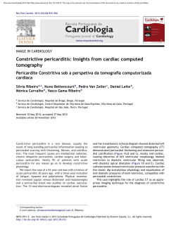
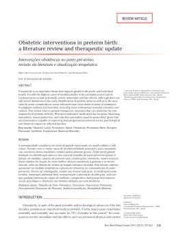
CASE REPORT
Braz J Cardiovasc Surg 2005; 20(2): 182-185
Surgical repair of left ventricular rupture after the
use of tenecteplase in the treatment of acute
myocardium infarction
Reparo de rotura de ventrículo esquerdo após uso de tenecteplase no tratamento do infarto agudo do
miocárdio
Marco Antonio Vieira GUEDES, Carlos Edson Campos CUNHA FILHO, Alexandre Ciappina HUEB, Sérgio
Almeida de OLIVEIRA
RBCCV 44205-750
Abstract
The use of thrombolytic agents in the treatment of acute
myocardium infarction represented an important impact in
the reduction of overall mortality. But this did not alter the
cardiac rupture rate. This complication must be recognized
and treated quickly. The authors report on a case of surgical
repair of an early left ventricle rupture, after the use of
tenecteplase in association with non-fractioned heparin for
the treatment of acute myocardium infarction.
Resumo
A utilização do trombolítico no tratamento do infarto
agudo do miocárdio apresentou um impacto importante na
diminuição da mortalidade geral, porém não alterou a
incidência da rotura cardíaca. Esta complicação deve ser
reconhecida e tratada rapidamente, necessitando um alto nível
de suspeição clínica. Os autores relatam um caso de reparo
cirúrgico de rotura precoce de ventrículo esquerdo, após
utilização de tenecteplase associado à heparina não
fracionada, no tratamento do infarto agudo do miocárdio.
Descriptors: Myocardial infarction. Thrombolytic therapy,
adverse effects. Heart rupture, post infarction.
Descritores: Infarto do miocárdio. Terapia trombolítica,
efeitos adversos. Ruptura cardíaca pós-infarto.
Work developed in the Instituto do Coração do Hospital das Clínicas,
Medical School of the University of São Paulo, São Paulo – SP.
Correspondence address:
Marco Antonio Vieira Guedes. Instituto do Coração do Hospital das
Clínicas da Faculdade de Medicina da Universidade de São Paulo.
Divisão de Cirurgia. Av. Dr. Enéas de Carvalho Aguiar, no 44. Cerqueira
César. São Paulo, SP. CEP 05403-000 Tel: (11) 3069-5000.
E-mail: [email protected]
182
Article received in February, 2005
Article accepted in April, 2005
GUEDES, MAV ET AL - Surgical repair of left ventricular rupture
after the use of tenecteplase in the treatment of acute myocardium
infarction
INTRODUCTION
Rupture of the left ventricular free wall or ventricular
septum is an important complication of acute myocardial
infarction (AMI), occurring in from 1% to 3% of all patients
who suffer from this problem.
Rupture is more frequently diagnosed after death than
in live patients. Nearly 20% of patients who die due to AMI
present with heart rupture making this the third highest
cause of death after cardiogenic shock and ventricular
arrhythmias [1].
In respect to the anatomoclinical findings of autopsies,
this complication is observed more frequently in over 60year-old women, with evidence of single arterial disease,
victims of their first infarction, suffering from hypertension
without ventricular hypertrophy and normally submitted to
thrombolytic therapy. The use of thrombolytic therapy in
the treatment of AMI, in spite of reducing the overall
mortality, seems to accelerate the evolution to heart rupture,
without necessarily increasing the incidence of this
complication. Reperfusion phenomena seem to favor early
heart rupture due to the transformation of ischemic infarction
to hemorrhagic infarction [2].
Rupture can occur some hours after of the beginning of
symptoms. Precocious and effective thrombolysis decreases
the risk of heart rupture, maybe by limiting the extent of
myocardial necrosis, thereby avoiding transmural infarction.
But, the impact of late thrombolytic therapy in heart rupture
remains unknown [2,3].
The aim of this work is to report on a case of surgical
repair of precocious left ventricle rupture, after the use of
tenecteplase associated with non-fractionated heparin, for
the treatment of AMI.
CASE REPORT
In May 2004 a 62-year-old female diabetics taking oral
hypoglycemiant and suffering from hypertension and
dyslipidemia presented with an episode of a moderate
burning precordial pain during resting. After three days
the patient consulted a physician and was diagnosed as
having AMI with positive deflection of the ST segment
in the anterior wall, evolving to Killip I. The
electrocardiogram (ECG) revealed: an inactive area in the
anterior and inferior wall, associated with the positive 2mm deflection of the ST segment in the anterior wall. A
transthoracic echocardiogram (ECO) revealed: ejection
fraction (EF) of 35% with important involvement of the
left ventricle systolic function due to an apical aneurysm
and basal paralysis of the inferior wall. The patient was
submitted to a heart catheterism (CAT) which showed
three-arterial coronary obstruction: the right coronary
Braz J Cardiovasc Surg 2005; 20(2): 182-185
artery occluded in the middle l/3, the anterior interventricular
artery occluded in the middle l/3, the circumflex artery (CxA)
with a lesion of 90% in the middle 1/3, the left marginal
arteries with a critical lesion in the proximal 1/3, grade III
collateral circulation to the right coronary artery, as well as
the inferior apical aneurysm. The patient was submitted to
the classical treatment for AMI without primary reperfusion
therapy, receiving hospital discharge on the 8th day after
hospital admission when an elective coronary artery bypass
grafting surgery was scheduled.
After fifteen days the patient presented with a new
episode of a strong precordial pain during rest associated
with dyspnea arriving at the Emergency Department of the
Heart Institute of São Paulo 40 minutes after the start of the
symptoms. At admission, the ECG revealed a positive 6-mm
deflection of the ST segment in the anterior wall associated
with an inactive area in the anterior and inferior wall and
another myocardial infarction in the anterior wall was
diagnosed with a positive deflection of ST segment. The
patient was submitted to thrombolytic therapy l hour 40
minutes after the start of the symptoms, using 40 mg
tenecteplase (TNK-tPA) via endovenous and 4500 units nonfractionated heparin via endovenous. After thrombolysis,
the patient was submitted to another CAT which did not
demonstrate any changes when compared to the first
coronary pattern.
After 4 hours of thrombosis, the patient evolved with
hemodynamic instability, pulmonary congestion, reduced
mental awareness, presenting with clinical signs of
cardiogenic shock and requiring vasoactive drugs. There
was no hemodynamic improvement and the therapeutic
option was the installation of an intra-aortic balloon
counterpulsation and emergency coronary artery bypass
grafting surgery was indicated.
In the trans-operative period, after opening the
pericardium, the presence of significant hemopericardium
was observed with adherence to the left ventricular anterior
wall of the pericardium. Without touching the region, a
cardiopulmonary bypass was established and only after
aortic clamping, removal of the adherence from the anterior
and apical region was initiated.
After removal, the presence of an anterior wall aneurysm
with left ventricular rupture and pseudo-aneurysm formation
was observed. Figure 1 demonstrates the intraoperative
aspect of the lesion. After resection of the pseudo-aneurysm
and repair of the edges of the aneurysm, endoaneurysm
repair was initiated utilizing a bovine pericardial patch with
reinforcement of the ventricular raffia using two strips of
Teflon (Figure 2). Myocardial revascularization was
performed, utilizing anastomosed saphenous vein graft for
the left marginal arteries and to the posterior interventricular
right coronary artery.
183
GUEDES, MAV ET AL - Surgical repair of left ventricular rupture
after the use of tenecteplase in the treatment of acute myocardium
infarction
Braz J Cardiovasc Surg 2005; 20(2): 182-185
COMMENTS
Fig. 1 - Aneurysm in anterior wall of the torn left ventricle
Fig. 2 - Intraoperative aspect of the repair of the left ventricular
rupture using the D’or technique. After the left ventricle remodeling
with a bovine pericardial patch, the ventricle is closed using
individual sutures anchored with two Teflon strips
The removal of the cardiopulmonary bypass was helped
by inotropic drugs, an intra-aortic balloon was uneventfully
inserted and the patient was moved to the surgical ICU. The
patient evolved with progressive hemodynamic
improvement, with removal of the intra-aortic balloon
performed on the 4th postoperative day. She was released
from the ICU on the 6th postoperative day with a CK-MB
mass peak at 64.7 ng/dL.
The patient received hospital discharge on the 17th
postoperative day taking acetylsalicylic acid 200 mg/day,
enalapryl 200 mg/day, carvedilol l00 mg/day, digoxin 0.25 mg/
day, sinvastatin 20 mg/day and glybenclamid 2.5 mg/day. An
ECHO at hospital discharge showed the Ejection Fraction
(EF) at 39% with apical akinesia and an aneurysm of the basal
region of the inferior septum and of the inferior-posterior wall.
184
Heart rupture after AMI occurs when the transmural
pressure is greater than the tensile force of the heart
conjunctive tissue. This occurs, generally, at the weakest site
of the ventricular wall, located in the non-complacent zone of
the infarcted area. The use of thrombolytic therapy in the
treatment of AMI has an important impact in reducing the
overall mortality, but the rupture rate remains unaltered [4].
Successful thrombolysis performed up to 6 hours from
the start of the symptoms, reduces the risk of rupture, maybe
by limiting the extent of the myocardial necrosis, reducing
the probability of transmural infarction occurring. After this
period, the impact of thrombolytic therapy remains uncertain.
The LATE (Late Assessment of Thrombolytic Efficacy)
Study was not able to conclusively demonstrate that late
use of thrombolysis increases the rupture rate, despite the
fact that rupture occurs earlier [2,4]. The reperfusion
phenomenon seems to favor early heart rupture due to the
transformation of the ischemic infarction into hemorrhagic
infarction [4].
Tenecteplase (TNK-tPA) is a variant of the native tissue
plasminogen activator (tPA), produced by means of
recombinant DNA technology. The substitution of some
amino acids grants a half life to the TNK-tPA greater than
alteplase (18 min and 4 min, respectively) greater specificity
to fibrin (14 times) and more resistance to PAI-I (80 times)
and can be utilized as a unique bolus dose. The TNK-tPA
presents the same efficacy as alteplase in a 90-minute
administration regimen, without increasing the rate of
intracranial bleeding, but reducing non-neurological
complications and the use of blood derivatives. Ventricular
rupture after the utilization of this thrombolytic therapy is a
rare complication, occurring in only 3.2% of the 6095 patients
involved in the ASSENT-3 study. This study evaluated the
efficacy and safety of the tenecteplase in association with
enoxaparin, abciximab or non-fractioned heparin [5].
The principles of the surgical repair are: to interrupt
bleeding, to anchor the repair in viable tissues and to
maintain the ventricular geometry. Several techniques can
be utilized to this end. Early recognition of the complication
and rapid intervention are important for therapeutic success.
The use of the intra-aortic balloon reduces the transmural
tension and can be an important adjunct therapy in the
control of these patients [4,6].
Heart rupture after utilization of the thrombolytic therapy
in acute myocardial infarction treatment is a severe
complication, which must be recognized and rapidly treated.
However, a high level of suspicion is necessary to identify
patients who may evolve with hemodynamic instability after
the use of thrombolytic therapy.
GUEDES, MAV ET AL - Surgical repair of left ventricular rupture
after the use of tenecteplase in the treatment of acute myocardium
infarction
BIBLIOGRAPHIC REFERENCES
1. Reddy SG, Roberts WC. Frequency of rupture of the left
ventricular free wall or ventricular septum among necropsy cases
of fatal acute myocardial myocardial infarction since introduction
of coronary care units. Am J Cardiol. 1989;63(13):906-11.
2. Becker RC, Charlesworth A, Wilcox RG, Hampton J, Skene A,
Gore JM et al. Cardiac rupture associated with thrombolytic therapy:
impact of time to treatment in the Late Assessment of Thrombolytic
Efficacy (LATE) study. J Am Coll Cardiol. 1995;25(5):1063-8.
3. Honan MB, Harrell Jr. FE, Reimer KA, Califf RM, Mark DB,
Pryor DB et al. Cardiac rupture, mortality and timing of
thrombolytic therapy: a meta-analysis. J Am Coll Cardiol.
1990;16(2):359-67.
Braz J Cardiovasc Surg 2005; 20(2): 182-185
4. Park WM, Connery CP, Hochman JS, Tilson MD,
Anagnostopoulos CE. Successful repair of myocardial free
wall rupture after thrombolytic therapy for acute infarction.
Ann Thorac Surg. 2000;70(4):1345-9.
5. Assessment of the Safety and Efficacy of a New
Thrombolytic Regimen (ASSENT-3) Investigators. Efficacy
and safety of tenecteplase in combination with enoxaparin,
abciximab, or unfractionated heparin: the ASSENT-3
randomised trial in acute myocardial infarction. Lancet.
2001;358(9282):605-13.
6. Kirklin JW, Barratt-Boyes BG. Aortic valve disease. In: Kirklin
JW, Barratt-Boyes BG, editors. Cardiac surgery. New
York:Wiley;2003. p.437-55.
185
Download