Journal of Photochemistry and Photobiology B: Biology 94 (2009) 25–31
Contents lists available at ScienceDirect
Journal of Photochemistry and Photobiology B: Biology
journal homepage: www.elsevier.com/locate/jphotobiol
Low-intensity red laser on the prevention and treatment of induced-oral
mucositis in hamsters
Cynthia M. França a, Cristiane M. França b, Silvia C. Núñez c,d, Renato A. Prates c, Elisângela Noborikawa b,
Miriam R. Faria b, Martha S. Ribeiro a,c,*
a
Professional Master Lasers in Dentistry, IPEN-CNEN/SP, Avenida Lineu Prestes, 2242, 05508-000 São Paulo, Brazil
Biodentistry Post-Graduation Program, Ibirapuera University, São Paulo, Brazil
c
Center for Lasers and Applications, IPEN-CNEN/SP, Avenida Lineu Prestes, 2242, São Paulo 05508-000, Brazil
d
Institute for Health Research – INPES/CETAO, São Paulo, Brazil
b
a r t i c l e
i n f o
Article history:
Received 28 March 2008
Received in revised form 28 July 2008
Accepted 24 September 2008
Available online 30 September 2008
Keywords:
Cancer
Chemotherapy
Cryotherapy
Radiotherapy
Ulcer
a b s t r a c t
Objective: The purpose of this study was to investigate the effects of laser phototherapy as preventive and
therapeutic regime on induced-oral mucositis in hamsters.
Design: The animals were divided into four groups: preventive cryotherapy, preventive laser, therapeutic
laser and therapeutic control group. Mucositis was induced in hamsters by intraperitoneal injection of 5fluorouracil (5-FU) and superficial scratching. All preventive treatment was performed on the right cheek
pouch mucosa. The left pouch mucosa was used for a spontaneous development of mucositis and did not
receive any preventive therapy. Laser parameters were: k = 660 nm, P = 30 mW, D = 1.2 J/cm2, Dt = 40 s,
spot size 3 mm2, I = 1 W/cm2. Cryotherapy was done positioning ice packs in the hamster mucosa
5 min before 5-FU infusion and 10 min afterward. To study the healing of mucositis, the left pouch
mucosa of each of the hamsters in the TLG received laser irradiation on the injured area. Irradiation
parameters were kept the same as abovementioned. The control hamsters in the TCG did not receive
any treatment. The mucositis degree and the animal’s body mass were evaluated. An assessment of blood
vessels was made based on immunohistochemical staining.
Results: The CG animals lost 15.16% of theirs initial body mass while the LG animals lost 8.97% during the
first 5 days. The laser treated animals had a better clinical outcome with a faster healing, and more granulation tissue. The quantity of blood vessels at both LG and CG were higher than in healthy mucosa.
Regarding the therapeutic analysis, the severity of the mucositis in the TLG was always lower than
TCG. TLG presented higher organization of the granulation tissue, parallel collagen fibrils, and increased
angiogenesis.
Conclusion: The results suggest that laser phototherapy had a positive effect in reducing mucositis severity, and a more pronounced effect in treating established mucositis.
Ó 2008 Elsevier B.V. All rights reserved.
1. Introduction
Mucositis is a significant clinical problem in patients receiving
cancer therapy. The manifestations may range from limited
patches of mildly sore erythematous mucosa to frank ulceration
and hemorrhage [1,2]. Reports from literature confirm the high
incidence of mucositis in patients receiving radiotherapy (RT), or
chemo-radiation therapy. In patients receiving standard chemotherapy, 5–15% develop mucositis. When the treatment protocol
involves 5-fluorouracil administration (5-FU), with or without leucovorin, this number rises up to 40% [3,4]. Approximately 75–85%
* Corresponding author. Address: Center for Lasers and Applications, IPEN-CNEN/
SP, Avenida Lineu Prestes, 2242, São Paulo, São Paulo 05508-000, Brazil. Tel.: +55 11
3133 9197; fax: +55 11 3133 9374.
E-mail address: [email protected] (M.S. Ribeiro).
1011-1344/$ - see front matter Ó 2008 Elsevier B.V. All rights reserved.
doi:10.1016/j.jphotobiol.2008.09.006
of the patients under conditioning regimen prior to bone marrow
transplantation present some degree of mucositis [5]. This treatment-induced complication causes oral discomfort and pain, and
can also lead to other complications such as poor nutrition due
to pain, delays in drug administration and increased medical costs.
It also may be a life-threatening condition due to infection (septicemia) [6].
Few interventions are of proven efficacy in reducing the severity
or duration of mucositis, and there are no universally accepted
treatment protocols [7]. Many agents and strategies have been
used, such as basic oral care, oral rinses, analgesics, antibiotics,
cryotherapy, local anesthetics, growth factors and cytokines, biologic mucosal protectants, and anti-inflammatory agents among
others. To date, there is not a single treatment capable of preventing or treating mucositis in an efficient way [5–8].
26
C.M. França et al. / Journal of Photochemistry and Photobiology B: Biology 94 (2009) 25–31
Some studies have reported beneficial effects of low-intensity
laser therapy (LILT) to promote tissue repair and reduce pain and
inflammation [9–11]. This therapy has been applied in clinical
practice to prevent and treat oral mucositis with some good results
been reported [12–15]. Recently, a clinical trial investigated the effects of a red laser on the prevention and reduction of the severity
of conditioning-induced-oral mucositis for hematopoietic stem cell
transplantation patients [16]. Those results indicated that pretreatment with laser irradiation was effective in reducing the incidence
of oral mucositis, although the mechanisms underlying laser effects are still poorly understood [17]. The present study aimed to
evaluate clinically and histologically the effects of LILT as a prophylactic tool as well as a treatment agent of 5-fluoracil induced-oral
mucositis in hamsters. In the prophylactic study, the effects of LILT
were compared with ice chips, a currently acceptable prophylactic
agent for oral mucositis [6].
2. Methods
Thirty female Golden Syrian hamsters with a mean of 150 g of
body mass, and 8 weeks old were used. All animals were acclimated to laboratory conditions for at least 5 days before being
dosed with 5-FU. During the acclimatizing period, the general
health condition of the hamsters was evaluated, and they were
fed with a standard laboratory diet and water ad libitum. During
the experimental period, all animals received humane care in compliance with the Ethical Principles of Animal Experimentation formulated by the Brazilian College for Animal Experimentation, and
in accordance with guidelines approved by the Council of the
American Psychological Society for the use of animal in
experiments.
2.1. Mucositis induction protocol
Before the beginning of the experiment, two animals were sacrificed in a CO2 chamber and excisional biopsies of the cheeks
pouch mucosa were performed and the specimens were routinely
processed for histology to serve as a normal mucosa control. The
rest of the animals were anesthetized with CopazineÒ (Xylazine)
0.075 mL/g and VetanarcolÒ (Ketamine) 0.3 mL/g, and the buccal
pouches were everted and photographed. Each animal received
an intraperitoneal injection of 100 mg/kg of 5-FU on day 1 and
65 mg/kg of 5-FU on day 3. The right and left cheek pouch mucosa
were irritated by superficial scratching with the tip of an 18-gauge
needle at days 4 and 5. The needle was dragged twice in a linear
movement across the everted cheek pouch once a day until erythematous changes were noted. This technique has been repeatedly used to produce ulcerative mucositis, which mimics the
human development of mucositis [18,19].
2.1.1. First phase: preventive analysis
Twenty eight hamsters, corresponding to n = 28 right cheek
pouch mucosas, were divided into two equally sized groups: (LG)
laser group (n = 14) and (CG) cryotherapy group (n = 14). All preventive treatment was performed only on the right cheek pouch
mucosa. The left side was kept untreated and unanalyzed, but with
the same mucositis induction protocol. Before any procedure, the
animals were anesthetized as previously described.
On the CG group, at days 1 and 3 (chemotherapy infusion days),
14 animals were submitted to applications of ice chips on the right
cheek pouch mucosa for 5 min before 5-FU injection and 10 min
afterward.
On LG group, 14 animals were irradiated daily from day 1 to day
4, before the scratching after 5-FU injection. The right cheek pouch
mucosa was irradiated with a GaAlAs laser (Kondortech, São Carlos,
SP, Brazil) emitting red light (k = 660 nm). The output power was
30 mW at a spot size of 2 mm in diameter corresponding to an irradiance of 1 W/cm2 per point. The tip of the laser device carefully
touched the mucosa for a total of Dt = 40 s per day scanning the total area of 1 cm2 in four different points with 10 S per point. The
total energy density (dose) was 1.2 J/cm2.
The clinical aspect of the right cheek pouch mucosa was observed by two blind-independent calibrated observers and the degree of mucositis was evaluated through oral mucositis assessment
scale (OMAS) modified for hamsters according Wilder-Smith et al.
[20] as follows: 0—no ulceration and no erythema; 1—ulceration <
4 mm2 and slight erythema; 2—ulceration of 4–9 mm2 and moderate erythema; 3—ulceration > 9 mm2 and severe erythema; 4—
appearance of pseudomembrane. The sores gave by the observers
were evaluated by Kappa’s test for inter-rater reliability.
The preventive treatment with cryotherapy and laser was performed until day 5, which corresponded to the onset of the mucositis. On day 5, the preventive protocol for CG and LG was ended,
but the clinical analysis followed through the 11 days, until the
end of the experimental procedure, without any further treatment.
The animals were euthanized on day 3 (n = 6), day 6 (n = 6), day 9
(n = 6), and day 11 (n = 10), and the right cheek pouch was collected for histological purposes.
The mucositis scores obtained during the 11 days were used to
plot a curve and areas under the curves were calculated using the
integrate function in Microcal Origin 7.0 software program (Northampton, MA) to provide an overview of the mucositis development
after preventive protocol. Statistical analysis was performed using
two-tailed t-test. The value of p < 0.05 was considered significant.
The data are presented as mean ± SEM.
The animal’s body mass was also evaluated during the first 5
days. A weight loss was expected due to discomfort and pain
[21]. The animals were weighed daily. The body mass was recorded
and statistical analysis was carried out using one-tailed t-test to
verify the intra-group performance as well as to compare the
weight loss values between groups. The data are presented as
mean ± SEM.
Histological analysis was performed on specimens from the
right cheek pouch mucosa to verify the influence of both treatments on the course of mucositis. After sacrifice, the areas of the
mucosa containing the wounds were collected and fixed by immersion in formaldehyde at 4 °C for 24 h. The specimens were dehydrated and thereafter embedded in Paraplast (Oxford, USA) to
provide transversal sections of the tissue. Five-micron sections
were stained with hematoxylin/eosin (HE). Stained sections were
observed and photographed with an optical microscope (Leica
DMLP, Germany).
For a quantitative assessment, blood vessels were counted after
immunohistochemical staining according to Dako EnVision
protocol.
The positive blood vessels localized at the ulcer area were
counted in five selected square areas measuring 10,000 lm2 each
using the Image J software and the total number of counted blood
vessels was divided by the number of squares used. At least three
slides from each animal were observed. Healthy mucosa was also
evaluated.
Statistical analysis of the number of blood vessels was performed. Values are given as means and bars are standard error bars
(SEM). Statistical comparisons between groups were carried out
with two-tailed t-test, which retains the overall significance level
at 5% (p < 0.05).
2.1.2. Second phase: therapeutic study
On day 5, the remaining animals (n = 22, since 6 were euthanized on day 3) were randomly regrouped; thus 22 animals were
alienated into two new groups with 11 animals per group and only
C.M. França et al. / Journal of Photochemistry and Photobiology B: Biology 94 (2009) 25–31
the left side was now manipulated to study the therapeutic effects
of the laser irradiation over the established mucositis.
The animals were divided into the groups TCG (no treatment
control group) and TLG (therapeutic laser group). Eleven left cheek
pouches mucosa from 11 animals on TCG did not receive any treatment and were used as a spontaneous healing control. Eleven animals from TLG received laser irradiation by contact of the laser tip
scanning the injured area at days 6–10. The laser parameters were
kept the same as mentioned above (k = 660 nm, output power
30 mW, total energy density 1.2 J/cm2, exposure time of 40 s [four
points for 10 S], spot size 3 mm2). During the therapeutic investigation, the left mucosa clinical aspect was evaluated and scored
as aforementioned. Areas under the curves were computed.
For both groups, three animals per day were killed on days 6
and 9. The remaining five animals of each group were euthanized
on day 11. Mucosa biopsies were carefully collected to include
the adjacent healthy mucosa and all the healed tissue in depth.
All biopsies were fixed in formalin 10% for 24 h. Thereafter, biopsies were included in paraffin and 5 lm thickness transversal sections were obtained. The specimens were stained with HE using
the same procedures described above as well as the immunohistochemical staining for the quantitative assessment of blood vessels.
Statistical analysis for the therapeutic study was achieved in the
same way as described in the preventive study.
27
with day 1, the mean weight in all the other moments where different; therefore a significant loss of body mass was detected from
day 1 and on. The percentage of body mass lost was 1.91% on day 1,
5.72% on day 2, 9.10% on day 3, 12.73% on day 4 and 15.16% on day
5.
As regards the weight loss on LG, a significant difference was
observed between day 0 and day 2 (p = 0.025). Comparing the
mean values of day 1 with day 2, a non-significant difference
(p = 0.37) was found. Comparing the mean weight on day 2 with
day 3, day 3 with day 4 and day 4 with day 5, no statistically significant differences were perceived. However, comparing with day
1, only following day 4 a significant difference was detected
(p = 0.047). Therefore a significant loss of body mass was detected
from day 4 and day 5. The percentage of body mass lost was 0.96%
on day 1, 1.91% on day 2, 3.04% on day 3, 4% on day 4 and 8.97% on
day 5.
3.1.2. Mucositis degree
The Kappa’s test score (0.69) between observers showed substantial agreement. Fig. 2A displays the mucositis course during
experimental period. It is possible to note that the obtained scores
3. Results
3.1. Preventive analysis
3.1.1. Body mass
The body mass analysis revealed weight loss for both preventive
groups (CG and LG) during the first five days of treatment (Fig. 1).
The weight loss of CG was significantly higher than in LG from day
2 to 5.
Regarding the weight loss on CG, a significant difference was
found comparing day 1 with the original mean weight on day 0
(p = 0.025). Comparing the mean values of day 1 with day 2, a significant difference (p = 0.038) was also noted. Comparing the mean
weight on day 2 with day 3, day 3 with day 4, and day 4 with day 5,
significant differences were not observed. However, comparing
Fig. 1. Hamster body mass in cryotherapy (CG) and laser groups (LG) during the
first five days of the experiment (preventive period). Note that before irradiation
(day 0) and on day 1 the groups presented similar body mass. From day 2 until day
5, the groups were considered statistically different, with a higher mean body mass
on LG. Data points are means and SEM. Significance was determined according to
one-tailed independent t-test.
Fig. 2. Mucositis development after preventive protocol during experimental
period. (A) Differences in the degree of induced-oral mucositis in hamsters between
CG and LG. Observe that on the 4th day, the degree of oral mucositis was
significantly lower for LG compared to CG. (B) Mean area under the curves obtained
for LG and CG. Note that laser phototherapy significantly diminishes the mucositis
severity. Data are means and SEM. Significance was determined according to twotailed independent t-test.
28
C.M. França et al. / Journal of Photochemistry and Photobiology B: Biology 94 (2009) 25–31
3.1.3. Histological analysis
Fig. 3A displays a healthy hamster’s mucosa showing normal
epithelium and connective tissue with absence of inflammatory
infiltrate. On the 3rd day, both groups presented mild epithelial
alteration, characterized by congested blood vessels and by a
greater amount of keratin. From day 6 until day 9 the differences
between the groups became more expressive. At day 6, both
groups showed epithelial atrophy, ulceration and mild to moderate
unspecific chronic inflammatory infiltrate in the connective tissue.
However, laser group presented areas with less inflammatory infiltrate; moreover, the LG presented a more pronounced granulation
tissue and the general characteristics indicated a more advanced
healing process. At day 9, both groups presented samples with ulcers and chronic infiltrate; all specimens had developed the granulation tissue. Once more, the LG specimens presented more
organized collagen fibers characterized by their level of parallelism; it was also noted an expressive angiogenesis (compare
Fig. 3B and C). At day 11, there was a complete re-epithelization
in the groups, and a scanty presence of lymphocytes and some
granulation tissue were also observed. LG showed abundant angiogenesis and mature fibrous connective tissue.
Fig. 4 displays the mean values and SEM of blood vessels during
preventive treatment (right mucosa). Healthy mucosa presented
an average of 3.75 vessels/area. Regarding comparison with the
healthy mucosa, for CG the mean of blood vessels was higher during all the course of mucositis, however, statistically significant differences were observed on days 3, 9 and 11 (p < 0.05). LG did not
show a significant difference when matched up with the healthy
samples on the 3rd and 6th days, but statistically significant differences were observed on days 9 and 11. In both groups, the number
of blood vessels increased on day 9 but there was no significant difference between CG and LG at this moment. On day 11, the number
of blood vessels continued to increase in the CG group while the LG
levels remained the same as day 9. At this moment, the number of
blood vessels for LG was significantly lower than CG (p = 0.029).
3.2. Therapeutic analysis–left cheek pouch mucosa
3.2.1. Mucositis degree
Kappa’s statistical measure of inter-rater reliability demonstrated a moderate agreement (0.55) between observers. The re-
Fig. 3. Photomicrograph from hamster right mucosa. (A) Healthy mucosa with
normal epithelium and no inflammatory infiltrate (original magnification—40).
(B) CG at day 9 showing areas of intense inflammatory infiltrate and granulation
tissue (original magnification—63). (C) LG at day 9 showing intense inflammatory
infiltrate (original magnification—63). The insert shows granulation tissue with
expressive angiogenesis (original magnification—100). HE.
were higher in the CG during all experimental period. The maximum degree obtained from cryotherapy group was 3.1 at day 8.
On the group that received preventive laser treatment, the highest
degree of mucositis was of 2.6 at day 7. At day 4, a statistically significant difference was observed between groups (p = 0.0268). LG
presented a mucositis degree significantly lower that CG.
To show the outcome of preventive therapy on the course of
mucositis, the areas under the curves were calculated (Fig. 2B). It
is possible to observe that LG shows an area under the curve significantly smaller than CG (p = 0.047). Therefore, in this model, LILT
shows a more positive effect on diminishing the mucositis severity
than cryotherapy.
Fig. 4. Number of blood vessels in hamster right mucosa during the course of the
preventive treatment for LG and CG. On the 11th day, LG presented a significantly
lower number of blood vessels compared to CG. Data points are means and SEM.
Significance was determined according to two-tailed independent t-test.
C.M. França et al. / Journal of Photochemistry and Photobiology B: Biology 94 (2009) 25–31
sults showed that following day 7, the degree of mucositis in the
laser group was significantly lower than the degree of mucositis
in control group. Laser group had a maximum degree of mucositis
of 2.5 at day 7, while control group showed a peak of 3.3 at day 7
and remained around this value until day 10. A noteworthy remark
was that until day 5, when the samples were redistributed, the degree of oral mucositis was equal for both groups on the left cheek
pouch, suggesting that the preventive intervention at the right
cheek pouch mucosa did not interfere with the left side (Fig. 5A).
Moreover, from day 7, the scores obtained from TLG were significantly lower than those obtained from TCG (p < 0.05). Grade 4
mucositis was not observed in the TLG through all experimental
period.
The areas under the curves for the therapeutic protocol are plotted in Fig. 5B. A statistically significant difference was detected between control and irradiated left cheek pouches (p = 0.00169).
These data suggest that LILT under the studied parameters has a
pronounced effect to improve mucosa healing since a minor area
size is observed.
3.2.2. Histologycal analysis
At day 6, both groups presented ulcers, acute inflammatory
infiltrate, areas of necrosis and granulation tissue (Fig. 6A and B).
Fig. 5. Mucositis development during therapeutic procedure. (A) Differences in the
degree of induced-oral mucositis in hamsters between TCG and TLG. Observe that
after the 7th day, the degree of oral mucositis was always significantly lower in TLG
compared to TCG (*p < 0.05). (B) Mean area under the curves obtained for TCG and
TLG. Note that LILT significantly improves mucosa healing. Data are means and
SEM. Significance was determined according to two-tailed independent t-test.
29
At day 9 there was a moderate chronic inflammatory infiltrate,
some organization on the granulation tissue with expressive angiogenesis and fibrogenesis in the laser group. At day 11, the control
group showed a more cellular connective tissue, with mild to moderate chronic inflammatory infiltrate. At the same period, the laser
group presented abundant blood vessels, scanty lymphocytes and
parallelism of the collagen fibrils and fibroblasts (compare Fig. 6C
and D).
Fig. 7 shows the mean values and SEM of blood vessels during
therapeutic analysis (left mucosa). Note that the number of vessels
for experimental groups is significantly higher than healthy mucosa on days 6, 9 and 11; however, despite TLG present a lower average of blood vessels than TCG on days 9 and 11, no significant
differences were observed between these groups throughout the
treatment.
4. Discussion
The effects of prophylactic and therapeutic laser irradiation on
mucositis establishment and development were evaluated in this
study. The results showed that under the conditions proposed in
this work, LILT has a positive effect in reducing mucositis severity,
and a more pronounced effect in treating established mucositis.
Chemotherapy and radiotherapy-induced-oral mucositis represents a major side effect, frequently encountered in cancer patients. This side effect causes significant morbidity and may
postpone the treatment plan. Due to its high incidence, a search
for methods that could prevent this side effect is a real necessity;
moreover the treatment compliance is also an important issue, owing to the general health conditions of the affected patients. Up to
now, a single effective intervention for the prophylaxis or even
management of oral mucositis has not been identified.
In this study, on the prophylactic assay, LILT was compared to
ice chips application. The idea behind cold application is to promote a temporary vasoconstriction on the oral mucosa reducing
the exposure of the oral epithelium cells to serum peak levels of
cytostatic agents with a relatively short half-life in plasma as 5FU [2,4,8]. Systematic reviews have pointed out the use of ice chips
or popsicles as a possible method to prevent oral mucositis [6].
The results suggest that LILT is more effective than the cold
application as prophylactic agent. The noteworthy data is the animals’ body weight. On the cryotherapy group, the animals lost on
the first 5 days approximately 15% of theirs initial body weight;
conversely, in LG group the animals lost approximately 9%. The significance of this weight maintenance may include less pain and
consequently less discomfort, which leads to better alimentary
conditions [21].
The effects of the red and near infrared laser radiation on oral
mucositis has been studied by several authors [11,12,16]. Many
mechanisms have been proposed to explain the clinical findings;
so far it seems that several different pathways may be involved
[22]. Clinical trials performed with low-intensity laser to prevent
or to treat oral mucositis had demonstrated via patient’s assessment of pain scores, morphine requirement and ability of swallow,
the analgesic effect and mucositis incidence reduction through LILT
[12,14,16]. Significant points in favor of LILT as a prophylactic
agent for oral mucositis are its compliance, as reported by Wong
[12], its low cost and the so far lack of reported side effects [23].
The histopathological results obtained from the preventive assay demonstrated that LILT acted, until certain point, as a protective agent since a less severe damage to the oral epithelium was
observed followed by a more advanced healing. According to Wong
[12], LILT does not alter the oral mucosa perfusion, thus the observed effects may not be linked with less 5-FU exposure, as it
seems to be the case with cryotherapy, therefore LILT effects are
30
C.M. França et al. / Journal of Photochemistry and Photobiology B: Biology 94 (2009) 25–31
Fig. 6. Photomicrograph from hamster left mucosa. (A) TCG at day 6 with areas of moderate inflammatory infiltrate with scanty granulation tissue (40). (B) TLG at day 6
showing few inflammatory cells and intense angiogenesis (40). (C) A detail of the granulation tissue from control group at day 11 (100). (D) Mucosa repair in TLG at day 11,
with less inflammatory infiltrate and more blood vessels (100). HE.
Fig. 7. Number of blood vessels in hamster left mucosa during the course of the
therapeutic analysis for TLG and TCG. No statistically significant differences were
observed between the groups at the investigated moments. Data points are means
and SEM. Significance was determined according to two-tailed independent t-test.
probably connected with cellular metabolism and host defenses
[24]. The blood vessels quantification enforces the histological
and clinical findings since in laser treated groups the smaller vascular reactions observed may indicate a less severe injury.
Regarding the therapeutic analysis, the histopathological observation showed that specimens from laser group presented higher
organization of the granulation tissue, parallel collagen bundles,
besides an increased angiogenesis. These findings are in agreement
with other reports, which observed differences in the healing process between non-irradiated and irradiated wounds [10,17].
Pathophysiologic theories have been classifying oral mucositis
as three phase pathology with an initial vascular-inflammatory
phase, an epithelial phase and ulcerative phase and a healing
phase. During the inflammatory phase, the injury induces the release of several tissue irritants as free radicals, pro-inflammatory
cytokines including interleukin-1b, prostaglandins and tumor
necrosis factor-a by epithelial, endothelial, and connective tissue
cells [23]. The free radicals importance in the mucositis course
seems to be more and more evident. In a recent study, Severin
et al. demonstrated that a reduced antioxidant capability of blood
plasma after radiotherapy had a significant relation with mucositis
severity. According to the authors, an improvement on the antioxidant status may counteract the damage caused by reactive oxygen
species (ROS) formed as a result of the chemo/radiotherapy [25].
The effect of the red radiation over antioxidant enzymes has
been investigated. It was demonstrated that under low pH, the cellular superoxide dismutase (Cu–Zn-SOD) is rendered inactive, but
under red laser irradiation this enzyme undergoes complete reactivation [26]. Superoxide dismutase reduces the concentration of
superoxide radicals, and as it is well known, ROS as superoxide
radicals represent a threat to biological systems due to theirs oxidative reactivity that may damage redox-sensitive components on
cells as proteins, lipids and nucleic acids [27]. In fact, studies have
demonstrated the action of the red laser on ROS and suggested ROS
as responsible or at least partially responsible for the effects observed in LILT studies [24].
As aforementioned, oral mucositis takes place probably via several and concomitant pathways. An important mechanism currently pointed out is the apoptotic route [28]. The tissue damage
observed during mucositiis course appears to be consequence of
C.M. França et al. / Journal of Photochemistry and Photobiology B: Biology 94 (2009) 25–31
cellular apoptosis rather than necrosis. Apoptotic changes have
been noted in fibroblasts and endothelial cells [28]. Therefore,
apoptosis and its regulation may be crucial factors in the development of oral mucositis. In the same context, in a recent clinical
study, Xanthinaki and collaborators demonstrated that patients
receiving head and neck radiotherapy showed an increased expression of pro-apoptotic protein and a decreased expression on antiapoptotic proteins [29].
The results of this study as well as the results of Desmet et al.,
that found that a light-emitting diode (LED) with a 670 nm central
emission may be an effective preventive countermeasure to the
development of oral mucositis in cancer patients, provided a further clue to investigate the role of red light on oral mucositis and
probably apoptosis prevention [30]. Besides, Liang et al. showed
the effects of a LED (670 nm central emission) over cyanide-induced apoptosis over visual cortical neurons [31]. The results of
their study showed that LED significantly decreases cyanide-induced apoptosis via several different pathways as the reduction
of ROS generation, down-regulation of apoptotic proteins and upregulation of anti-apoptotic proteins, as well as through the activation of cytochrome oxidase and energy metabolism. Their study
also showed that the prevention of apoptosis is not complete since
some degree of apoptotic cells was found on irradiated groups.
Therefore, in a parallel consideration with this study, LILT would
prevent a more severe damage due to apoptosis-induction by
mucositis, but it would not avoid the onset of it.
Several possible pathways may lead to the clinical findings reported on trials conducted with LILT to prevent or treat mucositis;
most likely several mechanisms may take place at the same time.
Even though control clinical studies are scarce in the literature,
the evidence suggests that LILT may be useful to control and/or
treat mucositis [5]. In this context, besides control clinical studies,
a clear explanation for this positive effect must be identified. Thus,
basic research has to be performed in this field to clarify the exact
scope of the LILT in this clinical condition. The experimental design
proposed in this study seems to be suitable for this end and the obtained data demonstrated that the results achieved in clinical trials
can be simulated with this model. To the best of our knowledge,
this is a pioneer study that may serve as one possible model to answer primary questions about the action of LILT on mucositis induced by 5-FU. Therefore, this study design may serve as a
reproducible research model that could be used for further testing
of light therapy and other therapeutic modalities for the prevention and healing of oral mucositis.
[5]
[6]
[7]
[8]
[9]
[10]
[11]
[12]
[13]
[14]
[15]
[16]
[17]
[18]
[19]
[20]
[21]
[22]
[23]
[24]
[25]
Acknowledgements
The authors kindly thank to Prof. Mavilde da Luz Pedreira for
critical reading of the manuscript and Prof. Sonia Lícia Baldochi
for the use of the light microscope.
[26]
[27]
[28]
References
[29]
[1] D.E. Peterson, Research advances in oral mucositis, Curr. Opin. Oncol. 11 (1999)
261–266.
[2] E. Avritscher, L. Cooksley, Scope and epidemiology of cancer therapy-induced
oral and gastrointestinal mucositis, Semin. Oncol. Nurs. 20 (2004) 3–8.
[3] A. Barasch, D.E. Peterson, Risk factors for ulcerative oral mucositis in cancer
patients: unanswered questions, Oral Oncol. 39 (2003) 91–100.
[4] A. Trotti, A.B. Lisa, J.B. Epstein, et al., Mucositis incidence, severity and
associated outcomes in patients with head and neck cancer receiving
[30]
[31]
31
radiotherapy with or without chemotherapy: a systematic literature review,
Radiother. Oncol. 66 (2003) 253–262.
M.T. Genot, J. Klastersky, Low-level laser for prevention and therapy of oral
mucositis induced by chemotherapy or radiotherapy, Curr. Opin. Oncol. 17
(2005) 236–240.
H.V. Worthington, J.E. Clarkson, Prevention of oral mucositis and oral
candidiasis for patients with cancer treated with chemotherapy: cochrane
systematic review, J. Dental Edu. 66 (2002) 903–911.
E.B. Rubenstein, D.E. Peterson, M. Schubert, et al., Clinical practice guidelines
for the prevention and treatment of cancer therapy-I induced oral and
gastrointestinal mucositis, Am. Cancer Soc. 100 (2004) 2026–2046.
P. Plevová, Prevention and treatment of chemotherapy- and radiotherapyinduced oral mucositis. A review, Oral Oncol. 35 (1999) 453–470.
G.K. Reddy, Comparison of the photostimulatory effects of visible He–Ne and
infrared Ga–As lasers on healing impaired diabetic rat wounds, Lasers Surg.
Med. 33 (2003) 344–351.
A.R.A.P. Medrado, L.S. Pugliese, S.R.A. Reis, et al., Influence of low level laser
therapy on wound healing and its biological action upon myofibroblasts,
Lasers Surg. Med. 32 (2003) 239–244.
D.M. Ferreira, R.A. Zangaro, A.B. Villaverde, et al., Analgesic effect of He–Ne
(632.8 nm) low-level laser therapy on acute inflammatory pain, Photomed.
Laser Surg. 23 (2005) 177–181.
S.F. Wong, Pilot study of laser effects on oral mucositis in patients receiving
chemotherapy, Cancer J. 8 (2002) 247–254.
R.J. Bensadoun, J.C. Franquin, G. Ciais, et al., Low-energy He Ne laser in the
prevention of radiation-induced mucositis – a multicenter phase III
randomized study in patients with head and neck cancer, Support Care
Cancer 7 (1999) 244–252.
D. Cowen, C. Tardieu, M. Schubert, et al., Low energy helium–neon laser in the
prevention of oral mucositis in patients undergoing bone marrow transplant:
results of a double blind randomized trial, Int. J. Rad. Oncol. Biol. Phys. 38
(1997) 697–703.
G.C. Jaguar, J.D. Prado, I.N. Nishimoto, et al., Low-energy laser therapy for
prevention of oral mucositis in hematopoietic stem cell transplantation, Oral
Dis. 13 (2007) 538–543.
H.S. Antunes, A.M. Azevedo, L.F.S. Bouzas, et al., Low-power laser in the
prevention of induced oral mucositis in bone marrow transplantation patients:
a randomized trial, Blood 109 (2007) 2250–2255.
A. Schindl, M. Schindl, H.P. Schön, et al., Low-intensity laser therapy. A review,
J. Invest. Med. 48 (2000) 312–326.
S. Sonis, T. Christine, G. Shkar, et al., An animal model for mucositis induced by
cancer chemotherapy, Oral Surg. Oral Med. Oral Pathol. 69 (1990) 437–443.
S. Sonis, A. Muska, J. Obrien, et al., severity and duration of chemotherapyinduced mucositis in hamsters by Interleukin-11, Eur. J. Cancer B Oral Oncol.
31B (1995) 261–266.
P. Wilder-Smith, M.J. Hammer-Wilson, J. Zhang, et al., In vivo imaging of oral
mucositis in an animal model using optical coherence tomography and optical
Doppler tomography, Clin. Cancer Res. 13 (2007) 2449–2454.
ST. Sonis, L. Lindquist, A. Van Vugt, et al., Prevention of chemotherapy-induced
ulcerative mucositis by transforming growth factor beta 3, Cancer Res. 54
(1994) 1135–1138.
T. Karu, Primary and secondary mechanisms of action of visible to near-IR
radiation on cells, J. Photochem. Photobiol. B: Biol. 49 (1999) 1–17.
W.J. Köstler, M. Hejna, C. Wenzel, et al., Oral mucositis complicating
chemotherapy and/or radiotherapy: options for prevention and treatment,
CA Cancer J. Clin. 51 (2001) 290–315.
Y.A. Vladimirov, A.N. Osipov, G.I. Klebanov, Photobiological principles of
therapeutic applications of laser radiation, Biochemistry 69 (2004) 81–90.
E. Severin, B. Greve, E. Pascher, et al., Evidence for predictive validity of blood
assays to evaluate individual radiosensitivity, Int. J. Radiat. Oncol. Biol. Phys.
64 (2006) 242–250.
Y.A. Vladimirov, E.A. Gorbatenkova, N.V. Paramonov, et al., Photoreactivation
of superoxide dismutase by intensive red (laser) light, J. Free Radic. Biol. Med.
5 (1988) 281–286.
D.B. Zorov, S.Y. Bannikova, V.V. Belousov, et al., Reactive oxygen and nitrogen
species: friends or foes?, Biochemistry 2 (2005) 215–221
S.T. Sony, Pathobiology of oral mucositis: novel insights and opportunities, J.
Support. Oncol. 5 (2007) 3–11.
A. Xanthinaki, O. Nicolatou-Galitis, P. Athanassiadou, et al., Apoptotic and
inflammation markers in oral mucositis in head and neck cancer patients
receiving radiotherapy: preliminary report, Support Care Cancer (2008). Epub
ahead of print.
K.D. Desmet, D.A. Paz, J.J. Corry, et al., Clinical and Experimental Applications of
NIR–LED photobiomodulation, Photomed. Laser Surg. 24 (2006) 121–128.
H.L. Liang, H.T. Whelan, J.T. Eells, et al., Photobiomodulation partially rescues
visual cortical neurons from cyanide-induced apoptosis, Neuroscience 139
(2006) 639–649.
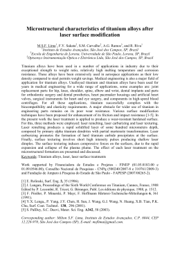
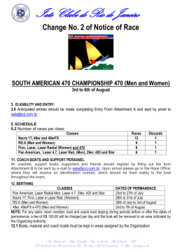
Download