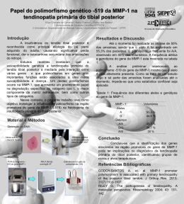
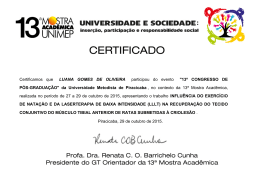
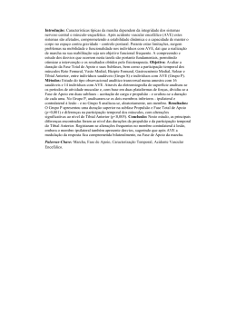
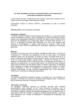
TRATAMENTO CIRÚRGICO DA DOR CRÔNICA NA LESÃO DE OSGOOD-SCHLATTER: RELATO DE DOIS CASOS
RELATO DE CASO / CASE REPORT
Tratamento cirúrgico da dor crônica na lesão
de Osgood-Schlatter: relato de dois casos*
Surgical treatment of chronic pain from
Osgood-Schlatter lesion: report of two cases*
GIOVANNINI CESAR FIGUEIREDO1, TATIANA DE OLIVEIRA MEDEIROS2,
LUCIMÁRIO DE OLIVEIRA VALE3, ALYSSON L.B. PEREIRA DE ASSIS3
RESUMO
ABSTRACT
Os autores realizaram estudo para determinar a freqüência de indicação cirúrgica no tratamento da lesão de
Osgood-Schlatter. De uma série de 28 pacientes (39 joelhos), dois pacientes (7,1%) ou dois joelhos (5,1%) apresentaram fragmento ósseo supratuberositário doloroso,
apesar da insistência do tratamento conservador. A ressecção cirúrgica do ossículo foi necessária para o alívio
dos sintomas em ambos os pacientes. Os autores consideram que essa pode ser forma recomendável de tratar a
dor crônica da lesão de Osgood-Schlatter.
The authors studied the frequency of surgical therapy for
Osgood-Schlatter lesion. Among 28 patients (39 knees), two
patients (7.1%), or two knees (5.1%) presented a painful bone
fragment above the tuberosity, despite conservative treatment.
Surgical removal of the ossicle warranted symptom relief in
both patients. The authors consider the surgical therapy an
adequate form to treat chronic pain from Osgood-Schlatter
lesion.
Key words – Osgood-Schlatter; knee; pain; osteochondritis; surgical
treatment
Unitermos – Doença de Osgood-Schlatter; joelho; osteocondrite; dor
articular; tratamento cirúrgico
INTRODUCTION
INTRODUÇÃO
A lesão de Osgood-Schlatter é considerada mais uma apofisite de tração do que uma verdadeira osteocondrite(1), que se
inicia durante a adolescência, segundo descrição de Osgood e
The Osgood-Schlatter lesion is considered more a traction
apophysitis than a true osteochondritis(1). It begins during
adolescence, according to Osgood and Schlatter’s description in 1903 [apud Glynn and Regan(2)]. The patellar ligament transmits to the tibial tuberosity, located at the proximal
* Trabalho realizado na Ortotrauma Campina Grande e Hospital Fundação
* From Ortotrauma Campina Grande and Hospital Fundação Assistencial
Assistencial da Paraíba (OCG-HFAP).
da Paraíba (OCG – HFAP), Brazil.
1. Mestre em Medicina; Professor Adjunto de Clínica Traumortopédica da Uni1. MSc in Medicine, Adjunct Professor of Clinical Traumatology and Orthopeversidade Estadual da Paraíba (UEPB); Membro Titular da Sociedade Brasidics, Universidade Estadual da Paraíba (UEPB); Full Membership of SBOT,
leira de Ortopedia e Traumatologia (SBOT), Sociedade Brasileira de CirurSBCJ, and SBOP.
gia de Joelho (SBCJ) e Sociedade Brasileira de Ortopedia Pediátrica (SBOP).
2. Orthopedic Surgeon; SBOT Full Member.
2. Membro Titular da SBOT.
3. Medical Student.
3. Acadêmico do 8o período da Faculdade de Medicina de Campina Grande,
PB.
Endereço para correspondência (Correspondence to): Giovannini Cesar Figueiredo, Rua Francisco Lima Neto, 146, Bodocongó – 58109-105 – Campina Grande, PB.
Fax: (83) 341-4666, E-mail: [email protected]
Recebido em (Received in) 29/5/02. Aprovado para publicação em (Approved in) 4/2/03.
Copyright RBO2003
Rev Bras Ortop _ Vol. 38, Nº 8 – Agosto, 2003
491
G.C. FIGUEIREDO, T.O. MEDEIROS, L.O. VALE & A.L.B.P. ASSIS
Schlatter em 1903 [apud Glynn e Regan(2)]. Na fisiopatologia
da afecção, o ligamento patelar transmite a intensa força gerada pelo quadríceps ao tubérculo tibial, que se localiza sobre
a lâmina epifisária tibial proximal. As contrações forçadas e
repetitivas do quadríceps, exigidas pelas atividades atléticas
freqüentes de correr e pular, irritam a apófise sob a tuberosidade tibial, que se torna sensível ao toque e dolorosa a qualquer contração forçada do quadríceps. Isso pode estimular a
apófise a produzir quantidade maior de osso, resultando na
reconhecida proeminência da tuberosidade da tíbia(3). Eventualmente, pode ocorrer fratura-avulsão da apófise, próxima
à inserção do ligamento patelar, causada por súbita e intensa
contração do quadríceps(4). O processo inflamatório e as mudanças histológicas de reparação podem levar à pseudartrose
do fragmento e à persistência dos sintomas(5).
Usualmente, a lesão resolve-se espontaneamente ou com
tratamento conservador(6), como a redução da prática desportiva, períodos repetidos de imobilização do joelho, uso de
antiinflamatórios, fisioterapia e, até, infiltrações locais com
lidocaína ou corticosteróides(3). Alguns casos, porém, não respondem ao tratamento conservador e a cirurgia é necessária
para aliviar a dor. A maioria dos autores recomenda a excisão
do ossículo livre e presente na porção distal do ligamento patelar(1,4,5).
O objetivo deste estudo é contribuir para o estabelecimento
da freqüência de indicação cirúrgica e descrever a técnica para
tratamento da dor crônica da lesão de Osgood-Schlatter pela
retirada do fragmento ósseo livre supratuberositário.
MATERIAL E MÉTODO
Foram estudados todos os prontuários disponíveis relativos aos pacientes atendidos na Ortotrauma Campina Grande
e no Hospital da Fundação Assistencial da Paraíba, com diagnóstico clínico e radiológico de enfermidade de OsgoodSchlatter, e cuja última consulta foi registrada entre fevereiro
de 1998 e março de 2002, para estabelecer a freqüência de
indicação cirúrgica.
RESULTADOS
De 28 pacientes, 22 (78,6%) eram do sexo masculino e seis
(21,4%) do feminino. A idade quando da última consulta variou entre 11 e 42 anos, com média de 16,82 (± 6,18) anos,
mediana de 15,5 anos e moda de 16 anos. A bilateralidade da
lesão ocorreu em 11 pacientes (39,3%), somente o joelho direito em 10 (35,7%) e somente o joelho esquerdo em sete
(25%).
492
tibial epiphysis, the huge force generated by the quadriceps
muscle. Forceful, repetitive quadriceps contractions seen in
frequent athletic activities involving running and jumping, irritate the apophysis under the tibial tuberosity, which becomes
tender on touch and painful at any forceful quadriceps contraction. This may stimulate the apophysis to produce more
bone, resulting in protuberance of the tibial tuberosity(3). There
may be an eventual avulsion fracture of the apophysis, near
to the patellar ligament insertion, caused by a sudden, intense quadriceps contraction(4). Inflammatory process and
reparation histological changes may lead to fragment pseudarthrosis, and symptoms persistence(5).
The lesion usually spontaneously resolves or by means of
conservative treatment(6), such as reduction in sports practice, repeated periods of knee immobilization, use of anti-inflammatory drugs, physical therapy, and even local lidocaine
and/or corticosteroid injections(3). Some cases, however, do
not respond to conservative treatment and the surgery is needed to alleviate the pain. Most authors recommend the excision of the loose ossicle present at the distal portion of patellar ligament(1,4,5). The aim of this study is to establish the
frequency rate of surgical indication and describe the technique for the therapy of chronic Osgood-Schlatter lesion by
removing the loose ossicle above the tuberosity.
MATERIAL AND METHODS
All available records from patients seen at Ortotrauma
Campina Grande and Hospital da Fundação Assistencial da
Paraíba, with clinical and radiological diagnosis of OsgoodSchlatter disease between February 1998 and March 2002,
were examined to determine the frequency of surgery indication.
RESULTS
Out of 28 patients, 22 (78.6%) were male and six (21.4%),
female. The age at the last visit ranged from 11 to 42 years,
mean 16.82 (± 6.18), median of 15.5 years, and mode 16 years.
Bilateral lesions were present in 11 (39.3%) patients, only in
the right knee in 10 (35.7%), and only the left knee in seven
(25%) patients.
The operative treatment indication with loose ossicle resection was found in two (7.1%) out of the 28 patients, corresponding to two knees in 39 (5.1%) of this series. Despite
conservative therapy, the patients went to surgery due to pain
persistence for a long period of time. One patient had pain
for 42 months and the other for 132 months. Their ages at the
Rev Bras Ortop _ Vol. 38, Nº 8 – Agosto, 2003
TRATAMENTO CIRÚRGICO DA DOR CRÔNICA NA LESÃO DE OSGOOD-SCHLATTER: RELATO DE DOIS CASOS
Fig. 2 – Caso 1.
Radiografia em
perfil do joelho
direito,
mostrando
fragmentos
ósseos livres
acima da
inserção do
ligamento
patelar na
tuberosidade
tibial.
Fig. 1 – Caso 1.
Adolescente de
14 anos de
idade, com
protuberância
visível em região
da tuberosidade
proximal tibial
direita.
Fig. 2 – Case 1.
Lateral
roentgenogram
of the right knee,
showing loose
bony fragments
above the
patellar ligament
attachment
at the tibial
tuberosity.
Fig. 1 – Case 1. A
14 year-old
adolescent, with
a visible bulge
over the right
proximal tibial
tuberosity.
A indicação do tratamento cirúrgico com ressecção do fragmento livre supratuberositário, nesta série, ocorreu em dois
dos 28 pacientes (7,1%), o que corresponde a dois joelhos em
39 (5,1%) com a lesão. Apesar do tratamento conservador, os
pacientes operados apresentaram a persistência da dor por um
longo período, até a data da cirurgia, um por 42 meses e o
outro por 132 meses. Um dos pacientes estava com 14 e o
outro 26 anos de idade na época da cirurgia. Os dois pacientes
apresentaram remissão completa da dor quando avaliados no
30o dia de pós-operatório. Na décima semana, o adolescente
continuava assintomático em relação às atividades da vida
diária. Com o adulto não mais tivemos contato.
Caso 1
RAE, 14 anos de idade, do sexo masculino, praticante de
futebol como lazer, queixava-se de dor na face anterior do
joelho direito, havia três anos, mais precisamente na tuberosidade anterior da tíbia (figura 1). Nesse período, foi submetido
a tratamento conservador, com restrição da prática desportiva
intensa, imobilização do joelho em períodos programados e
fisioterapia. Fez uso, também, de “tira subpatelar” para contenção da força de tração do ligamento patelar no tubérculo
tibial. Nos últimos seis meses, vinha relatando dor constante
no local. No exame radiográfico, visualizou-se a presença de
Rev Bras Ortop _ Vol. 38, Nº 8 – Agosto, 2003
moment of surgery were, respectively, 14 and 26 years old.
Both presented a complete pain remission by the 30th postoperative day. The adolescent remained asymptomatic when
performing daily activities at ten weeks. We lost contact with
the adult.
Case 1
RAE, male, 14 years old, a leisure soccer player, complained
of anterior knee pain for three years, precisely at the anterior
tibial tuberosity (figure 1). During this period, he had been
submitted to conservative treatment, by restriction of intense
sports practice, knee immobilization during programmed periods of time, and physical therapy. He also wore a “subpatellar band” for traction strength contention of patellar ligament. During the last six months he had been complaining of
a constant, local pain. Loose bone fragments within the tuberosity were seen on X-rays (figure 2). Axial view of CT scan
showed the fragment and the epiphyseal lamina still open (figure 3). The adolescent and his family agreed to proceed with
surgery.
Patient was placed supine, under rachis anesthetic. A tourniquet was inflated, with the knee flexed at 15° and positioned
on a sandbag. A 4 cm longitudinal skin incision was made
above the patellar ligament above the tibial tuberosity. The
493
G.C. FIGUEIREDO, T.O. MEDEIROS, L.O. VALE & A.L.B.P. ASSIS
Fig. 4 – Caso 1.
O tendão patelar
foi dividido
longitudinalmente
e o fragmento
ósseo livre
é retirado.
Fig. 4 – Case 1.
The patellar
ligament
has been
longitudinally
divided and the
loose ossicle
was removed.
Fig. 3 – Caso 1. Visão coronal de tomografia computadorizada do
tubérculo tibial mostrando o fragmento ósseo livre.
Fig. 3 – Case 1. Coronal CT scan of the tibial tuberosity showing
the loose ossicle.
fragmentos ósseos livres da tuberosidade (figura 2). Na tomografia computadorizada, em corte axial, observou-se o fragmento e a lâmina epifisária ainda aberta (figura 3). O adolescente e a família concordaram com o tratamento cirúrgico.
O paciente foi colocado em decúbito dorsal, sob anestesia
raquimedular. Foi usado torniquete, com o joelho posicionado sobre coxim, fletido a 15o. Foi feita incisão longitudinal de
4cm na pele sobre o ligamento patelar, acima da tuberosidade
tibial. O retináculo ligamentar foi aberto longitudinalmente.
A seguir, o ligamento patelar foi incisado longitudinalmente
na linha mediana. O ossículo foi removido do tecido ligamentar com bisturi de lâmina 15 (figura 4). A superfície proximal da tíbia foi suavemente curetada. O ligamento patelar
foi suturado com três pontos de vicryl 5ø. O retináculo foi
suturado com categute simples 4ø e a pele com mononylon
4ø.
O paciente iniciou exercícios isométricos já no pós-operatório imediato. Utilizou muletas por uma semana e foi imobilizado com um brace. A flexão do joelho foi permitida até 45o
na segunda semana, 90o na terceira e liberada da quarta semana em diante. O paciente caminhou na primeira semana, nadou na quarta e fez bicicleta na sexta semana. Na décima semana, afirmou estar sem dor e satisfeito com o tratamento.
Caso 2
Homem de 26 anos de idade, recepcionista de hospital, atendido pela primeira vez em nosso serviço, queixando-se de
tumoração dolorosa em face anterior do joelho esquerdo, que
494
ligament retinaculum was longitudinally opened. Next, the
patellar ligament was longitudinally opened at the median
line. The ossicle within ligament tissue was removed with a
number 15-blade scalpel (figure 4). The proximal tibial surface was gently curetted. Patellar ligament was sutured with
three stitches of 5-0 vicryl. Retinaculum was sutured with 4-0
simple catgut, and skin with 4-0 mononylon.
Isometric exercises were started at the immediate postoperative period. He used crutches for one week, along with a
brace. Knee flexion was allowed to 45° on the second week,
90° on the third week, swimming during the fourth week, and
bicycle riding on the sixth week. The patient claimed to be
painless and satisfied with the treatment at the tenth week
follow-up visit.
Case 2
A 26-year-old man who worked as a hospital receptionist
was first seen in our service complaining of a painful bulge,
worse when climbing stairs, on the left knee anterior aspect.
On the past history, he reported episodes of painful bouts since
15 years old, and had used medications and physical therapy
during several occasions (as stated). He presented a prominent tibial tuberosity on physical examination (figure 5). Radiological exam showed a loose ossicle above the patellar
ligament insertion (figure 6). The patient agreed with the surRev Bras Ortop _ Vol. 38, Nº 8 – Agosto, 2003
TRATAMENTO CIRÚRGICO DA DOR CRÔNICA NA LESÃO DE OSGOOD-SCHLATTER: RELATO DE DOIS CASOS
Fig. 7 – Caso 2.
O ossículo é
ressecado
através de
incisão
longitudinal
do ligamento
patelar.
Fig. 5 – Caso 2. Adulto de 26 anos, com tuberosidade tibial esquerda muito proeminente.
Fig. 5 – Case 2. A 26 year-old man with a prominent bulge over
the left tibial tuberosity.
Fig. 7 – Case 2.
The ossicle is
removed by a
longitudinal
incision of
the patellar
ligament.
Fig. 6 – Caso 2. O ossículo é visualizado em radiografia em perfil
do joelho esquerdo.
Fig. 6 – Case 2. The ossicle is seen on the lateral roentgenogram
of the left knee.
piorava ao subir escadas. Na história pregressa, relatou episódios de crises dolorosas desde os 15 anos de idade, tendo feito
uso de medicamentos e fisioterapia em várias oportunidades
(sic). Ao exame físico, apresentava tuberosidade tibial proeminente (figura 5). O exame radiográfico mostrou ossículo
livre acima da inserção do ligamento patelar (figura 6). O paciente concordou com o tratamento cirúrgico. O ossículo foi
removido através de incisão longitudinal do ligamento patelar (figuras 7 e 8). Após 30 dias da cirurgia, o paciente conseguia subir escadas sem dor e não mais retornou para seguimento.
Fig. 8 – Caso 2. O ossículo ressecado comparado em tamanho
com uma lâmina de bisturi no 23.
Fig. 8 – Case 2. Size of resected ossicle compared with a number23 surgical blade.
gical treatment. The ossicle was removed through a longitudinal incision of the patellar ligament (figures 7 and 8). After
30 days of surgery, the patient painlessly climbed stairs and
did not return anymore for follow-up.
DISCUSSÃO
DISCUSSION
O tratamento cirúrgico da dor crônica na lesão de OsgoodSchlatter não é freqüentemente necessário, tornando-se difí-
Surgical treatment of chronic pain from Osgood-Schlatter
lesion is not often needed. Hence, it is hard to find a large
Rev Bras Ortop _ Vol. 38, Nº 8 – Agosto, 2003
495
G.C. FIGUEIREDO, T.O. MEDEIROS, L.O. VALE & A.L.B.P. ASSIS
cil encontrar uma larga série de casos e, mais ainda, comparar
técnicas cirúrgicas diferentes. Mital et al(4) indicaram tratamento cirúrgico com ressecção do ossículo em 14 entre 118
pacientes (11,9%), ou seja, 15 entre 151 joelhos (9,9%). Dos
joelhos operados, 14 (93,3%) tiveram remissão completa da
dor. Binazzi et al(1), em 1993, mostraram 93% de excelentes e
bons resultados com a excisão do ossículo, associada ou não
à técnica de Thomson-Ferciot (ressecção da proeminência do
tubérculo tibial), contra 72% do grupo submetido a enxertia
para cura da pseudartrose, reinserção e fixação interna do fragmento e perfuração da tuberosidade. Flowers e Bhadreshwar(7)
recomendaram a excisão do tubérculo tibial como tratamento
de escolha nos casos de falência do tratamento conservador.
O alívio da dor, nesta série, foi de 95%. Trail(8), em 1988, não
encontrou melhores resultados no grupo submetido à ressecção da tuberosidade tibial em relação ao que teve tratamento
conservador. Orava et al(9), em 2000, apontaram 90,3% de
excelentes e bons resultados em 62 pacientes submetidos à
ressecção do ossículo livre supratuberositário.
A idade dos pacientes operados em nossa casuística mostra
que o tratamento cirúrgico pode tornar-se necessário na idade
adulta, como já referiram outros autores(10,11,12). Os bons resultados relatados na literatura e a simplicidade da técnica
cirúrgica induzem a considerar a indicação da ressecção do
ossículo livre supratuberositário como opção segura no tratamento da dor crônica na lesão de Osgood-Schlatter, conduta
que está indicada em qualquer idade.
series of cases, and specially comparing different surgical
options. Mital et al(4) advocated surgical treatment with ossicle resection for 14 among 118 patients (11.9%), or 15 among
151 knees (9.9%). Of the operated knees, 14 (93.3%) had a
complete pain remission. Binazzi et al(1) in 1993 reported 93%
of excellent and good outcomes with ossicle excision associated or not to Thomson-Ferciot technique (tibial tuberosity
protuberance excision), as compared to 72% from the group
submitted to bone fragment pseudarthrosis grafting, reinsertion, internal fixation, and tibial tuberosity drilling. Flowers
and Bhadreshwar(7) recommended the excision of tibial tuberosity as the treatment of choice in cases of conservative
therapy failure. They obtained 95% rate of pain relief. Trail(8),
in 1988, did not find better results in the group who suffered
tibial tuberosity resection in comparison to the group receiving conservative treatment. Orava et al(9), in 2000, reported
90.3% of excellent and good outcomes from 62 patients submitted to resection of loose ossicle above the tuberosity.
The age group of operated patients from our series demonstrates that surgical treatment may be warranted at adult age,
as previously stated by other authors(10,11,12). Good outcomes
reported in literature and simple surgical technique lead to
consider the indication of loose ossicle resection as a safe
option for the treatment of chronic pain from Osgood-Schlatter lesion at any age.
REFERÊNCIAS / REFERENCES
1. Binazzi R., Felli L., Vaccari V., Borelli P.: Surgical treatment of unresolved
Osgood-Schlatter lesion. Clin Orthop 289: 202-204, 1993.
2. Glynn M.K., Regan B.F.: Surgical treatment of Osgood-Schlatter’s disease.
J Pediatr Orthop 3: 216-219, 1983.
3. Kaeding C.C., Whitehead R.: Musculoskeletal injuries in adolescents. Prim
Care 25: 211-223, 1998.
4. Mital M.A., Matza R.A., Cohen J.: The so-called unresolved Osgood-Schlatter lesion: a concept based on fifteen surgically treated lesions. J Bone Joint
Surg [Am] 62: 732-739, 1980.
5. Robertsen K., Kristensen O., Sommer J.: Pseudoarthrosis between a patellar tendon ossicle and the tibial tuberosity in Osgood-Schlatter’s disease.
Scand J Med Sci Sports 6: 57-59, 1996.
6. Elias N., Teixeira J.C.M., Fernandes Junior N.O., Oliveira L.P.: Tratamento
conservador da doença de Osgood-Schlatter. Rev Bras Ortop 26: 216-218,
1991.
496
7. Flowers M.J., Bhadreshwar D.R.: Tibial tuberosity excision for symptomatic Osgood-Shlatter disease. J Pediatr Orthop 15: 292-297, 1995.
8. Trail I.A.: Tibial sequestrectomy in the management of Osgood-Schlatter
disease. J Pediatr Orthop 8: 554-557, 1988.
9. Orava S., Malinen L., Karpakka J., Leppilahti J., Rantanen J., Kujala U.M.:
Results of surgical treatment of unresolved Osgood-Schlatter lesion. Ann
Chir Gynaecol 89: 298-302, 2000.
10. King A.G., Blundell-Jones G.: A surgical procedure for the Osgood-Schlatter lesion. Am J Sports Med 9: 250-253, 1981.
11. Orava S., Virtanen K.: Osteochondroses in athletes. Br J Sports Med 16:
161-168, 1982.
12. Hogh J., Lund B.: The sequelae of Osgood-Schlatter’s disease in adults. Int
Orthop 12: 213-215, 1988.
Rev Bras Ortop _ Vol. 38, Nº 8 – Agosto, 2003
Download