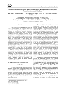
Anim. Reprod., v.11, n.4, p.511-516, Oct./Dec. 2014
Laparoscopy of the genitourinary tract of small ruminants
D.P. Vrisman1, E. Choaire1, F. Strucher1,M.S. Oliveira1, T.M.B. Ribas1, L.N. Coutinho2,
R.S.G. Mariano2, M.G. Oliveira1, W.R.R. Vicente2, M.A.M. Silva3, P.P.M. Teixeira1,2,4
1
Veterinary Medicine Department,Middle-West State University (UNICENTRO), Guarapuava, PR, Brazil.
Faculty of Agricultural Sciences and Veterinary (FCAV), São Paulo State University “Júlio de Mesquita Filho”
(UNESP), Jaboticabal, SP, Brazil.
3
School of Agronomy and Veterinary Medicine, University of Passo Fundo (FAMV/UPF), Passo Fundo, RS, Brazil.
2
Abstract
Laparoscopic endosurgery is an important tool
for small ruminant breeding especially due to its wide
versatility, both in reproductive biotechnologies and
therapeutic approach. The purpose of this review is to
present the applicability and advantages of endosurgery
for routine use in small ruminants. Several endosurgical
techniques of the genitourinary tract of small ruminants,
including artificial insemination, embryo transfer, and
oocyte retrieval for IVF, ovariectomy, cystotomy and
renal biopsy are approached. The endoscopic techniques
can improve the development of potential zootechnical
indexes on the short-term period in comparison to
traditional approaches.
Keywords: endosurgery treatment, goats, reproductive
biotechnologies, sheep.
Introduction
The evident expansion of sheep and goat breeding
lead to the need for the development of reproductive
biotechnologies for maximizing productivity of livestock,
aiming at the production of high genetic pattern and
productive animals (Simplício et al., 2002). Laparoscopy is
a minimally invasive technique that allows the exploration
of the abdominal and pelvic organs using a transabdominal
endoscope and other adapted equipment for endosurgery,
allowing it to work with these organs made for both
therapeutic and for reproductive biotechnologies (artificial
insemination, embryo transfer and laparoscopic ovum
pick-up) particularly in small ruminants (Bouré, 2005;
Teixeira et al., 2013).
Artificial insemination (AI) is an important
reproductive management aspect in sheep breeding due
to the specie’s short reproductive cycle and calving
intervals. Using biotechnologies, high genetic value
offspring production can be reached (Bettencourt,
1999). With the employ of embryo transfer (ET),
besides the same advantages of AI, it is possible to
expand the genetic value and production potential of
females, while avoiding mating, and thus, minimizing
the transmission of reproductive infectious diseases
_________________________________________
4
Corresponding author: [email protected]
Phone: +55(42)9912-5588
Received: April 1, 2014
Accepted: August 7, 2014
(Varago et al., 2009). The ovum pick up (OPU) for in
vitro production of embryos is an efficient
biotechnology, which allows for repeated follicle
aspiration sessions at short intervals in adult and
prepubertal females following several ovarian
stimulation protocols (Wani, 2002; Valasi et al., 2009;
Baldassare, 2012; Blash et al., 2012; Crocomo et al.,
2012; Teixeira et al., 2011b, 2012).
Although laparoscopy for ovariectomy is not
routinely performed, it is used for herd management,
weight gain of the animals, improvement of the quality
of carcass, recovery of gametes and for researches on
reproductive technology (Teixeira et al., 2011a).
Laparoscopy is also used for laparoscopic-assisted
cystotomy, presenting benefits such as short duration of
anesthesia, fast convalescence and avoidance of
urethrostomy/urethrostomy (Gheller, 2008). Moreover,
laparoscopic-assisted renal biopsy is useful in studies on
pharmacokinetics and toxicology (Toutain et al., 1985).
Based on such relevant facts, the aim of the
current study was to review the applicability and
advantages of the endosurgery applied to the
genitourinary tract of small ruminants.
Artificial insemination
Improvement of the herd is one of the main
goals of sheep and goat breeding farms. Regardless of
the technique used, the knowledge of ovulation time is
critical in order to obtain a success full insemination
program. The ovum is usually viable for about 12-24 h
(Ax et al., 2004; Ferra and Sereno, 2006). In
laparoscopic IA, frozen semen is thawed and deposited
directly into the uterus following needle puncture,
unlike cervical or trans-cervical AI (Donovan et al.,
2004). In order to optimize the technique,
synchronization of the estrous cycle of the animals is
crucial. Protocols using progestins are usually optimal
(Silva, 2009).
For laparoscopic-assisted AI the animals are
submitted to general anesthesia and positioned in dorsal
recumbence, on a 45º head tilting, which is known as
Trendelenburg position (Rabassa et al., 2007; Teixeira
et al., 2011b), as shown in Fig. 1 and 2.
Vrisman et al. Genitourinary laparoscopy in small ruminants.
A two-port technique is used. It is recommended
to perform local anesthesia on the two port insertion sites,
both caudolateral to the umbilicus. Following a 1-2 cm
skin incision, a 5-mm or 10-mm trocar with sharp tip
obturator is inserted through the abdominal wall, followed
by the establishment of a 5-mmHg CO2
pneumoperitoneum. Afterwards, a second trocar is
inserted using laparoscopic guidance. An insemination
pipette with a needle tip is introduced through the
second trocar cannula to penetrate the uterine horns and
deposit the semen in contact with the uterine mucosa. It
is essential to locate the most cranial aspect of the
uterus, which presents less blood supply, to insert the
needle perpendicularly to the uterus wall. Semen is
injected within the uterine lumen. The laparoscope and
the cannulas are removed and the abdominal incisions
are closed routinely. Procedure time may take as short
as 2-5 min, depending on the experience of the surgeon
(Carneiro et al., 2011).
Semen doses should contain about 150-200 x
106 viable spermatozoa. It means that about 15 to 30
sheep can be inseminated with a single ejaculate
(Bicudo et al., 2005). In commercial herds, the use of
laparoscopic AI is still limited due to the high cost of
implementation, learning curve and team formation
(Kershaw et al., 2005).
Figure 1. Figure showing the Trendelenburg position used for artificial insemination (AI), embryo transfer (ET) and
laparoscopic ovum pick up (LOPU). Observing the 45º slope angle position, side view (B) and front view (A and C).
Also showing the two or three videolaparoscope portals, according to the triangular form and facing the inguinal
region. Source: personal file.
512
Anim. Reprod., v.11, n.4, p.511-516, Oct./Dec. 2014
Vrisman et al. Genitourinary laparoscopy in small ruminants.
Figure 2. Schematic draw (A) and picture (B) of ovine patient for lateral recumbency position by video laparoscopy.
(A) Sites of portals for videolaparoscopy, introduction of Veress needle and laparotomy. (1) Caudal border of the rib
cage; (AV) Veress needle introduction site; (P1) First portal site for introduction of the endoscope; (P2) Second
portal for introduction of instruments; (LP) Laparotomy site to perform blind biopsy. (RD) Right kidney. (B) Same
positioning, but performed the first entry port blind, Positioning Even though the first entry portal performed blind,
without the use of a Veress needle. Adapted from Néspoli et al., 2010.
Embryo transfer(ET)
The main goal of embryo transfer is to promote
a wide spread of desirable genetic values of the herd,
while assuring biosecurity to the herd as natural mating
is avoided (Varago et al., 2009).
Superovulation is required, which is usually
performed by exogenous administration of FSH in
decreasing doses (Traldi, 2006; Varago et al., 2009). In
the goat, insemination must be carried out 12-24 h after
detection of estrus (Medeiros et al., 2012). In the sheep,
AI was recommended 54-56 h after vaginal sponge
removal (Rizzo et al., 2009). Laparoscopic insemination
should be performed once or twice in a 12 h interval,
which increases the rate to around 40 to 70% of success
with the use of frozen semen (Varago et al., 2009).
Laparoscopy is a safe and accurate technique for
embryo collection. The same port placement and patient
positioning of AI should be used (Fonseca, 2006).
The two-port technique of intrauterine
laparoscpic ET was first described by Schiewe et al.
(1984). Embryos are collected from donors by washing
the uterine horns between the 7 and 8th days following
the onset of estrus in goat and the 6 and 7th day in sheep
(Traldi, 2006), using a 37°C phosphate buffered saline
(PBS) and 1% of fetal bovine serum (FBS) solution per
uterine horn (Zanetti, 2009). Two-port access on the
linea alba is usually performed, placed between the
umbilicus and the mammary gland (Fonseca, 2006).
One of the major advantages of laparoscopy
use in embryo transfer is the lower incidence of uterine
adhesions and easy deposition of the embryo near the
utero-tubal junction (Varago et al., 2009). ET success
Anim. Reprod., v.11, n.4, p.511-516, Oct./Dec. 2014
also depends on the selection of good recipients and
donors. Selection criteria should be focused on
reproductive tract health, body score and maternal
ability (Simplício et al., 2002; Fonseca, 2013).
On average 5 viable embryos are recovered for
harvest, both in laparoscopy, laparotomy or by cervix.
The laparoscopy advantage compared to laparotomy for
the largest number of repetitions, laparotomy is limited
to three procedures, as laparoscopy does not have a
procedure limit described and can be used for several
repetitions in the same female. Transcervical embryo
collection technique is limit by the large number of
animals with limited cervix passage (Fonseca, 2013;
Teixeira et al., 2013).
Ovum pick-up for in vitro production
The laparoscopic ovum pick-up (LOPU) is the
initial step for in vitro embryo production (IVP), with
ovulation induction and ovarian stimulation essential for
best results of this biotech, especially in females in
anestrus or prepubertal. The use of different doses of
FSH or eCG is essential to obtain the highest production
of follicles. The high hormone doses and the interval
between ovarian stimulation and aspirations will be
responsible to determine oocyte quality (Baldassarre
and Karatzas, 2004; Teixeira et al., 2011b, 2013;
Baldassarre, 2012).
Classically, ovaries obtained in slaughterhouses
were used to recover oocytes for research purpose, but
this method is being replaced by LOPU (Baldassarre,
2012). This new approach allows repeated aspirations
and optimal sanitary control (Wani, 2002; Teixeira et
513
Vrisman et al. Genitourinary laparoscopy in small ruminants.
al., 2011b; Baldassarre, 2012).
Different treatments were tested for LOPU and
"one-shot" is the treatment of choice, because it
decreases the interval between the use of the hormone
and aspiration. Usually, 70-80 mg of NHI-FSH P1 and
300UI of eCG are administered in a single dose regimen
in order to achieve optimal in vitro maturation (IVM) of
oocytes (Morton et al., 2005). Follicles must be 2-8 mm
in diameter for aspiration and present greater cumulus
layer for improved fertilization (Crozet et al., 1995;
Mermillod et al., 1999, 2008; Baldassarre, 2012;
Crocomo et al., 2012).
As for laparoscopic AI and ET, LOPU
requires general anesthesia. The most widely used preanesthetic protocol in animals submitted to
laparoscopy is xylazine 0.05 mg/kg (IM) alone,
acepromazine 0.05 mg/kg alone or in combination
with midazolam 0.1 mg/kg (IV) and chlorpromazine
1.0 mg/kg (IM) or tramadol 2 mg/kg (Teixeira et al.,
2011b).
For
anesthetic
induction,
ketamine
hydrochloride 2 mg/kg (IV), propofol 3-6 mg/kg (IV)
or thiopental 10.5 mg/kg (IV) are used. Anesthetic
maintenance should be performed using either
halothane or isoflurane vaporized in oxygen (Teixeira
et al., 2012) or continuous rate infusion of a mixture of
propofol (0.5 mg/k/min, IV) and lidocaine
hydrochloride (1 mg/kg/min, IV; Teixeira et al., 2013).
LOPU is well described using a two-port
technique, for insertion of the telescope and a grasping
forceps, or three-port access, for the telescope and two
forceps. The use of three ports has reduced significantly
surgical time in other studies (Teixeira et al., 2011b,
2012).
After aseptic preparation, the patient is
positioned in Trendelenburg head tilt, as described for
laparoscopic AI and ET. The first trocar is inserted
either by open minilaparotomy or blind technique and
pneumoperitoneum is established. Veress needle may be
used, however increased risk of rumen perforation is
advised (Teixeira et al., 2013).
A traumatic graspers, such as Babcock forceps,
are used for manipulation of the reproductive tract,
including ovaries and uterine horns. For follicle
punctures and OPU, a simple vaccum system using a
16G, 18G or 20G needle is more efficient and less
traumatic to the cumulus/oophorus complex. The
suction pressure of 50-70 mmHg also contributes to
oocyte quality (Wieczorek et al., 2010; Crocomo et al.,
2012; Padilha et al., 2014).
The LOPU for IVP is still limited due to the
need for investment in laboratories. However, it is
important to emphasize the applicability also in
prepubertal animals and the improvement of herd genetic
gain inherent to such technique. In small ruminants,
recovery of more than five oocytes per donor is usually
achieved, and at the end of the process (LOPU and IVP),
achieving 1.2 pregnancies per week (Baldassarre and
Karatzas, 2004; Teixeira et al., 2012, 2013).
514
Ovariectomy
Ovariectomy results in many advantages for
animal production, such as (1) improving management
by breeding males along with females in the same
environment; (2) increasing live weight gain; (3)
production of high quality carcass. Furthermore, this
technique has also been used to isolate oocytes of
animals bearing high genetic and productive
performance for assisted reproduction purposes.
Laparoscopic ovariectomy requires general anesthesia,
aseptic preparation and positioning similar to those for
LOPU (Teixeira et al., 2011a, c).
Laparoscopic-assisted ovariectomy requires
one or two ports. The use of bipolar coagulation and
cutting forceps (Lina Tripol PowerBlade® - WEM &
Vivamed) and use of pre-tied ligature was reported for
ovariectomy in goats. A relevant factor on laparoscopic
ovariectomy is the reduced surgical time compared to
the conventional procedure and significantly decreased
postoperative pain (Teixeira et al., 2011b, c; Barros et
al., 2012).
Cystotomy and cystostomy
Laparoscopic
or
laparoscopic-assisted
cystotomy and cystostomy are feasible in patients
presenting obstructions of the urinary tract. Obstructions
of the lower urinary tract lead to urinary retention,
bladder distention, abdominal pain and urethral or
bladder rupture, which usually lead to death by uremia
or sepsis (Gheller, 2008). Rupture of the urethra or
bladder may occur in 2-3 days following untreated acute
obstruction. Other clinical signs include depression,
anorexia, pain on palpation of the penile area,
tachycardia, tachypnea, anuria or dysuria and hematuria
(Riet-Correa et al., 2008).
The obstruction episodes usually occur in goat
and sheep during exposures due to energy-rich diet,
such as grain and their products, leading to
calcium/phosphorus imbalance. Young orchiectomized
lambs are in increased risk due to poor urethra
development. The site of highest obstruction probability
is the sigmoid flexure, ischial curvature and urethral
process. Urinary obstructions are not common in
females due to the shorter and larger diameter of the
urethra in comparison to males (Gheller, 2008; RietCorrea et al., 2008).
Surgical preparation includes fasting for 24 h,
wide shaving and aseptic preparation of the abdomen
(Franz et al., 2009). The patients are submitted to
general anesthesia and subsequently placed in supine
position (Gheller, 2008).
A 0.5 cm incision is made 4 cm cranially to
umbilicus for optical port placement, while the second
portal was made 3 cm lateral right to foreskin and 10 cm
distal to umbilicus. The second port is generally placed
using laparoscopic guidance, through the prepubic
abdominal area, which penetrates the bladder lumen.
Anim. Reprod., v.11, n.4, p.511-516, Oct./Dec. 2014
Vrisman et al. Genitourinary laparoscopy in small ruminants.
Keeping the cannula inside the bladder, a 18-gauge Foley
catheter is inserted into the bladder through the trocar
cannula and its cuff is inflated to prevent urine leakage.
The cannula is then removed, leaving only the Foley
catheter into the urinary bladder. The laparotomy is
expanded and the bladder is exposed by pulling the Foley
catheter for subsequent cystotomy. The bladder is washed
with sterile saline solution and routinely sutured using 3-0
monofilament suture material. The bladder is finally
replaced into the abdominal cavity and the abdominal
wall is routinely closed (Gheller, 2008; Franz et al.,
2009).
Postoperative care includes patient monitoring,
routine wound care and antibiotic therapy for 7-10 days.
The prognosis of the return of urinary and reproductive
functions is favorable as the laparoscopic approach
maintains the integrity of the urethra and bladder
sphincter (Gheller, 2008).
Renal biopsy
Kidney diseases in small ruminants are poorly
diagnosed in sheep. However, renal biopsies are useful
in studies of pharmacokinetics and toxicology (Toutain
et al., 1985).
For laparoscopic-assisted renal biopsy the
animals should be submitted to fasting for 12 h. If a
right renal biopsy is desired, the animal should be
positioned in left lateral recumbence and vice-versa.
After abdominal shaving and aseptic preparation, 2%
lidocaine hydrochloride is infiltrated at the port site, 10
min prior to incision. A Veress needle is introduced into
the abdominal cavity, dorsal and cranially on the flank
at the end of the last rib (Fig. 2). A 7-10 mmHg
pneumoperitoneum is established and a 6-11 mm trocar
is introduced into the abdomen through the skin
incision, halfway between the last rib and the coxal
tuberosity, for insertion of the telescope. The second
port is placed 5-10 cm beside the first trocar, for
insertion of laparoscopic scissors. A window is opened
on the parietal peritoneum/renal capsule. The scissors
are replaced by a Blakesley biopsy forceps and one or
more renal samples are taken. Complications such as
hemorrhage and fibrosis may occur because of this
technique (Néspoli et al., 2010).
Conclusion
Even though laparoscopic techniques still have
limited commercial use, clinical and reproductive gains
brought by endosurgery are extremely relevant for goat
and sheep production. Such biotechnologies should be
employed in rural properties, which may improve
breeding and cost-effectiveness.
References
Ax RL, Dally MR, Didion BA, Lenz RW, Love CC,
Anim. Reprod., v.11, n.4, p.511-516, Oct./Dec. 2014
Varner DD, Hafez B, Bellin ME. 2004. Inseminação
artificial. In: Hafez B, Hafez ESSE (Ed.). Reprodução
Animal. 7th ed. São Paulo: Manole. pp. 381-394.
Baldassarre H, Karatzas CN. 2004. Advanced assisted
reproduction technologies (ART) in 33 goats. Anim
Reprod Sci, 82/83:255-266.
Baldassarre H. 2012. Practical aspects for
implementing in vitro embryo production and cloning
programs in sheep and goats. Anim Reprod, 9:188-194.
Barros FFPC, Teixeira PPM, Silva MAM, Coelho
CMM, Lopes
MCS, Kawaname
AE,
Chung
DG, Coutinho LN, Padilha LC, Vicente WRR. 2012.
Single-port laparoscopic ovariectomy in Santa Ines
ewes using pre-tied loop ligature. Reprod Domest Anim,
47 (suppl. 1):76. (abstract).
Bettencourt EMV. 1999. Caracterização de
parâmetros reprodutivos nas raças ovinas Merina
branca, Merina preta e Campaniça. Lisboa:
Universidade Técnica de Lisboa, Faculdade de
Medicina Veterinária. 106 pp. Dissertation.
Bicudo SD, Azevedo HC, Silva Maia MS, Sousa DB,
Rodello L. 2005. Aspectos peculiares da inseminação
artificial em ovinos. Acta Sci Vet, 33(suppl.1):127-130.
Blash S, Schofield M, Echelard Y, Gavin W. 2012.
Update on the first cloned goats. Nat Biotechnol,
30:229-230.
Bouré L. 2005. General principles of laparoscopy. Vet
Clin North Am Food Anim Pract, 21:227-249.
Carneiro GF, Medeiros LRD, Gomes NO. 2011.
Inseminação artificial em pequenos ruminantes. Portal
ASCAPO. Available on: www.aspaco.org.br/materiais.
Accessed on: Apr. 19, 2013.
Crocomo LF, Marques FWC, Landim FCA, Bicudo
SD. 2012. Peculiaridades da coleta de oócitos para
produção in vitro de embriões ovinos. Rev Bras Reprod
Anim, 36:25-31.
Crozet N, Ahmed-AliM, Dubos MP. 1995.
Developmental competence of goat oocyte from
follicles of different size categories following
maturation, fertilization and culture in vitro. J Reprod
Fertil, 103:293-298.
Donovan A, Hanrahan JP, Kummen E, Duffy P,
Boland P. 2004. Fertility in the ewe following cervical
insemination with fresh or frozen - thawed semen at a
natural or synchronised oestrus. Anim Reprod Sci,
84:359-368.
Ferra JC, Sereno JRB. 2006. Inseminação Artificial
em Ovinos. Planaltina, DF: Embrapa Cerrados. p.10-11.
(Documentos, 156).
Fonseca JF. 2006. Alguns aspectos da transferência de
embriões em caprinos. Acta Sci Vet, 34 (suppl. 1):65-70.
Fonseca JF. 2013. Inseminação Artificial e Transferência
de Embriões em Caprinos e Ovinos. Coronel Pacheco,
MG: Embrapa Gado de Leite. 21 pp.
Franz S, Dadak AM, Schoffmann G, Khol JL,
Baumgartner W, Dupre G. 2009. Laparoscopicassisted cystotomy: an experimental study in male
sheep. Vet Med, 54:367-373.
515
Vrisman et al. Genitourinary laparoscopy in small ruminants.
Gheller V. 2008. Abordagens laparoscópicas em pequenos
ruminantes. Ciênc Vet Trop, 11(supl. 2):52-55.
Kershaw CM, Khalid M, Mc Gowan MR, Ingram K,
Leethongdee SG, Scaramuzzi RJ. 2005. The anatomy
of sheep cervix and its influence on the transcervical
passage of an inseminating pipette into the uterine
lumen. Theriogenology, 64:1225-1235.
Medeiros CHN, Monteiro AWU, Medeiros ALN,
Freitas VJF, Salles MGF, Araújo AA. 2012. Efeito de
sucessivos tratamentos superovulatórios com pFSH
sobre a resposta ovulatória e produção de embriões de
cabras Boer e posterior sobrevivência embrionária após
transferência em receptoras sem raça definida. Rev Bras
Hig San Anim, 6:15-23.
Mermillod P, Oussaid B, Cognié Y. 1999. Aspects of
follicular and oocyte maturation that affect the
developmental potential of embryos. J Reprod Fertil,
54:449-460.
Mermillod P, Dalbiès-Tran R, Uzbekova S. 2008.
Factors affecting oocyte quality: who is driving the
follicle? Reprod Domest Anim, 43:393-400.
Morton KM, De Graaf SP, Campbell A. 2005.
Repeated ovum pick-up and in vitro embryo production
from adult ewes with and without FSH treatment.
Reprod Domest Anim, 40:422-428.
Néspoli PB, Gheller VA, Peixoto PV, Carvalho AU,
França TN, Facury Filho EJ, Araújo DKG, Bordin
AI. 2010. Avaliação de técnicas de biópsia renal em
ovinos. Pesq Vet Bras, 30:260-266.
Padilha LC, Teixeira PPM, Pires-Buttler EA,
Apparício M, Motheo TF, Savi PAP, Nakaghi EYO,
Alves AE, Vicente WRR. 2014. In vitro maturation of
oocytes from Santa Ines ewes subjected to consecutive
sessions of follicular aspiration by laparoscopy. Reprod
Domest Anim, 49:243-248.
Rabassa VR, Tabeleão VC, Pfeifer LFM, Schneider A,
Ziguer EA, Schossler E, Severo NC, Del Pino FA,
Corrêa MN. 2007. Efeito das técnicas transcervical e
laparoscópica sobre a taxa de prenhez de ovelhas
inseminadas em tempo-fixo. Ciênc Anim Bras, 8:129-131.
Riet-Correa F, Simões SVD, Vasconcelos J. 2008.
Urolitíase em caprinos e ovinos. Pesq Vet Bras, 28:319322.
Rizzo H, François D, Fassier T, Guitton E, Baril G,
Cognié J, Fatet A, Guignot F, Mermillod P, Petit JP,
Beckers JF, Remy B, Gilles FF, Meyer G. 2009.
Transferência de embriões como ferramenta para
formação de rebanho experimental ovino. Ciênc Anim
Bras, (supl. 1):814-820.
Schiewe MC, Bush M, Stuart LS, Wildt DE. 1984.
Laparoscopic embryo transfer in domestic sheep a
preliminary. Theriogenology, 22:675-682.
Silva TG. 2009. Inseminação artificial em ovinos (Ovis
aries) por laparoscopia: revisão de literatura. Curitiba,
PR: Universidade Federal do Paraná. Monography.
Simplício AA, Salles HO, Santos DO. 2002.
Transferência de embriões nos pequenos ruminantes
516
domésticos. Rev Bras Reprod Anim Supl, 5:17-23.
Teixeira PPM, Padilha LC, Motheo TF, Silva MAM,
Oliveira MEF, Silva ASL, Barros FFPC, Coutinho
LN, Flôres FN, Lopes MCS, Rodrigues LFS, Vicente
WRR. 2011a. Ovariectomy by laparotomy, a videoassisted approach or a complete laparoscopic technique
in Santa Ines sheep. Small Rumin Res, 99:199-202
Teixeira PPM, Padilha LC, Oliveira MEF, Motheo
TF, Silva ASL, Barros FFPC, Coutinho LN, Flôres
FN, Lopes MCS, Bandarra MB, Silva MAM,
Vasconcelos RO, Rodrigues LFS, Vicente WRR.
2011b. Laparoscopic ovum collection in sheep: gross
and microscopic evaluation of the ovary and influence
on oocyte production. Anim Reprod Sci, 127:169-175.
Teixeira PPM, Silva MAM, Oliveira MEF, Padilha
LC, Silva ASL, Barros FFPC, Medeiros RM, Coelho
CMM, Rossi A, Lopes MCS, Vicente WRR. 2011c.
Ovariectomia total vídeolaparoscópica em ovelhas prépúberes. Vet Zootec, 18:493-496.
Teixeira PPM, Silva MAM, Padillha LC, Silva ASL,
Barros FFPC, Coutinho LN, Medeiros RM, Kirnew
MD, Conceição EM, Vicente WRR. 2012. Evaluation
of serum fibrinogen and total protein in prepubertal
sheep subjected to laparoscopic ovum pick-up. Reprod
Domest Anim, 47(suppl. 5):95-95.
Teixeira PPM, Padilha LC, Mariano RSG, Coutinho
LN, Barros FFPC, Silva, MAM, Silva ASL. 2013.
Aspiração folicular. In: Oliveira MEF, Teixeira PPM,
Vicente WRR. Biotécnicas Reprodutivas em Ovinos e
Caprinos. São Paulo, SP: MEDVET. vol.1, pp. 147-155.
Toutain PL, Pomyers H, Larrieu G, Periquet B,
More J. 1985. An in vivo model for pharmacokinetic
studies in the kidney. J Pharmacol Methods, 14:1-11.
Traldi AS. 2006. Biotécnicas aplicadas em reprodução
de pequenos ruminantes. In: III Feira Internacional de
Caprinos e Ovinos (FEINCO), 2006, São Paulo, SP. São
Paulo, SP: FEINCO. 11 pp.
Valasi L, Fthenakisa GC, Prassinosa NN,
Menegatosb I, Grigoropoulouna V, Deligiannisc C,
Vainasd E, Amiridisa GS. 2009. The effect of repeated
follicular aspiration on the onset of puberty and growth
rate of winter or autumm born lambs. Small Rumin Res,
84:35-40.
Varago FC, Moustacas VS, Cruz BC, Lagares MA,
Henry MRJM. 2009. Biotécnicas da reprodução
aplicadas a pequenos ruminantes. Cienc Anim Bras,
8(supl. 1):1-17.
Wani NA. 2002. In vitro maturation and in vitro
fertilization of sheep oocytes. Small Rumin Res, 44:89-95.
Wieczorek J, Kosenyuk Y, Cegła M, Ryńska B. 2010.
A new concept on laparoscopic ovum pick-up (OPU) in
sheep: efficiency of method and morphology of
recovered oocytes. Ann Anim Sci, 10:39-48.
Zanetti ES. 2009. Protocolos de superovulação em
veado-catingueiro (Mazama gouazoubira). Jaboticabal,
SP: Universidade Estadual Paulista, Faculdade de
Ciências Agrárias e Veterinárias. Thesis.
Anim. Reprod., v.11, n.4, p.511-516, Oct./Dec. 2014
Download