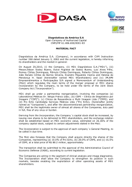
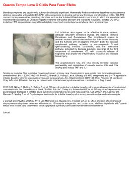
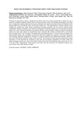
PMLNNLNO
pï ÉÉí=ëóåÇêçã É=~ë=íÜÉ=éêÉëÉåíáåÖ=ëóã éíçã =çÑ=Ü~áêó=ÅÉää=äÉì âÉã á~
Volume 15 Number 2
February 2009
DOJ
Contents
Sweet syndrome as the presenting symptom of hairy cell
leukemia
Filipa Ventura MD1, Joana Rocha MD1, Teresa Pereira MD1,
Herlander Marques MD2, Fernando Pardal MD3, Celeste Brito
MD1
Dermatology Online Journal 15 (2): 12
1. Dermatology and Venereology Department
2. Oncology Department
3. Pathology Department
Hospital de São Marcos, Braga, Portugal. [email protected]
Approximately 20 percent of patients with Sweet syndrome have
an associated cancer [1]. Hematologic malignancies, most commonly
acute myelogenous leukemia, account for 85 percent of the associated
malignancies [1, 2, 3]. The most common solid tumors are those of the
genitourinary tract [4]. We report the case of a patient with Sweet
syndrome as the presenting symptom of hairy cell leukemia.
Case Report
Figure 1
Figure 2
Figure 1. Violaceous papules and plaques on the back
Figure 2. The skin biopsy showed a diffuse neutrophilic
infiltrate within the upper dermis with involvement of several
follicular units (H&E, x100)
A 62-year-old caucasian man,
otherwise healthy, presented with a
3-week history of a progressive skin
eruption asymetrically distributed on
the upper extremities and trunk.
Dermatologic findings were
accompanied by fever (39ºC),
asthenia, anorexia, and weight loss.
He was not taking any medication.
There was no family history of skin
Figure 3
disorders. Physical examination
revealed asymptomatic, violaceous, Figure 3. Bone marrow
ÇÉêã ~íçäçÖóKÅÇäáÄKçêÖLNRMOLäÉííÉêëLÜ~áêóÅÉääLî Éåíì ê~KÜíã ä
NLP
PMLNNLNO
pï ÉÉí=ëóåÇêçã É=~ë=íÜÉ=éêÉëÉåíáåÖ=ëóã éíçã =çÑ=Ü~áêó=ÅÉää=äÉì âÉã á~
round, or irregularly shaped papules biopsy showed hypercellular
and plaques, covered with a fine
infiltrated with malignant
scale. The lesions varied in diameter lymphocytes (H&E, x400)
from 0.5 to 4 cm (Fig. 1). The patient
had no adenopathy; there was no mucous or ocular involvement. Skin
biopsy was taken and sent for histopathology and cultures.
Histopathology showed a diffuse neutrophilic infiltrate within the upper
dermis with involvement of several follicular units, prominent
leukocytoclasis, and absence of necrotizing vasculitis. Culture for
bacteria, mycobacteria, and fungi were negative. The diagnosis of Sweet
syndrome was made. Laboratory investigations revealed a macrocytic
anemia (10.2 g/dL), leukopenia (2.5 x 10⁹ /L, with neutropenia and
lymphopenia), and thrombocytopenia (96 x 109 /L). Some of the
lymphocytes were atypical. Therefore, a bone marrow biopsy (Fig. 2)
and the immunophenotype of bone marrow aspirate were performed.
They were compatible with the diagnosis of hairy cell leukemia. Chest
and abdominal CT scan showed axillary and mediastinal
lymphadenopathies and a pulmonary involvement characterized by
several micronodules. Systemic prednisolone 60 mg daily had been
initiated however patient's condition deteriorated and he died one month
later due to a sepsis.
Discussion
The pathogenesis of Sweet syndrome remains to be definitively
determined. Indeed, it may be multifactorial and many etiologies, not
necessarily exclusive, have been postulated. A hipersensitivity reaction
to an eliciting bacterial, viral, or tumor antigen may promote the
development of Sweet syndrome is one of the hypothesis [5, 6].
Hairy cell leukemia (HCL) is an uncommon, chronic B cell
lymphoproliferative disorder characterized by pancytopenia,
splenomegaly, and the presence of atypical lymphocytes in the bone
marrow and peripheral blood. Termed "leukemic reticuloendotheliosis"
in 1958 and renamed hairy cell leukemia in the 1960s to describe the
cytoplasmic projections observed on the surface of the malignant cells.
HCL is usually an indolent disorder whose course is dominated by
pancytopenia and recurrent infections. This disease affects
predominantly middle-aged men [7]. As for other types of leukemia, the
cause of HCL is not known. Patients can be asymptomatic at
presentation or present with a variety of clinical signs that include
anemia, bleeding, and life-threatening sepsis [8]. Approximately onequarter of patients present with fatigue, weakness, and weight loss [8],
just like our patient.
The association between Sweet syndrome and malignancies is very
significant. Recommendations for the initial malignancy workup in newly
diagnosed Sweet syndrome patients without a prior cancer were
proposed by Cohen and Kurzrock in 1993 [1]. Malignancy-associated
Sweet syndrome is most commonly related to acute myelogenous
leukemia. Our patient presented Sweet syndrome associated with hairy
cell leukemia, which has rarely been reported. This is the eighth case of
this association reported in literature [8].
References
1. Cohen PR, Kurzrock R. Sweet syndrome and cancer. Clin Dermatol.
1993;11:149-157. [PubMed]
ÇÉêã ~íçäçÖóKÅÇäáÄKçêÖLNRMOLäÉííÉêëLÜ~áêóÅÉääLî Éåíì ê~KÜíã ä
OLP
PMLNNLNO
pï ÉÉí=ëóåÇêçã É=~ë=íÜÉ=éêÉëÉåíáåÖ=ëóã éíçã =çÑ=Ü~áêó=ÅÉää=äÉì âÉã á~
2. Cohen PR et al. Malignancy-associated Sweet's syndrome: Review
of the world literature. J Clin Oncol. 1988;6:1887-1897. [PubMed]
3. Haverstock C, Libecco JF, Sadeghi P, Maytin E. Tender
erythematous plaques in a woman with acute myelogenous leukemia.
Arch Dermatol. 2006;142:235-240. [PubMed]
4. Hussein K, Nanda A, Al-Sabah H, Alsaleh QA. Sweet's syndrome
(acute febrile neutrophilic dermatosis) associated with adenocarcinoma
of prostate and transitional cell carcinoma of urinary bladder. J Eur
Acad Dermatol Venereol. 2005;19:597-599. [PubMed]
5. Cohen PR. Sweet's syndrome - a comprehensive review of an acute
febrile neutrophilic dermatosis. Orphanet Journal of Rare Diseases.
2007;2:34-62. [PubMed]
6. Honigsmann H, Cohen PR, Wolff. Acute febrile neutrophilic
dermatosis (Sweet's syndrome). (Chapter 94). In Fitzpatrick's
Dermatology in General Medicine 6th edition. Edited by: Freedberg IM,
Eisen AZ, Wolff K, Austen KF, Goldsmith LA, Katz SI, Fitzpatrick
TB. New York: McGraw-Hill Health Professions Division;2003:949955.
7. Tallman MS, Hakimain D, Peterson LC. Hairy cell Leukemia. In
Clinical Oncology 2th edition. Camden NJ. Churchill
Livingstone;1999:2564-2578.
8. Levy RM, Junkins-Hopkins JM, Turchi JJ, James WD. Sweet
syndrome as the presenting symptom of relapsed hairy cell leukemia.
Arch Dermatol. 2002;138:1551-1554. [PubMed]
© 2009 Dermatology Online Journal
ÇÉêã ~íçäçÖóKÅÇäáÄKçêÖLNRMOLäÉííÉêëLÜ~áêóÅÉääLî Éåíì ê~KÜíã ä
PLP
Baixar

![(Microsoft PowerPoint - Curso de Ver\343o mspo 2013 [1].ppt](http://s1.livrozilla.com/store/data/000822103_1-1d7d29af114ca4d1538a6317d5129437-260x520.png)