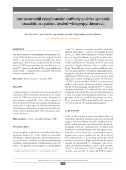
Lincoln Sakiara Miyasaka, Ary de Andrade Junior,
Carlos Eduardo Bueno, Alvaro Nagib Atallah
Relapsing Polychondritis
Internal Medicine Division of Hospital São Paulo, Escola Paulista de Medicina 01
Universidade Federal de São Paulo - São Paulo, Brazil
Purpose: This article describes a clinically-diagnosed case 01relapsing polychondritis (RP), attended at the Hospital São Paulo, and
presents a literature review 01the subject. Source of research: The literature review was made via Medline (1990-96), Lilacs (198096). textbooks 01rheumatology, and some articles about the history 01the disease. In Medline, 113 articles Irom 1990 to 1996 were
lound, and there were 23 articles lrom 1980 to 1996 in Lilacs. Research procedure: We reviewed the articles available at BIREME
(Biblioteca Regional de Medicina) with the primary locus being on the disease in questiono Summary: RP is arare disease 01unknown
etiology described initially by Jackson-Wartenhorst in 1923 and characterized by a recurrent and acute inllammatory process that
causes the collapse 01the cartilaginous structures and their subsequent replacement by librous connective tissue. The cartilage most
commonly attacked is that 01the auricle 01the ear and nasal septum, while the cartilage 01 the trachea, larynx, epiglottis, ribs, and
articulations may also be involved. Ocular inflammations and systemic reactions with leve r are also described. In 1976, McAdam
presented a complete prospective study 01 23 patients, reviewed the 136 cases. described up until that time, and then proposed
diagnostic criteria which were later expanded by Damiani and Levine. Currently, more than 500 cases have been described. Conclusion:
Although arare disease, better knowledge 01it is needed, as RP may be lethal with tracheal collapse and obstruction 01 respiratory
pathways, making precise diagnosis and adequate therapeutic intervention necessary.
UNITERMS: Relapsing polychondritis.
CASE REPORT
A
White Brazilian male, aged 31, fireman, native
of Terra Boa, Parana, and resident of Poa, São
Paulo, was interned on July 3, 1996. The patient
came to the hospital with a history of chest pain for one
month.
Eleven months before this, with no prior symptoms,
the patient began to feel stabbing pains localized in the
region between the shoulder blades, which worsened with
movement
and physical
force. Ten months before
Address for correspondence:
Lincoln Sakiara Miyasaka
Rua Teodoro Sampaio, 972 - casa 18
São PaulolSP - Brasil - CEP 05406-050
MIYASAKA, L.S.: ANDRADE
Relapsing polychondri1is
JUNIOR,
A.; BUENO, C.E.; ATALLAH, A.N. -
internment, pain and redness in the Ieft eye appeared. The
patient then consulted a physician who said it was an
allergy and prescribed prednisone 20 mg. During this
period, the patient received a light trauma to the nose
which provoked an intense, but painless edema. It was
observed that, with the use of the prescribed medication,
alI of the symptoms disappeared, but left as a sequeI a
depression in the nose. Two months before internment,
the patient stopped taking the medication and immediately
the pain reappeared in his back at a high intensity. and
also appeared in the wrists and knees. One month before
internment, in addition to the pain in the back, wrists and
knees, the patient began to experience chest pain which
was aggravated by palpation and deep breathing, a daily
fever of 38.S - 39.0 "C, sweating, chilIs. lack of appetite,
prostration, weight loss (7 kg in one month, 13 kg total),
São Paulo Medical JournallRPM
116(1): 1637-1642, 1998
1638
dry cough and dyspnea when coughing. The patient denied
having addictions to tobacco or alcohol.
Physical examination showed the patient to be in
regular general condition, fever (38°C), slimmed, eupneic,
anemic, HR= 94, arterial blood pressure 11/8 em of Hg.
On his face, a depression in the nose was observed
(photo). A decrease of vesicular murmur in both lungs
without abnormal rale was noted. There was no alteration
in the heart, nor in the abdomen. There was pain in the
articulations of the knees, wrists, and costo-condral
articulations without local heat or redness.
LABORATORY FINDINGS
Creatinin = 0.5 mg/dl; Potassium = 4.5 mEqll; sodium
= 143 mEqll; Bilirrubin T= 0.4, D = 0.2, I = 0.2 GOT = 9
UIl, GPT = 11UIl; Erythrocyte count =4.4 x 10 I mm';
Hbe 12.2 g/dl; Ht = 38%; MCH = 27.73 pg; MCHC =
32.11 g/dl; MCV
86.36; platelet, 394,000; leukocyte
= 14,100 (neut = 76, eos = 3, bas = O, limph = 12, mono =
=
=
9); Alb = 3.1 g/dl; Urinalysis: pH = 6.5, D = 1020, prot (), gluc(-), leukocytes = 12,000/mI, erythrocytes = 800,0001
ml dismorphism (-), calcium oxalate crystals = few;
calcium = 284 mg in 1,620 ml urine in 20 h; uric acid =
729 mg in 1,620 ml of urine in 20 h.
Antinuclear antibody (-), anti native DNA antibody
(-), anti ENA antibody (UI-RNP, anti-Sm, anti SS-AlRo,
anti-SS-BlLa) = (-);CH50 = 188 U(130-330); Rheumatoid
factor (-); cryoglobulin (-); anticardiolipin antibody (-);
ANCA (-); C reactive protein = 1/16; LE cels (-);
Mucoprotein = 5.3; anti streptolisin O < 200 Ul/rnl; VDRL
(-); urine protein excretion (-); creatinin clearance = 124
ml!min/1.73m; urine culture (-).
Total protein = 6.7 (6-8 g/dl), Alb = 2.91 (3.2 - 5.6);
alpha 1 = 0.21 (0.1 - 0.4); alpha 2 = 1.27 (0.4 - 1.1); beta
= 1.13 (0.1 - 1.2); gamma = 1.18 (0.5 - 1.6); Erythrocyte
Figure - "Saddle" aspect 01the nose.
São Paulo Medical
JournallRPM
116(1): 1637·1642,
1998
MIYASAKA,
L.S.; ANDRADE
JUNIOR,
A.; BUENO, C.E.; ATALLAH, A.N. Relapsing polychondritis
1639
=
sedimentation
rate
53 (1-7 mm/h); acid alpha 1
glucoprotein
395 (43-130 mg/dl).
Prothrombin Time = 80.8 (>70%); RNI = 1.19; PTT
= 1.0 «1.25); Bleeding Time = 1 (1-3 min); coagulation
time = 7 (4-9).
X ray examination of facial bones showed the nasal
bone without alteration,
and cartilage
hipoplasia.
Ophthalmologic
examination showed episcleritis in the
left eye.
Renal biopsy results were: optic and immunofluorescence microscopy normal.
=
EVOLUTION
Diagnosis of relapsing polychondritis
(RP) was
made clinically on July 9, 1996, and treatment
with
prednisone 1 mg/kg (60 mg per day) started. There was a
prompt subsidence of articular pain, fever and general
condition, with only hoarseness remaining.
The patient is now being followed up as an outpatient
receiving 40 mg of prednisone per day.
LlTERATURE
REVIEW
Epidemiology
The prevalence of RP is equal among men and
women. It has been described in all races but is more
frequent among Caucasians (17).
Clinical Findings
(6"2)
RP is characterized
by acute inflammatory
episodes of cartilaginous
structures that might recover
spontaneously,
but that frequently
relapse
and
culminate with the destruction of its structure. It may
present systemic involvement with fever, weight loss,
and other organs such as central nervous
system,
kidneys, respiratory tract, and blood vessels.
The most frequent clinical features of 2 literature
reviews with 159 and 112 cases were summarized
by
Andres J Politi (Table I).
The most frequent clinical feature in both series is
auricular chondritis. The auricular pavilion is swollen,
MIYASAKA, LS.; ANDRADE
Relapsing polychondritis
JUNIOR,
A.; BUENO,
C.E.; ATALLAH,
A.N ..
Table 1
Clinical features in the course of relapsing
pofychondritls'": comparison of 2 series
Me Adam
n = 159
Auricular Chondritis
Arthritis
Nasal Chondritis
Ocular Manifestation
Larynx- Tracheal
Inner Ear
Cardiopathy
Vasculopathy
Nephropathy
n
=
88.6%
81%
72.4%
65.4%
55.9%
45.9%
11.9%
16.3%
NR
Isaak
n
=
112
85%
52%
54%
51%
48%
43%
6%
14%
26%
number of patients
painful and angry-red in colour. In 95% of cases it is
bilateral. It may provoke the narrowing of the external
auditive canal. The ear lobe is preserved. The process may
subside spontaneously,
or it may destroy the cartilage,
leaving a "cabbage-like"
appearance. It may present as a
poly or oligoarthritis.
Nasal chondritis
leads to the destruction
of the
cartilaginous portion of the nasal septum, giving the nose
a "saddle-Iike" appearance.
The ocular alterations
are unspecific,
such as
conjunctivitis,
uveitis, retinopathies,
optical neuritis,
palpebral edema, and proptosis. Episcleritis is the most
frequent, occurring in 39 per cent of cases (21).
Respiratory
manifestations
are hoarseness,
dyspnea, a choking sensation, wheezing, and pain in
the laryngotracheal
articulations.
These can provoke
obstruction
through three mechanisms:
edema of lhe
glottis, trachea and bronchi; formation of a fibrous mass
in the airways; and collapse of the tracheal cartilage.
The obstruction is complicated
by infection.
Hearing loss may be experienced due to obstruction
of the external auditory canal, middle ear otitis, or by
affliction of the auditory branch of the VIII cranial nerve
due to vasculitis. Nausea, dizziness, vomiting, and ataxia
by neuritis of the cochlear branch due to vasculitis may
also be present.
Vasculitis
is also found in some cases of RP.
According to Michet, systemic vasculitis is the most
frequent associated inflammatory syndrome (17) but this
was not Politi's finding in his summary of 271 cases of
polychondritis (6). There are cases described of aortal and
mitral insufficiency, but these are rare.
São Paulo Medical JournallRPM
116(1): 1637-1642,
1998
, 1640
Histopathology
Table 2
Diseases associated with relapsing polychondritis
Rheumatoid Arthritis
Sjogren's Syndrome
Systemic Lupus Erithematous
Systemic Sclerodermia
Psoriatic Arthritis
Raynaud's Syndrome
Thyroiditis
Ulcerative Colitis
Glomerulonephritis
Disgammaglobulinemia
Pernicious Anemia
Neoplasm
Other
5%
3.1%
1.25%
1.25%
1.25%
1.25%
5%
1.88%
1.25%
1.25%
0.62%
3.1%
6.9%
In a review of 129 patients with RP, 29 presented
affected
kidneys.
Mesangial
expansion,
cellular
proliferation, and necrotizing glomerulonephritis
were
observed. Electron microscopy detected dense deposits
at the mesangial leveI, and direct immunofluorescence
revealed C3 and 19M in the mesangio (6).
Neurological
manifestations
incIude ophthalmic
neuritis of the oculomotor, facial and auditive-vestibular
neuritis,
headaches,
encephalopathy,
convulsions,
hemiparesias and ataxias.
Associated Oiseases
Approximately 25 percent of RP cases are associated
with other diseases, with the following being the most
common according to Politi.
Complementary Exams
No complementary exam is diagnostic for RP; ali
are only slightly sensitive and specific. Their utility is only
to discard other pathologies and recognize other ailments.
The erythrocyte sedimentation rate and reactive C
protein are the best exams to accompany the evolution of
the disease. Tadaki et aI studied the composition of urinary
glucosaminoglican
and found an elevation of dermatan
sulphate and hyaluronic acid, both of skin origin in the
active phases of the disease and not the 4-6 sulphate
condroitin that would be expected in cases of cartilage
destruction (24). So further studies are necessary in this area.
Antibodies to type Ir collagen have been reported to
correlate with the activity of the disease (211).
São Paulo Medical JournallRPM
116(1): 1637-1642,
1998
The histopathological
firrdings are diagnostico
During the active phase, there is a loss of cartilage
metacromasi s, chondral
and perichondral
necrosis,
infiltration
by the polymorphonucIear
neutrophils,
plasmacytes,
Iymphocytes,
apoptotic
bodies,
and
perichondral hemorrhage. In the advanced stage, there is
cicatrical
repair with lymphohistiocytic
infiltration,
fibrosis, deposit of hemosiderin and loss of elastic tissue.
Studies with direct immunofluorescence
revealed
positivity for 19A, 19M, 19G and C3, suggesting
the
presence of circulating immune comp\exes (22,23).
Etiopathogenesis
The etiopathogenesis
is not clear, but there are indications of auto-irnmune
involvement.
RP is associated
with auto-imrnune diseases in 25% of cases; imrnunoglobulins and lesion comp\ements
are encountered,
as are
type II anticollagen antibodies in the serum of patients
with active RP. Clinical improvement occurs with immunosuppression therapy.
OifferentialOiagnosis
Considering the general clinical situation , the most
difficult
differential
diagnosis
is with Wegener's
granulomatosis (WG) and other systemic vasculitis. WG
is manifested as a systemic vasculitis with collapse of the
nasal wall, arthritis, and aff1iction of the respiratory
system. WG can be differentiated
by the presence of
granulomas, involvement of lungs and kidneys, and the
lack of auricular pavilion involvement.
The "saddle" nose can also be found in WG and
leishmaniosis,
advanced
congenital
syphilis,
lupus,
sarcoidosis, the lepromatous type of leprosy, basocellular
epitheliomas,
cocaine
inhalation,
aspergilosis
and
paracoccidioidomicosis.
Prognosis
The five-year survival rate is 74 per cent (14) The
most frequent
reason for death is infection
of the
respiratory tract, secondary to the bronchial obstruction
and the use of corticosteriods.
Other causes are systemic
vasculitis,
acute respiratory
insufficiency
due to
respiratory
collapse,
and renal insufficiency
due to
glomerulonephritis.
Treatment
The treatment
of RP consists
nonsteriod
anti-inflammatories
MIYASAKA,
L.S.; ANDRADE
JUNIOR,
of lhe use of
and low dose
A.; BUENO, C.E.; ATALLAH, A.N. Relapsing polychondritis
1641
corticosteroids
in situations
of mild auricular/nasal
chondritis
or arthritis.
For cases
with serious
manifestations
such as laryngotracheal
or ocular
symptoms, inner ear inflammation,
severe auricular or
nasal chondritis,
systemic
vasculitis,
aortitis
or
glomerulonephritis,
prednisone at a dose of 1 mg/kg/
day is indicated. The use of immunosuppressors
such
as azathioprine and cyclophosphoamide
is reserved for
those patients refractory to steroid therapy (13). The use
. of dapsona,
colchicina
(15), anti-CD4
monoclonal
antibody, D-penicillamine
and antimalarials
has also
been described. The increasing number of successful
reports of cyclosporin
A in a dose of 15 mg/kg/day
suggests that it may be the best tolerated and potentialIy
most reliable drug for the corticosteroid-resistant
patient (14,16), Nebulized racemic ephedrine is an option
used successfully in cases of acute subglotic edema (19).
Continuous
positive
airway pressure
masks may
provi de relief of severe dyspneic symptoms (17),
DISCUSSION
RP is arare
disease seldom described
in the
literature. Only 500 cases have been published worldwide
up until now. In Brazil, only four articles were encountered
from 1990-96 (search by Lilacs).
Histological
diagnosis
is made with difficulty
because cartilage material cannot be obtained for analysis,
or because findings are uncharacteristic,
principalIy in the
advanced stages when there is destruction of the structures
and their replacement
by fibrosis. When there is no
possibility of a histological diagnosis, it must be made
clinicalIy. According to Damiani and Levine, this clinical
diagnosis can be confirmed by:
1) Three of McAdam 's criteria: bilateral auricular
chondritis,
polychondritis,
nasal chondritis,
ocular inflammation
(conjunctivitis,
scleritis, .
episcleritis, uveitis or queratitis), chondritis of
the respiratory tract or involvement
of the VII
cranial
nerve,
even without
histological
confirmation;
2) One or more of McAdam 's criteria
with
histological
confirmation;
3) Two or more affected sites with response to
treatment with corticoids or dapsona.
The case in question presents four of McAdam's
criteria: nasal chondritis,
polychondritis,
episceritis,
and chondritis of the respiratory
tract, in addition to
the immediate response to corticotherapy.
In spite of
not having histological
confirmation,
we were able to
make the diagnosis
clinicaIly.
The hematuria
with
negative dysmorphism
and elevated urinary calcium
was interpreted as being due to urinary microca\culi,
which improves the prognosis.
The patient is being folIowed as an outclinic patient,
due to the possibility of relapses, renal involvement, and
airway obstruction,
all of which would require rapid
intervention.
RESUMO
Objetivo: O propósito deste artigo e a descrição de um caso com diagnostico clinico de Policondrite Recorrente atendido no
Hospital São Paulo e realizar uma revisão da literatura sobre o tema. Fonte de dados: A pesquisa foi feita através do
Medline(1990-96) e Lilacs (1980-96) buscando os temas Policondrite Recorrente, e Policondrite Recidivante, alem de livros
textos de reumatologia e artigos mais antigos sobre a historia da moléstia. Seleção dos estudos: No Medline foram encontrados 113 artigos publicados em inglês, espanhol, e português entre 1990 e 1996. No Lilacs foram encontrados 23 artigos entre
1980 e 1996. Coleta dos dados: Foram analisados os artigos disponíveis na BIREME cujo enfoque primário era a moléstia
em questão. Síntese dos dados: Policondrite recorrente é uma doença rara de etiologia desconhecida descrita inicialmente
por Jacksch-Wartenhorst em 1923 e caracterizada por um processo inflamatório agudo e recorrente de cartilagens que
provoca o desabamento das estruturas cartilaginosas e sua substituição por tecido conjuntivo fibroso. As cartilagens mais
comumente acometidas são do pavilhão auricular e septo nasal, embora as cartilagens da traquéia, laringe, epiglote, arcos
costais e articulações também possam estar envolvidas. Inflamações oculares e reações sistêmicas como febre também são
descritas. Em 1976, Mc Adam apresentou um estudo prospectivo completo de 23 pacientes e revisou os 136 casos descritos
até aquela época, propondo critérios diagnósticos que foram loqo-arnpliados por Damiani e Levine. Atualmente já foram
descritos mais de 500 casos. Conclusão: Mesmo sendo uma doença rara, há necessidade de maior divulgação na classe
médica desta afecção, pois a policondrite recorrente pode se tornar letal por colapso traqueal e obstrução das vias respiratórias, sendo que um diagnóstico preciso e uma intervenção terapêutica adequada se fazem necessários.
MIYASAKA, L.S.; ANDRADE
Relapsing polychondritis
JUNIOR,
A.; BUENO,
C.E.; ATALLAH,
A.N ..
São Paulo
Medical
JournallRPM
116(1): 1637·1642,
1998
1642
-,
REFERENCES
1. Person CM, Kline HM, Victor D. Newcomer. Relapsing
Polychondritis. The New England Journal of Medicine
1960;263:51-58.
2. Kaye RL, Sones DA. Relapsing Polychondritis. Clinical
and Pathologic Features in Fourteen Cases. Annals of
Internal Medicine 1964;60(4):653-664.
3. Dolan DL, Lemmon GB, Teitelbaum SL. Relapsing
Polychondritis. Analytical Literature Review and Studies
on Pathogenesis.
American Journal of Medicine
1966;41:285-297.
4. Retamar RA, Leon AR, Spiner RE, Magnin PH. Estado
Actual de Ia Policondritis Cronicas Recurrente dos Nuveas
Observaciones. Rev Arg Derm 1993;74:85-96.
5. Michet CJ. Vasculitis and Relapsing Polychondritis.
Rheumatic Disease Clinics of North America 1990;
16(2):441-444.
6. Poli ti AJ. Polycondritis Recidivantes Revisión Crítica.
Archivos Argentinos de Dermatología 1993;T.XLIll:73-91.
7. Pereira MD, Araujo CM, Silva RC, Azevedo JRGM,
Ferreira MAT, Resende PS, Amaral MJCS, Pedroso ERP.
Associação entre Policondrite Recidivante e Tuberculose
Tratada com Corticóide e Ciclofosfamida - Relato de Um
Caso. Rev Méd Minas Gerais 1994;4(4):58-60.
8. Self J, Hammarsten JF, Lyne B, Peterson DA, Paul ST.
Relapsing Polychondritis.
Arch Intern Med 1967;
120:109-112.
9. Parrochia EB. Policondritis Atrofiante. Boi Hosp S J de
Dios 1994;41 (5):330-331.
10. Gibson GJ. Rare Rheumatic Disorders.
Relapsing
Polychondritis. Baillieres Clin Rheumatol 1993;7: 179-181.
11. Freedman SF, Amedee RG. Relapsing Polychondritis. J La
State Med Soe 1992;144:131-133.
São Paulo Medical
Journal/RPM
116(1): 1637-1642,
1998
-,
12. Perez MS, Lario AB, Lopez AJ, Valdinelso AJL. Acta
Otorrinolaringol Esp 1992;43:21:26.
13. Karpman WJ. Arthritis and Allied Condition. A Textbook
ofRheumatology, 13th ed. 1996;1595-1603.
14. Klippel JH, Dieppe PA. Rheumatology, 1994;6.31.1-6.31.4.
15. Goihman- Yahr M. Relapsing Polychondritis Responding
to Colchine. Int J Derrnatology 1994;33(6):448-9.
16. Ormerod AD, Clark LJ. Relapsing Polychondritis
treatment with cyclosporin
A. British Journal of
Dermatology 1992; 127:300-301.
17. Michet CJ. Relapsing Polychondritis of Koopman WJ
Arthritis
and Allied Conditions.
A Textbook of
Rheumatology, 13'" ed. 1997;82: 1595-1603.
18. Valenzuela R, Cooperrider PA, Gozate P, et aI. Relapsing
Polychondritis. Human Pathol 1980; 11: 19-22.
19. Gaffney RJ, Harrison M, Path FRC, Blayney AW. Nebulized
Racemic Ephedrine in the Treatment of Acute Exacerbation
of Laryngeal Relapsing Polychondritis. J Laryng Otol
1992; 106:63-64.
20. Foidart JM, Abe S, Martin GR et aI. Antibodies to type II
collagen in relapsing polycondritis. New England Journal
of Medicine 1978;299: 1203-1207.
21. Issak BL, Liesegang TJ, Michet CJ. Ocular and Systemic
Findings in Relapsing Polychondritis. Ophthalmology
1986;93:681-689.
22. Foidart JM, Katz SJ. Subtlevc lues to diagnosis by
immunopathology:
relapsing polychondritis.
Am J
Dermathopathol 1979; 1:257-260.
23. Bergfeld WF. Relapsing Polychondritis with positive direct
immunofIuorescence. Arch Dermatol 1978; 114: 127.
24. Tadaki T, Aiba S, Igarashi M, Tagami H. Analysis of
increased urinary acid glycosaminoglicans in a patient with
relapsing polychondritis.
Acta Derm Venereol 1987;
67:441-445.
MIYASAKA,
L.S.; ANDRADE
JUNIOR,
A.; BUENO,
C.E.; ATALLAH, A.N. -
Relapsing
polychondritis
Download