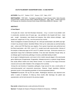
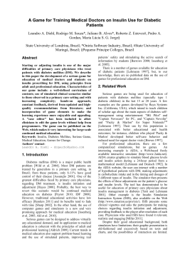
52
Note
Rev Iberoam Micol 2002; 19: 52-56
Zygomycosis: A report of eleven
cases and a review of the Brazilian
literature
Luiz Carlos Severo1, Flávio de Mattos Oliveira2, Rodrigo Dreher3, Paulo
Zimermann Teixeira4, Nelson da Silva Porto4 e Alberto Thomaz Londero5
Pesquisador do CNPq; Faculdade de Medicina, Universidade Federal do Rio Grande do Sul (UFRGS);
Laboratório de Microbiologia Clínica, Instituto Especializado em Pesquisa e Diagnóstico (IPD), Santa Casa,
Porto Alegre, RS, Brasil; 3Médico Residente, Santa Casa - Fundação Faculdade Federal de Ciências Médica de
Porto Alegre; 4Pavilhão Pereira Filho, Santa Casa; 5Universidade Federal de Santa Maria, Santa Maria, RS,
Brasil
1
2
Summary
Key words
Eleven cases of zygomycosis (mucormycosis) observed throughout an eighteen
year period (1982–2000) have been reviewed. The most important demographic
and clinical data of seven patients were tabulated. The remaining four are related as illustrative cases. Seven patients presented with the pulmonary form of
the disease; two patients presented with pulmonary manifestations associated
with sinusitis; and two patients presented with the rhinocerebral form.
Predisposing conditions, in decreasing order of frequency, were diabetes mellitus (6), renal transplantation (2), associated with pancreas-kidney transplantation
and diabetes (1), bone marrow aplasia (1), and chronic obstructive lung disease
treated with corticosteroids (1). The diagnoses were based on the detection of
characteristic zygomycetous hyphae in tissue. The causative organism was isolated and identified in only four cases; three were due to Rhizopus arrhizus, and
one to Absidia corymbifera. In addition the Brazilian literature on zygomycosis is
reviewed.
Zygomycosis, Mucormycosis, Zygomycetes, Rhizopus arrhizus,
Absidia corymbifera
Zigomicosis: informe de once casos y revisión de la
literatura brasileña
Resumen
Palabras clave
Dirección para correspondencia:
Dr. L.C. Severo
Laboratório de Microbiologia Clínica
IPD - Santa Casa, Annes Dias, 285
Porto Alegre 90020-090, RS, Brazil
Fax: +55 51 3214 8435
E-mail: [email protected]
Aceptado para publicación el 4 de Febrero de 2002
©2002 Revista Iberoamericana de Micología
Apdo. 699, E-48080 Bilbao (Spain)
1130-1406/01/10.00 Euros
Se revisaron once casos de zigomicosis (mucormicosis) observados durante un
periodo de dieciocho años (1982-2000). Los datos demográficos y clínicos más
importantes de siete pacientes fueran tabulados. Los cuatro restantes fueron
reportados como casos ilustrativos. Siete pacientes presentaron la forma pulmonar de la enfermedad, dos pacientes presentaron manifestaciones pulmonares
asociadas con sinusitis; y dos pacientes presentaron la forma rinocerebral.
Fueron condiciones predisponentes, en este orden de frecuencia: diabetes mellitus (6), trasplante renal (2), trasplante pancreas-riñón en paciente con diabetes
(1), aplasia de médula ósea (1) y enfermedad pulmonar obstructiva crónica tratada con corticosteroides (1). El diagnóstico fue hecho por la detección de las
hifas características en el tejido; sin embargo, el aislamiento y la identificación
del organismo causal fue hecha solamente en cuatro casos: Rhizopus arrhizus
(3) y Absidia corymbifera (1). Se comenta la revisión de la literatura brasileña
sobre zigomicosis.
Zigomicosis, Mucormicosis, Zigomicetes, Rhizopus arrhizus,
Absidia corymbifera
Zygomycosis is a group of opportunistic infections
caused by species of zygomycetes in the order Mucorales.
The agents of zygomycosis (or mucormycosis) normally
reside in the environment. The abundant airborne sporangiospores may be inhaled or contaminate wounds or burns
of patients with predisposing conditions, such as diabetes
or immunosuppression, or trauma, and cause a spectrum
of diseases. Zygomycosis results from the invasion of the
major blood vessels by the fungus, which grows in their
walls and lumen causing thromboembolism, ischemia, and
necrosis of the tissues. The infection is usually acute and
rapidly progressive; however, chronic forms and colonization of some the sinuses and other anatomic sites have
been reported [1,2].
Zygomycosis in Brazil
Severo LC, et al.
53
Table 1. Clinical data on seven cases with zygomycosis.
Case No.
Patient
Age, Sex
1
2
44, M
69, M
3
41, F
4
5
55, F
25, M
6
62, F
7
48, M
Clinical form/Lesions
Predisposing conditions
Agent
Therapy /Outcome
Pulmonary/Lobar consolidation
Pulmonary/ Lobar consolidation
and pleural effusion
Pulmonary/Lobar consolidation
and pleural effusion
Lung transplantation
Chronic obstructive pulmonary
disease and corticoids
Diabetes mellitus
Zygomycetes
Rhizopus arrhizus
None/Died
Amphotericin B /Died
Rhizopus arrhizus
Amphotericin B /Died
Pulmonary/Multiple masses
Rhinocerebral/Cephalic
structures
Rhinocerebral/Cephalic
structures
Pulmonary/Lobar consolidation
and paranasal sinus
Diabetes mellitus
Diabetes mellitus
Zygomycetes
Zygomycetes
Amphotericin B /Alive
Amphotericin B and surgery/Alive
Diabetes mellitus
Zygomycetes
Amphotericin B /Alive
Politraumatism
Zygomycetes
Amphotericin B /Died
In the paper we present the data of 11 cases of
zygomycosis collected throughout an eighteen year period
in Porto Alegre, RS (Brazil). We also review the Brazilian
literature on the mycosis.
CLINICAL MATERIALS AND METHODS
Eleven patients in our records fulfilled the histological criteria for a diagnosis of zygomycosis: the finding
of broad, nonseptate hyphae, branching at right angles in
smears or in cut sections of tissues. Data on seven of these
cases are presented in Table 1. The remaining four cases
are related as illustrative cases.
REPRESENTATIVE CASES
Case 8. A six year-old Caucasian boy with bone
marrow aplasia had been treated with prednisolone for
four weeks. His blood findings showed a leukocyte count
of 1000/mm3 and an absolute neutrophil count of 500.
Complaining about fever, fatigue and epistaxis, he was
hospitalized, received many blood transfusions and wide
spectrum antibiotics (doxycycline and cefuroxime). After
some days of improvement, he became febrile and presented nasal obstruction. An X-ray revealed opacity of the
maxillary sinus and the ethmoidal cells. A punctiform skin
lesion appeared near the right mammilla, which was initially erythematous but enlarged and became necrotic in
three days, measuring 5 cm in diameter. An hematic bubble involved the lesion (Figure 1). A needle aspiration
specimen, obtained from the lesion, microscopically revealed many wide nonseptate hyaline hyphae with right
angle branching (Figure 2). Absidia corymbifera was
identified in the isolated cultures. A Chest roentgenogram
showed an extensive consolidation in the right upper lobe
(Figure 3). The patient received amphotericin B (0.5
mg/kg increased to 1 mg/kg) and vancomycin. In spite of
this treatment the pulmonary lesion worsened and a consolidation in the left lung appeared (Figure 4). The skin
lesion increased abruptly with destruction of the pectoral
muscles exposing the ribs. The patient presented then
hemoptysis, necrotic sputum, signs of pulmonary insufficiency, shock, and died.
Comments: Several cases of cutaneous zygomycosis lesions have been described. The majority of which
were due to local factors (traumatic implantation, especially in patients with burns). The remaining cases were
associated with additional forms of zygomycosis, among
which pulmonary disease was less common [3].
Figure 1. Case 8, necrotic lesion (5cm ) on the thoracic wall of a patient
with bone marrow aplasia.
Figure 2. Case 8, showing broad, nonseptate hyphae branching at right
angles from the skin lesion of the figure 1.
Figure 3. Case 8, chest X-ray showed an extensive consolidation in the
right upper lobe.
54
Rev Iberoam Micol 2002; 19: 52-56
Case 9. This patient, a 44 year-old man, with an
acute myelogenous leukemia had been treated with daunorubicin, cytarabine, and prednisone. He sought medical
attention with fever and oral candidosis. An abdominal
examination disclosed enlargement of the liver and spleen. White blood cell count 2x10⁴/mm3 (90% blasts), platelets 1.5x10³; bone marrow aspirate was hipercellular with
100% blast cells. A chest roentgenogram had showed consolidation with a cavitating lesion in the right lower lobe
(Figure 5). A transbronchial biopsy by fiberoptic bronchoscope had been performed. Hyphae consistent with
those of a zygomycete were detected in the direct examination of biopsied tissue but no growth was obtained in
culture. Fluconazole, administered for candidosis, was
changed to intravenous amphotericin B and antibacterial
therapy (ceftriaxone, gentamicin, and oxacillin) were
added.
The patient serum contained a high titer of specific
antibody to zygomycete antigens (1:800, measured by
enzyme immunoassay). After clinical and radiological
improvement, the patient was submitted for a lobectomy.
Macroscopic examination of the excised lobe revealed
parenchymal abscesses and infarctions; the subsegmental
bronchus and artery were partially necrotic; subsegmental
branch of the basal artery was occluded by a thrombus.
Microscopic examination of cut sections stained with
hematoxylin-eosin (H&E) and Gommori metenamine silver (GMS) disclosed extensive tissue invasion by wide,
nonseptate hyphae, branching at right angles, especially in
the wall and lumen of the thrombosed artery. Fungal culture of the surgical specimen was negative. Treatment
with amphotericin B continued after the surgery (total
dose 1.6 g). The patient was discharged in good conditions but died by leukemia.
Comments. This case illustrates the importance of
the association of antifungal therapy with surgical procedures [4].
Case 10. Two years ago (from 1995), a 55 year-old
man was submitted to a renal transplant, and he was using
immunosuppressive drugs (prednisone and ciclosporine).
The patient was also a diabetic and used insulin. He was
admitted to a hospital complaining about cough with
purulent expectoration, fever, cyanosis, nausea, and vomiting. He received amikacin and cephalexin. Six days later
the patient was transferred to our hospital in diabetic ketoacidosis.
The admission laboratory findings were as follow:
glycemia 474 mg/dl; uremia 77 mg/dl; qualitative ketonenmia positive. The serologic test for cytomegalovirus
was positive. Chest X-ray showed, in the right upper lobe,
consolidation, in which CT scan revealed a central cavitation (Figure 6). A biopsy specimen from a necrotic area of
the right upper lobe was obtained by fiber optic bronchoscopy. Broad non-septate hyphae invading the bronchial
wall were seen. Rhizopus arrhizus was isolated in culture.
Doses of the immunosuppressive drugs were lowered and
amphotericin B administration was started. Fifteen days
later, when the patient would be went to surgery, he presented a massive hemoptysis and died.
Autopsy findings included intense hemorrhagic
aspect of the lung, respiratory tree fully of coagula, the
posterior segmental bronchus of the upper right lobe communicated with a large cavitation (7x5 cm) with necrotic
aspect and alveolar hemorrhage. Hyphae of the zygomycete were detected microscopically in the thrombosed vessels and in the wall of the cavitation.
Comments. In this renal transplanted, diabetic
patient with ketoacidosis, immunosuppressive therapy,
Figure 4. Case 8, pulmonary lesion worsened, appearing consolidation in
the left lung.
Figure 5. Case 9, chest roentgenogram showing consolidation with cavitating lesion in the right lower lobe.
Figure 6. Case 10, chest CT scan showing consolidation with central cavitation in the right upper lobe.
Figure 7. Case 11, the CT-guided fine needle transthoracic biopsy.
Zygomycosis in Brazil
Severo LC, et al.
55
Table 2. Summary data of data from 27 reported cases of zygormycosis in Brazil (1959-1996).
Ref.
[11]
[11]
[12]
[13]
[14]
[15]
[16]
[17]
[18]
[19]
[20]
[21]
[21]
[22]
[23]
[24]
[25]
[26]
[27]
[28]
[29]
[30]
[31]
[32]
[33]
[34]
[35]
Year
59
59
70
73
75
77
80
81
82
82
83
85
85
85
86
86
86
87
87
87
88
89
90
93
95
96
96
Patient
Age
Sex
Clinical form
Predisposing
Diagnosis
Isolated agent
10m
1y
23y
8m
38y
46y
15y
35y
50y
29y
15y
57y
49y
61y
44y
38y
20y
9y
47y
39y
23y
39y
34y
61y
19y
6y
62y
F
F
M
M
F
M
M
F
M
F
F
F
M
M
M
M
F
M
M
M
F
F
M
M
M
M
M
Gastrointestinal
Gastrointestinal
Rhinocerebral
Meningoencefalitis
Gastric
Rhinocerebral
Rhinocerebral
Gastric
Gastric
Rhinocerebral
Rhinocerebral
Rhinocerebral
Rhinocerebral
Subcutaneous
Disseminated
Gastric colonization
Disseminated
Cutaneous
Cerebral
Rhinocerebral
Rhinocerebral
Chronic sinusitis
Disseminated
Nasal septum
Cutaneous*
Rhinocerebral
Pulmonary
Malnutrition
Malnutrition
Diabetes
Diabetes
Diabetes
Corticoids
Pacemaker
Diabetes
Diabetes
Diabetes
Diabetes
Diabetes
Diabetes
Gastric ulcer
Acidosis
Diabetes
Alcoholism
Diabetes
Corticoids
Leukemia
Diabetes
Leukemia
Leukemia
Necropsy
Necropsy
Necropsy
Necropsy
Necropsy
Surgery
Surgery
Surgery
Necropsy
Biopsy
Biopsy
Biopsy
Biopsy
Biopsy
Surgery
Biopsy
Biopsy
Biopsy
Necropsy
Surgery
Biopsy
Surgery
Biopsy
Biopsy
Biopsy
Biopsy
Surgery
Rhizopus arrhizus
Rhizopus arrhizus
Rhizopus arrhizus
Rhizopus arrhizus
Rhizopus arrhizus
Rhizomucor pusillus
Absidia corymbifera
Cunninghamella bertolletiae
y = year, m = month, M = male, F = female, * 15 months previously the patient presented pulmonary zygomycosis
and concomitant cytomegalovirus infection was predisposed to bronchovascular zygomycosis. The bronchoscopic
biopsy providied a prompt diagnosis. The surgery, an
urgent problem [5] unfortunately was delayed.
Case 11. An associated pancreas-kidney transplantation was performed in a 33 year old woman at the end
stage of a renal disease secondary to diabetic nephropathy.
The post operative course was complicated by Candida
albicans peritonitis, cytomegalovirus esophagitis and
Gram negative bacilli pneumonia in the left upper lobe.
After two week-treatment with fluconazole, ganciclovir
and imipenem the patient had improved. Twenty days
later a chest roentgenogram did not show any pulmonary
lesion in the left upper lobe; however, it revealed a solitary nodule adjacent to the pleura at the right upper lobe.
A CT guided fine needle transthoracic biopsy obtained
(Figure 7), and from this specimen, hyphae of a zygomycete were detected. Amphotericin B was started. The
patient was discharged from the hospital but continued to
use amphotericin B as an out patient.
Comments. CT scans revealed significant unsuspected abnormalities in 26% of patients with zygomycosis
[6]. The CT-guided fine needle transthoracic biopsy may
provide additional benefit in early management of the
mycosis.
This case has emphasized the importance of a surgery. As other similar cases have been healed with AMB
and surgery or only with a lobectomy [7].
DISCUSSION
The main clinical forms of zygomycosis are rhinocerebral, pulmonary, cutaneous, gastrointestinal and disseminated [1,2,8-10]. However, the disease may also
manifest itself as an isolated involvement of internal
organs: brain, kidney, and heart. Mucorales may also be a
colonizer of anatomic sites such as stomach, vagina,
external ear, sinus, or they be cause of allergic disease [1].
In the period 1959-1996, 27 cases of infection by
Mucorales could be gathered in the Brazilian literature
(Table 2). Eleven cases are added in the present report
(Table 1 and cases 8-11). All the major clinical forms of
the mycosis have been observed in Brazil. However, cases
of the rhinocerebral form predominated in earlier reports,
and the pulmonary form was more common in the present
series. Diabetes has been the most frequent predisposing
condition to the mycosis. The infection of isolated
organs/sites have been reported rarely. However, two of
these cases deserve to be pointed out: 1) an ulceration of
the nasal septum [32]; and 2) a case of chronic sinusitis
caused by an association of a Mucoraceae and an
Aspergillus spp. [30].
The diagnosis of zygomycosis was based on histopathological examination of surgical or biopsied specimens. Isolation and identification of the agent was
obtained in 12 of the 38 observed cases. As usual, the
rapidly growing, thermotolerant Rhizopus arrhizus was
most frequent. The pulmonary form of the mycosis is difficult to diagnose due to the several non-specific radiological patterns presented and the usual necessity of an
invasive approach to obtain clinical material to examine
[4,6,36].
We are grateful to the kindness of Dr. Leo Kaufman (CDC,
Atlanta, GA, USA) for performing the serologic test in case 2.
56
Rev Iberoam Micol 2002; 19: 52-56
References
1.
2.
3.
4.
5.
6.
7.
8.
9.
10.
11.
12.
13.
14.
Ribes JA, Vanover-Sams R, Sams CL,
Baker D. Zygomycetes in human diseases. Clin Microbiol Rev 2000; 13: 236301.
Rinaldi MG. Zygomycosis. Infect Dis Clin
North Am 1989; 3:19-41.
Adam RD, Hunter G, DiTomasso J,
Comerci Jr. G. Mucormycosis: emerging
prominence of cutaneous infections. Clin
Infect Dis 1994; 19: 67-76.
Tedder M, Spratt JA, Anstadat MP, et al.
Pulmonary mucormycosis: results of
medical and surgical therapy. Ann Thorac
Surg 1994; 57:1044-1050.
Brown RB, Jhoson JH, Kessinger JM,
Sealy NC. Bronchovascular mucormycosis in the diabetic: an urgent surgical problem. Ann Thorac Surg 1992; 53:
854-855.
McAdams HP, Christenson MR, Strollo
DC, Patz EF. Pulmonary mucormycosis:
radiologic findings in 32 cases. Am J
Radiol 1997; 168: 1541-1548.
Gribetz AR, Chuang MT Burrows L,
Teirstein AS. Rhizopus lung abscess in
renal transplant patient successfully treated by lobectomy. Chest 1980; 77: 102104.
Espinel-Ingroff A, Oakley LA, Kerkering
TM. Opportunistic zygomycotic infectious.
A literature review. Mycopathologia 1987;
97: 33-41.
Ferguson BJ. Mucormycosis of the nose
and paranasal sinuses. Otolaryngol Clin
North Am 2000; 33: 349-365.
Lee FYW, Mossad SB, Adal KA.
Pulmonary mucormycosis. The last 30
years. Arch Intern Med 1999; 159:13011309.
Montenegro MR, Brito T, Lombardi J,
Lacaz CS. Mucormicose intestinal.
Registro de dois casos. Rev Hosp Clin
1959; 14: 59-64.
Franco MF, Iriya K. Ficomicose órbitorino-cerebral associada `a cetacidose
diabética. Registro de um caso. Rev Inst
Med Trop São Paulo 1970; 12: 354-363.
Wittig EO, Cat I, Abdla H, Kasting G.
Meningencefalite a mucormicose. Arq
Neuro-Psiquiatria 1973; 31: 151-155.
Guidugli Neto J, França LCM,
Szpiskowski L, Timoner J. Ficomicose
gástrica: relato de um caso. Rev Méd
IAMPSE 1975; 6: 38-40.
15. Grellet M, Colafemina JF Dias FF, et al.
Mucormicose do seio paranasal. Rev
Bras Oto-Rino-Laringol 1977; 43: 18-25.
16. Guerreiro CAM, Nobrega JPS, Carvalho
MP. Ficomicose (Mucormicose) orbitorino-cerebral. Arq Neuro-Psiquiatria
1980; 38: 100-103.
17. Haron ES, Leirner AA, Carneiro PC,
Pereira VG. Mucormicose Gástrica associada a antibiótico e corticoesteroidoterapia. Rev Hosp Clin Fac Med S Paulo
1981; 36: 89-90.
18. Guidugli Neto J, Mendonça JS, Sucena
MAS. Endocardite por mucormicose:
complicaçao em um caso com marca
passo cardíaco de demanda. Rev Méd
IAMPSE 1982; 13: 25-29.
19. Pereira VG, Pereira MAA, Cruz JOB,
Haron ES. Mucormicose rino-orbitária:
relato de um caso. Rev Hosp Clín Fac
Med S Paulo 1982; 37: 140-146.
20. Zanon U, Andrade L. Lições de um caso
de mucormicose. Arq Bras Med 1983;
57: 11-14.
21. Costa JML, Costa MRSR, Bonfim ML,
Albarelli AL. Mucormicose órbito-rinocerebral associada ã cetoacidose diabética. Registro de dois casos. Rev Soc
Bra Med Trop 1985; 18: 263-265.
22. Telles Filho FQ, Coelho, A, Porto E,
et al. Subcutaneous mucormycosis caused by Rhizopus oryzae probable nosocomial infection. Rev Inst Med Trop Sao
Paulo 1985; 27: 201-206.
23. Porto E, Lacaz CS, Silva LM, et al.
Zigomicose sistémica (mucormicose)
provocada por Rhizopus oryzae.
Registro de um caso em paciente diabético. An Bras Dermatol1986; 61:97-102.
24. Santos W, Londero AT, Rempel W, et al.
Mucormicose gástrica. Relato de um
caso. Arq Bras Med 1986; 60: 295-298.
25. Gonçalves AJR, Oliveira F, Catão ML,
et al. Mucormicose. Consideraçioes
sobre a forma septicémica aguda com
endocardite e lesões cutâneas observadas no puerpério imediato. Arq Bras Med
1986; 60: 289-294.
26. Cirino CG. Mucormicose. Rev Bras Clin
Terap 1987; 16:137-138.
27. Braga FM, Ferraz FAP, Vituzzo RM,
Stavalle N. Mucormicose cerebral.
Relato de caso. Arq Bras Neurocirurg
1987; 6: 141-145.
28. Cúndari MC, Campana DR, Cedin AC,
et al. Mucormicose rino- órbito cerebral.
Apresentação de um caso. Rev Bras
Otorrinolaringol 1987; 53: 96-102.
29. Gonçalves AJR, Londero AT,
Rozembaum R, et al. Mucormicose
(zigomicose) rinocerebral por Rhizopus
oryzae - relato de um caso. Arq Bras
Med 1988; 62: 339-342.
30. Severo LC, Guindani C, Geyer GR.
Chronic sinusitis caused by zygomycosis
and aspergillosis. Eur J Clin Microbiol
Infect Dis 1989; 8: 317-318.
31. Severo LC, Job F, Mattos TC. Systemic
zygomycosis: nosocomial infection by
Rhizomucor pusillus. Mycopathologia
1990; 113: 79-80.
32. Gonçalves AJR, Rozenbaum R Mesquita
M, et al. Mucormicose do septo nasal.
Arq Bras Med 1993; 61: 13-14.
33. Lopes JO, Pereira DV, Streher LA, et al.
Cutaneous zygomycosis caused by
Absidia corymbifera in a leukemic
patient. Mycopathologia 1995; 130: 8992.
34. Ventura LO, Travassos SB, Lira VMC.
Zigomicose: relato de um caso com comprometimento naso-óculo-cerebral. Rev
bras Oftalmol 1996; 55: 933-938.
35. Lopes JO, Pereira DV, Alves SH, et al.
Pulmonary zygomicosis due to
Cunninghamella bertholletiae: Report of
the first case in Brazil and review. Rev
Iberoam Micol 1996; 13: 29-30.
36. Rubin SA, Chaljub G, Winer-Muram HT,
Flicker S. Pulmonary zygomycosis: a
radiographic and clinical spectrum.
J Thorac Imaging 1992; 7: 85-90.
Download