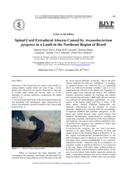
r e v b r a s o r t o p . 2 0 1 5;5 0(6):739–742
www.rbo.org.br
Case report
Fungal spondylodiscitis due to Candida albicans: an
atypical case and review of the literature夽
Álynson Larocca Kulcheski a,b,∗ , Xavier Soler Graells a,b , Marcel Luiz Benato a,b ,
Pedro Grein Del Santoro a,b , André Luis Sebben a,b
a
b
Orthopedics and Traumatology Service, Hospital de Clínicas, Universidade Federal do Paraná (UFPR), Curitiba, PR, Brazil
Hospital do Trabalhador, Universidade Federal do Paraná (UFPR), Curitiba, PR, Brazil
a r t i c l e
i n f o
a b s t r a c t
Article history:
Spondylodiscitis due to Candida is a rare complication from hematogenic dissemination
Received 18 August 2014
of infection caused by this fungus. We present an atypical case of spondylodiscitis caused
Accepted 14 November 2014
by this germ that occurred after chest contusion and progressed with necrotizing fasciitis
Available online 18 October 2015
of the anterior region of the chest and osteomyelitis of the sternum. Through contiguity,
it also affected the upper thoracic spine. The patient evolved with neurological alterations
Keywords:
and recovered satisfactorily after appropriate treatment with surgical decompression of the
Candida albicans
spinal cord and specific antibiotic therapy.
Discitis
© 2015 Sociedade Brasileira de Ortopedia e Traumatologia. Published by Elsevier Editora
Ltda. All rights reserved.
Spinal diseases
Espondilodiscite fúngica por Candida albicans: um caso atípico e revisão
da literatura
r e s u m o
Palavras-chave:
A espondilodiscite por Candida albicans é uma rara complicação da disseminação
Candida albicans
hematogênica da infecção por esse fungo. Apresentamos um caso atípico de
Discite
espondilodiscite por esse germe ocorrido após trauma contuso torácico que cursou
Doenças da coluna vertebral
com fasceíte necrotizante da região anterior do tórax, osteomielite de esterno e, por
contiguidade, afetou a coluna vertebral torácica alta. O paciente evoluiu com alteração neurológica e recuperou-se satisfatoriamente após tratamento adequado com descompressão
medular cirúrgica e antibioticoterapia específica.
© 2015 Sociedade Brasileira de Ortopedia e Traumatologia. Publicado por Elsevier Editora
Ltda. Todos os direitos reservados.
夽
Study carried out at the Orthopedics and Traumatology Service, Hospital de Clínicas, Universidade Federal do Paraná (UFPR) and
Hospital do Trabalhador, Universidade Federal do Paraná (UFPR), Curitiba, PR, Brazil.
∗
Corresponding author.
E-mails: [email protected], alynson [email protected] (Á.L. Kulcheski).
http://dx.doi.org/10.1016/j.rboe.2015.10.005
2255-4971/© 2015 Sociedade Brasileira de Ortopedia e Traumatologia. Published by Elsevier Editora Ltda. All rights reserved.
740
r e v b r a s o r t o p . 2 0 1 5;5 0(6):739–742
Fig. 1 – Initial aspect of the sternum lesion.
Introduction
Spinal cord infections are rare and comprise approximately
1% of bone infectious involvement.1 Most of these infections are of pyogenic or tuberculous origin. Fungal infections
are increasing, but are still extremely rare and occur
more as opportunistic infections in immunocompromised
individuals.2 Despite the increased frequency, infection by
Candida albicans is not common.3 We report an unusual case of
thoracic spondylodiscitis caused by C. albicans. The literature
was reviewed, aiming to better understanding the subject.
Case report
The patient was a 39-year-old homeless, chronic alcoholic
male individual. He fell two meters to the ground in October
2012. He was treated in a trauma hospital, where he showed
signs of septic shock, hyperemia and crackles in the sternal region, with 10 cm in diameter. Chest radiography and
computed tomography (CT) showed pre-sternal subcutaneous
emphysema and signs of sternum fracture, and culminated
with a diagnosis of anterior chest wall necrotizing fasciitis
(Fig. 1). Surgical debridement was performed in this region.
The result of the of sternum soft tissue culture was positive for
Fig. 3 – Cobb angle in the preoperative period between T2
and T7.
multisensitive Escherichia coli and the result of the sternal bone
fragment culture for C. albicans was positive. Treatment with
fluconazole (6 mg/kg/day) and Ciprofloxacin (400 mg 12/12 h)
was started and drug use was scheduled for six months, initially intravenously and, after clinical improvement, by oral
route. The patient developed vertebral osteomyelitis signs,
with decreased height of the vertebral bodies and discs at
the thoracic spine levels of T4–T5–T6 (Fig. 2). The patient was
paralyzed, with altered sensitivity at the T4 level, compatible
with Frankel B. Initial Cobb angle of 68◦ (Fig. 3) was observed.
The patient underwent thoracotomy, which disclosed a spinal
abscess and a large amount of purulent secretion. A corpectomy was performed from T4 to T6 with autologous iliac graft
replacement and comprehensive spinal decompression in T4.
There was improvement of pain complaints in the thoracic
spine, with fever disappearance and improvement to Frankel
Fig. 2 – CT scans in the sagittal, axial and coronal sections.
r e v b r a s o r t o p . 2 0 1 5;5 0(6):739–742
741
Fig. 4 – postoperative AP and profile radiographies.
C. At a second procedure, he was submitted to posterior fixation and arthrodesis with pedicle screws at the level of the
thoracic spine from T3 to T7 (Fig. 4).
Postoperatively, he showed improvement of 13◦ of kyphosis in the Cobb angle and remained at 55◦ (Figs. 4 and 5). After
eight months of the diagnosis, the patient showed improvement of the neurological level to Frankel D at the T4 level.
Upon assessment at 12 months after the first diagnosis, the
wounds were healed and he showed significant improvement
in the thoracic kyphosis (Fig. 5). The patient was well, communicative, independent in relation to self-care, and managed
to perform his activities without assistance or difficulty. During hospitalization the Oswestry Disability Index 2.0 was
applied preoperatively and after the definitive surgical procedure. Preoperatively, he scored 70% and was classified as
disabled. Postoperatively, the index was 25%, which showed
good results in the pain/disability item.
Discussion
Despite the increase in the frequency of fungemia, infection by
C. albicans is also a rare cause of spinal infection.3 The main
risk factors are: prior antibiotic therapy, ICU stay, long-term
catheter use, corticosteroids, intravenous drugs, transplants
and chemotherapy.1,2,4,5 In our case, the patient was alcoholic,
homeless and immunocompromised.
The most common location of spondylodiscitis by Candida
is in the lumbar spine, and the presence of neurological deficit
is infrequent.2
In 2001, Miller6 described 59 cases of spinal infection by
Candida, 33 affecting the lumbar spine, 17 the chest, three the
cervical and six both the thoracic and lumbar spine.
In our case, the upper thoracic region was affected and
there was neurological deficit, in contrast to the literature. This
Fig. 5 – Clinical evolution 12 months after the initial diagnosis.
742
r e v b r a s o r t o p . 2 0 1 5;5 0(6):739–742
condition is usually insidious. The most useful clinical finding is pain in the affected area, both bone and paravertebral
types.7 The paraplegia was noteworthy in our case. An association was observed between chest trauma and the spinal
injury, a fact validated by literature.8
When C. albicans affects the spine, it usually causes disk
narrowing, destruction of the endplates and the subjacent vertebral bone.4 These imaging findings are consistent with what
we found in our case.
The optimized management of spinal infections by Candida remains unclear. Case reports such as this one help to
increase the experience in the management and treatment of
this disease.
Surgical treatment is not required in spondylodiscitis by
Candida. However, it should be performed in cases where
there is neurological deficit and vertebral instability.4,5 In the
present report, the patient had neurological deficit (Frankel
B) and vertebral instability, characterized by kyphosing of the
thoracic spine.
Clinical treatment is carried out with antifungal drugs,
using amphotericin B or fluconazole. One proposed treatment
consists of 6–10 weeks of Amphotericin B IV at a dose of
0.5–0.6 mg/kg/day.9 Studies have shown that Fluconazole is as
effective as amphotericin, showing higher safety and tolerability. In our institution, we chose to carry out the treatment
with fluconazole.
Studies have documented that diagnostic delay is
common.10 That is attributed to the rare occurrence and
difficulty in cultivating the microorganisms. It has been
suggested that a delay in the start of antifungal therapy is
associated with a worse outcome, particularly the neurological one.10 We believe that our success was due to the
early diagnosis and confirmation by biopsy and the sternum
bone culture, as well as the identification of spinal cord
compression. The treatment was promptly carried out with
spinal decompression, rapid microbiological results and start
of specific antifungal treatment.
Spondylodiscitis by Candida should be considered in
immunocompromised patients. The definitive diagnosis is
achieved through isolation of C. albicans in blood or cultures.
The antifungal treatment often results in the cure, even in
cases of delayed diagnosis. When there is neurological instability or deficit, surgical treatment should be considered.
Conflicts of interest
The authors declare no conflicts of interest.
references
1. Ghanayem AJ, Zdeblick TA. Cervical spine infections. Orthop
Clin North Am. 1996;27(1):53–67.
2. Broner FA, Garland DE, Zigler JE. Spinal infections in the
immunocompromised host. Orthop Clin North Am.
1996;27(1):37–46.
3. Johnson MD, Perfect JR. Fungal infections of the bones and
joints. Curr Infect Dis Rep. 2001;3(5):450–60.
4. Gathe JC Jr, Harris RL, Garland B, Bradshaw MW, Williams TW
Jr. Candida osteomyelitis. Report of five cases and review of
the literature. Am J Med. 1987;82(5):927–37.
5. Almekinders LC, Greene WB. Vertebral Candida infections. A
case report and review of the literature. Clin Orthop Relat Res.
1991;267:174–8.
6. Miller DJ, Mejicano GC. Vertebral osteomyelitis due to
Candida species: case report and literature review. Clin Infect
Dis. 2001;33(4):523–30.
7. Smith AS, Blaser SI. Infectious and inflammatory processes of
the spine. Radiol Clin North Am. 1991;29(4):809–27.
8. Graells XS, Zaninelli EM, Collaço IA, Nasr A, Cecílio WAC,
Borges GA. Thoracic injuries and spinal trauma: a complex
association. Coluna/Columna. 2008;7(1):8–13.
9. Rex JH, Walsh TJ, Sobel JD, Filler SG, Pappas PG, Dismukes WE,
et al. Practice guidelines for the treatment of candidiasis.
Infectious Diseases Society of America. Clin Infect Dis.
2000;30(4):662–78.
10. Frazier DD, Campbell DR, Garvey TA, Wiesel S, Bohlman HH,
Eismont FJ. Fungal infections of the spine. Report of eleven
patients with long-term follow-up. J Bone Joint Surg Am.
2001;83(4):560–5.
Download