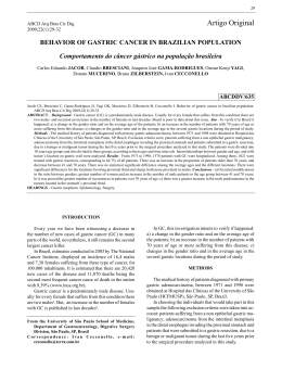
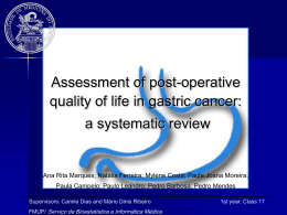
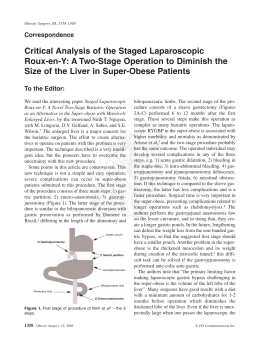
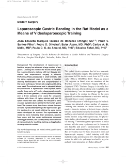
Artigo Original
Revista Brasileira de Física Médica. 2011;5(2):161-4.
Gastric assessment by images processing
of ultrasound in LabVIEW platform:
preliminary results
Avaliação gástrica pelo processamento de imagens de
ultrassom na plataforma LabVIEW: resultados preliminares
T. Córdova1,2, M. Sosa1, J. J. Bernal1, A. Hernandez1, G. D. Gutiérrez1, D. Rodriguez1, S. Solorio3,
M. A. Hernandez3, M. Vargas1, I. Delgadillo1, G. Moreno1, J. G. Villalpando2 and C. R. Contreras2
Departamento de Ingeniería Física, Universidad de Guanajuato, Campus León – México.
Facultad de Ingeniería en Computación y Electrónica, Universidad De La Salle Bajío – México.
3
Unidad Médica de Alta Especialidad, Clínica T1-León, Instituto Mexicano del Seguro Social – México.
1
2
Abstract
Nowadays, the gold technique in gastric evaluations still is scintigraphy in spite of ionization radiation dose per patient undergoing this procedure.
Gastro images with ultrasound technique are controversial, because the stomach is a hollow cavity filled with gas in basal conditions or in fast state.
Fortunately, a stomach with food is recommended in gastric motility and gastric emptying assessment. So, a lack of air in stomach contributes in this
kind of study and in recordings of excellent images by ultrasound. In this study, a digital image processing of gastric ultrasound is presented. Whole
automated routine and implemented filters are described in order to use this procedure in gastric peristalsis and gastric emptying evaluations. Ten
volunteers were recruited and required to attend the measurements about dominant frequency, with values of at least 3 cpm. Although the behavior
stomach activity is observed in dynamic graph, an analysis in frequency space is performed.
Keywords: gastric, peristalsis, ultrasound, emptying, LabVIEW.
Resumo
Hoje em dia, a técnica de ouro nas avaliações gástricas ainda é a cintilografia apesar da dose de radiação de ionização por paciente submetido a esse
procedimento. As imagens gástricas com a técnica do ultrassom são controversas, pois o estômago é uma cavidade oca preenchida com gás em
condições basais ou em estado rápido. Felizmente, um estômago com comida é recomendado em motilidade gástrica, como também na avaliação do
esvaziamento gástrico. Portanto, falta de ar no estômago contribui para este tipo de estudo e para gravações de excelentes imagens por ultrassom.
Neste estudo, o processamento da imagem digital do ultrassom gástrico é apresentado. Uma rotina totalmente automatizada e filtros implementados
estão descritos para usar este procedimento no peristaltismo gástrico e nas avaliações de esvaziamento gástrico. Dez voluntários foram avaliados
em relação à frequência dominante, com valores de no mínimo 3 cpm. Embora a atividade estomacal comportamental seja observada em gráfico de
dinâmica, uma análise de frequência espacial é realizada.
Palavras-chave: gástrico, peristaltismo, ultrassonografia, esvaziamento, LabVIEW.
Introduction
The gastrointestinal system evaluation is, currently, as
important as other clinical procedures, like heart monitoring. If patients are not adequately treated, they may
die. This is especially important for some kinds of patients, for instance, diabetes patients with problems
of gastroparesis. The scintigraphy technique is now
the gold standard in this evaluation, despite ionizing
radiations that undergo the persons1. There are other
imaging techniques for this study, like the ultrasound,
that has been an alternative for assessment and monitoring of the gastric activity in the last years2-6, although
it has still not taken off, which could be due to a lack
of conclusive results and proper procedure, leading to
results highly correlated with the gold standard technique, scintigraphy.
A routine for processing ultrasound images of
stomach, implemented in LabVIEW platform, is presented. This has been used to perform evaluations of
Corresponding author: Teodoro Córdova – Departamento de Ingeniería Física – DCI, Universidad de Guanajuato, campus León – Loma del Bosque, 103 –
Lomas del Campestre – León, GTO, Mexico – E-mail: [email protected]
Associação Brasileira de Física Médica®
161
Córdova T, Sosa M, Bernal JJ, Hernandez A, Gutiérrez GD, Rodriguez D, Solorio S, Hernandez MA, Vargas M, Delgadillo I, Moreno G, Villalpando JG, Contreras CR
the peristaltic activity and gastric emptying in preprandial and postprandial conditions in healthy subjects
and patients.
Procedure
Results and discussion
The above description is appropriated for gastro evaluations. The peristaltic information is seen from first measurement (Figure 7) in time domain, in which about three
contractions are shown. Nevertheless, physicians are
interested in the exact value of the dominant frequency,
An ultrasound equipment model Medison SONOACE
8000 SE was used in this study. A group of ten healthy
volunteers with no history of gastrointestinal diseases
and two patients were recruited and required to attend
the measurements after a night of fast. Each subject
swallowed 300 mL of water previous to first measurement; then, a solid test meal was ingested in order to
estimate the gastric area. Each subject was in supine
position during the auscultation and along the data acquisition time, that lasted one minute. It is important to
point out that this work was carried out according to
the Helsinki agreement for studies in human.
Protocol
Once the gastric area was identified, a video of 1740
frames was recorded in B Mode and in M modes (Figure
1). Then, the M mode video was split in each one of its
frames and the image processing was performed.
From the M mode video, a representative frame is
selected and the region of interest (RI) is studied (Figure
2); the superior and inferior bands are identified. In the
RI of the frame, the points x1,y1, x2 and y2 are determined and the same action is automatically executed
in each of the 1740 frames. Simultaneously, a new file
(Figures 2 and 3) of this new outage image is created in
order to perform the imaging processing over all of it.
Imaging processing
The upper and lower bands, in the image of the Figure
4, correspond to the upper and lower walls of the
stomach, respectively.
Here, a series of filters were implemented during image processing in order to reduce the area of
each band to a single line (IMAQ MathLookup +
IMAQ GrayMorphology + IMAQ MathLookup + IMAQ
BCGLookup + IMAQ LowPass + IMAQ Convolute +
IMAQ RejectBorder). When the filtering process is over,
one distance from inferior to superior band is measured through a subroutine of find Vertical Edge of the
IMAQ sofware; this leads to determine only one couple
of coordinates, selected from each one of the edge
points of the band. With that couple of coordinates,
the distance from one to other side of the stomach is
measured (Figure 5).
It is important to point out that only one distance
is measured from each couple of coordinates. If we
have 29 frames in one second, then, this corresponds
to have a rate frequency of 29 samples per second.
These points are simultaneously plotted (Figure 6).
162
Revista Brasileira de Física Médica. 2011;5(2):161-4.
Figure 1. A frame showing the B and M mode.
Figure 2. Representative frame.
Figure 3. LabVIEW Screen for the RI.
Gastric assessment by images processing of ultrasound in LabVIEW platform: preliminary results
x 107
Power spectral density [a.u.]
3.5
3
2.5
2
1.5
1
0.5
0
-0.5
Figure 4. Outage image of the represented frame.
Dominant Frequency 2.46 cpm
2
4
6
8 10 12 14
Frequency [cpm]
16
18
20
Figure 8. PSD of the gastric mechanical activity.
so a FFT (fast Fourier transform) is obtained from above
signal in order to present this value (Figure 8).
Figure 5. The measurement of the stomach wall can be obtained from de filtering of the left image.
Conclusions
This new ultrasound gastric imaging processing is presented as a powerful technique, with relatively easy
implementation, because the ultrasound equipments
are very common in hospitals and the LabVIEW routine
can be executed in any personal computer.
Acknowledgment
Figure 6. The left figure shows the superior and inferior band; the
right figure presents the square and gastric activity along the time.
Authors want to thank DAIP grant No. 000017/10 and
UDLSB grant of 2010.
Amplitude [a.u.]
References
25
1.
20
2.
15
3.
10
4.
5
0
5.
0
10
20
30
Time [Sec]
Figure 7. Gastric mechanical activity.
40
50
60
6.
Odunsi ST, Camilleri M. Selected interventions in nuclear medicine:
gastrointestinal motor functions. Semin Nucl Med. 1999;39(3):186-94.
Kusunoki H, Haruma K, Hata J, Ishii M, Kamada T, Yamashita N, et al.
Efficacy of Rikkunshito, a traditional japanese medicine (Kampo), in
treating functional dyspepsia. Inter Med. 2010;49(20):2195-202.
Newell SJ, Chapman S, Booth IW. Ultrasonic assessment of gastric
emptying in preterm infant. Arch Dis Child. 1993;69(1):32-6.
Gilja OH, Hausken T, Degaard S, Berstad A. Gastric emptying measured
by ultrasonography. World J Gastroenterol. 1999;5(2):93-4.
Bateman DN, Whittingham TA. Measurements of gastric empyting by
real-time ultrasound. Gut. 1982;23:524-7.
Holt S, McDicken WN, Anderson T, Stewart IC, Heading RC. Dynamic
imaging of the stomach by real-time ultrasound–a method for study
of gastric motility. Gut. 1980;21(7):597-601.
Revista Brasileira de Física Médica. 2011;5(2):161-4.
163
Download