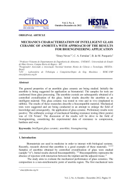
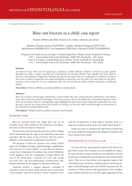
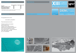
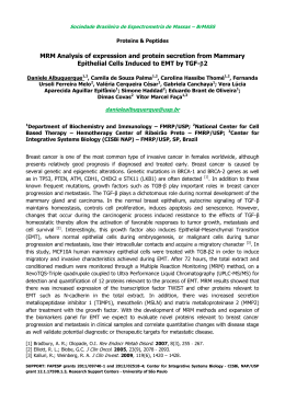
REDUCTIONS IN GROWTH FACTORS IN THE FRACTURE HEMATOMA OF DIABETIC PATIENTS
Gandhi, A; Gebauer, G; Berberian; W S; +Lin, S S
University of Medicine & Dentistry – New Jersey Medical School, Newark, NJ
Table 1. * indicates significance (p < 0.05)
PDGF concentration (pg/ml)
The diabetic and non-diabetic groups were matched for age, time to
surgery and fracture type and location. The diabetic group has a
significantly higher HbA1c levels indicating poor glucose control.
Growth Factor Quantitation:
PDGF:
1600
1400
1200
1000
800
600
400
Plasma
*
Hematoma
#
200
0
non-diabetic
;
diabetic
TGF-β:
TGF-beta1 concentration (pg/ml)
Introduction: The association between diabetes mellitus (DM) and
impaired bone healing has been documented in both clinical and
experimental settings. Although there is a paucity of literature
concerning diabetic fracture healing, previous work suggests that the
effects of diabetes begin at a very early stage in the fracture healing
process. Cellularity in the callus during the proliferative phase of
healing is reduced by 40 percent in untreated diabetic animals due to
impairment in proliferation, migration or both. Despite these advances,
little work has been done to describe the critical factors affecting cellular
proliferation in fracture healing, such as mitogenic growth factors and/or
inflammatory cytokines. Growth factors, which are locally produced
within the fracture environment, play a critical role in cellular
chemotaxis, cellular proliferation, extra-cellular matrix production and
angiogenesis. Two of these growth factors are platelet-derived growth
factor (PDGF) and transforming growth factor-β (TGF-β). The purpose
of this study was to determine whether the expression of growth factors,
PDGF and TGF-β, are altered in DM versus non- DM fracture
hematoma.
Materials and Methods: Patient Population:At this Level I trauma
center, patients undergoing open reduction internal fixation (ORIF)
surgery were enrolled in an IRB-approved study. To minimize
variability in this study the following inclusion criterion were used: 1.
closed fracture requiring ORIF, 2. patient’s age between 18 and 72 years
old, and 3. the time from injury to surgery < 20 days. Sample Collection/
Processing: Fracture hematoma and blood samples were obtained from
patients undergoing surgical treatment. One set of blood samples were
collected for testing of HbA1C levels which is an indicator of blood
glucose control. The other blood samples were centrifuged for ten
minutes at 2,000 RPM, and the supernatant was collected and stored at –
80° C until testing. The fracture hematoma samples were collected
during surgery, flash frozen, and then homogenized using a
biopulverizer to release the cellular contents. These contents were then
diluted by 4 mg/ml with a protease inhibitor cocktail (Sigma, St. Louis,
MO) and stored at –80° C until testing. Growth Factor and Total Protein
Quantitation: For both PDGF and TGF-B quantitation, Quantikine
ELISA kits (R&D Systems, Minneapolis, MN) were used. The ELISA
assay used a quantitative sandwhich enzyme technique. Using a
spectrophotometer and a standard curve, the reflected wavelength of
each sample was correlated to a specific concentration. These growth
factor levels were then normalized using a total protein assay (Pierce St.
Louis, MO). Statistical Analysis: Statistical analysis was performed
using 1-way ANOVA and correlation analyses using StatView (SAS,
Kerry, NC).
Results: Patient Population:
Avg Age
Time to Surgery
HbA1C
(yrs)
(days)
Non-diabetic
38.6 ± 15.8
12.8 ± 4.0
3.15±.0.68
(n=23)
Diabetic
42.8 ± 11.0
17.0 ± 6.1
7.61±.0.97*
(n=7)
18000
16000
14000
12000
10000
8000
6000
4000
2000
0
*
Plasma
#
Hematoma
non-diabetic
diabetic
Figures 1 and 2. * and # indicates statistical significance (p < 0.05) between the
diabetic and non-diabetic plasma and hematoma groups, respectively.
Protein Content:
The protein content in the fracture hematoma (314 Vs. 385 mg/ml) and
plasma (513 Vs. 594 mg/ml) were not statistically different between the
DM and non-DM groups.
Statistics:
PDGF
PDGF
TGF-β
TGF-β
Plasma
Hematoma
Plasma
Hematoma
DM vs non-DM
0.005
0.048
0.047
0.021
Table 2. Summary of p-values
Discussion: Little work has been done to describe the role of the
inflammatory phase, specifically the fracture hematoma, in the normal
and DM fracture healing process. Grundness et al removed the fracture
hematoma in a Sprague-Dawley rat fracture model during stabilization,
which impaired the initial phase of fracture healing. Removal of the
fracture hematoma at days 2 and 4 significantly decreased bending
moment, bending rigidity and fracture energy, compared to control
fractures. However, the removal of the fracture hematoma 30 minutes
following fracture had less of an impact on the mechanical properties.
They concluded that removal of an organized fracture hematoma two or
four days after fracture impairs fracture healing more acutely than
immediate removal. It is inferred that the early fracture hematoma
develops critical factors or role in the development of the fracture callus.
Analysis of the non-DM human fracture hematoma confirm this
observation with the presence of two critical factors TGF-β and PDGF.
While both growth factors, PDGF and TGF-β, are released from
platelets during the earliest phase of fracture healing leading to the
hematoma formation, the growth factor levels quantitated in this study
(over 12 days after injury) probably reflect local production and nonplatelet derived etiology.
Growth factors PDGF and TGF-β, which are locally
produced within the fracture environment, play a critical role in cellular
chemotaxis, cellular proliferation, extra-cellular matrix production and
angiogenesis. Andrew et al demonstrated that PDGF is expressed during
normal human fracture repair and likely to be an important local
regulator in this process. Our recent study using DM BB Wistar rat
femur fracture model, confirmed decreased PDGF and TGF-β levels
using immunohistochemical and quantitative mRNA methods at days 2,
4, and 7 post-fracture.
This study noted similar findings with significant reduction
in the early growth factor levels (PDGF, TGF-β) in the human DM
compared to non-DM fracture hematoma. However, circulating levels of
these growth factors were significantly higher in the DM group
compared to the non-DM group. The clinical implication of our findings
lies within its confirmation of decreased human growth factor levels in
DM fracture hematoma. This reduction of critical growth factors may
contribute to the delayed healing documented in diabetic patients. The
increased levels of circulating growth factors may be related with
complications associated with DM. This study justifies the potential use
of growth factors localized to the fracture site in the amelioration of
impaired DM fracture healing.
References: 1. Andrew JG et al Bone 16:455-60, 1995; 2. Grundness et
al Acta Orthop Scand, 64:340-42, 1993; 3 Loder Clin Orthop, 232:21016, 1988;. 4. Joyce ME et al Prog Clin Biol Res, 365:391-416, 1991.
49th Annual Meeting of the Orthopaedic Research Society
Poster #0541
Download