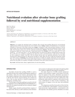
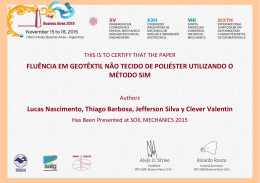
Int. J. Morphol.,
30(3):1173-1176, 2012.
Anthropometric Measurements in Toothed and
Toothless Maxillaries and its Consequences
in Human Alveolar Bone Resorption
Mediciones Antropométricas en Maxilares con y sin Dientes
de Humanos y sus Consecuencias en la Reabsorción Ósea Alveolar
*
Jaciel Benedito de Oliveira; **Andrelle Nayara Cavalcanti Lima de Almeida; ***Carla Cabral dos Santos Accioly Lins;
***
Adelmar Afonso de Amorim Júnior & ****Zélia Albuquerque Seixas
DE OLIVEIRA, J. B.; DE ALMEIDA, A. N. C. L.; LINS, C. C. S. A.; JÚNIOR, A. A. A. & SEIXAS, Z. A. Anthropometric
measurements in toothed and toothless maxillaries and its consequences in human alveolar bone resorption. Int. J. Morphol., 30(3):11731176, 2012.
SUMMARY: This study proposes to measure and compare anthropometric measurements between toothed and toothless maxillas.
26 human skulls were used with: 13 toothed and 13 toothless maxillas. The measurements were made between the distances of the
alveolar ridge (AR): the anterior nasal spine (ANS), to the incisive foramen (FI) and the palatine foramen, greater (FPMA) and lower
(FPME) on both sides by two evaluators. The data obtained showed that the average of the measurements were correspondingly higher
in toothed maxillas than in toothless, for all measurements in any one of the evaluators. The AR-ANS (p = 0.001), IF-AR (p = 0.006),
AR-FPMA right (p = 0.001) and AR-FPMA left (p <0.001) and AR-FPME on both sides (p = 0.001) demonstrating significant differences
between the two types of maxilla. Thus we verify that, as the tooth loss occurs, irreversible anatomical changes take place in the maxilla
bone, which must be taken into account in the design and manufacture of prostheses and osseointegrated implants.
KEY WORDS: Anatomy; Maxilla; Total prosthesis; Alveolar bone loss; Dental implantation.
INTRODUCTION
The growing number of elderly in the population has
caused a significant increase in studies that propose to
investigate the phenomena of human aging (Unfer et al.,
2006). Within this context, the oral health has an important
role, because there is a close relationship between aging and
loss of teeth as the teeth of the elderly are exposed for a
longer period to factors that lead to its loss, such as caries,
periodontal disease, endodontic complications, trauma and
iatrogenic dental treatment (Jorge et al., 2009; Ribeiro et
al., 2009). In addition, there are systemic factors related to
bone loss as low calcium intake, osteoporosis,
hyperparathyroidism, hyperthyroidism, diabetes and the use
of corticosteroids (Paganini-Hill, 2007; Jorge et al.).
In modern dentistry, the lack of bone in the alveolar
ridges has been a major problem in aesthetic and functional
recovery of patients who have suffered tooth loss (Marzola, 2008;
Astudillo Silva, 2010). The vertical resorption of the maxilla
is, on average, four times higher than that of the mandible,
with an estimated average annual vertical bone resorption
of 0.1 mm after tooth loss (Magini, 2006; Astudillo Silva).
Therefore, this study proposed to conduct an
investigation of the anatomical changes that occur in the
maxillary alveolar bone ridge after loss of teeth, measure
and compare anthropometric measurements between toothed
and toothless maxillas, checking for possible differences
between them.
MATERIAL AND METHOD
It were used 26 human skulls, 13 toothed and 13
toothless maxillas, belonging to the Department of Human
Anatomy of the Centre for Biological Sciences, Federal
*
Graduated in Dentistry, Federal University of Pernambuco, Recife-PE, Brazil.
Graduate Student Course in Dentistry, Federal University of Pernambuco, Recife-PE, Brazil.
***
PhD Professor of the Departament of Human Anatomy, Federal University of Pernambuco, Recife-PE, Brazil.
****
PhD Professor of the Departament of Prosthodontics and Oral-Facial Surgery, Federal University of Pernambuco, Recife-PE, Brazil.
**
1173
DE OLIVEIRA, J. B.; DE ALMEIDA, A. N. C. L.; LINS, C. C. S. A.; JÚNIOR, A. A. A. & SEIXAS, Z. A. Anthropometric measurements in toothed and toothless maxillaries and its consequences
in human alveolar bone resorption. Int. J. Morphol., 30(3):1173-1176, 2012.
University of Pernambuco,
Recife-PE, Brazil. The aspects
of race, age and sex were not
considered. The measurements
were performed with calipers
(accuracy 0.05 mm) according
to Mandarim-de-Lacerda
(1995), between the distance of
the crest of the alveolar ridge
(AR): to the anterior nasal
spine (ANS), to the incisive
foramen (FI) and the palatine
foramen, greater (FPMA) and
lower (FPME) on both sides
(right [R] and left [L]), by two
evaluators so in toothed
maxillas (Fig. 1), as in
toothless maxillas (Fig. 2).
Fig. 1. Measurement of distances studied in toothed maxillas. A. Alveolar ridge (AR) to the anterior nasal spine (ANS), B. Alveolar ridge (AR) to the incisive foramen (IF), C. Alveolar ridge
(AR) to the greater palatine foramen (FPMA) and D. alveolar ridge (AR) to the lesser palatine
foramen (FPME).
Data analysis was
obtained from to the average,
median, standard deviation, the
concordance correlation
coefficient with a confidence
interval of 95.0%. Data were
entered into an Excel
spreadsheet and statistical
calculations were obtained
using SPSS (Statistical Package for Social Sciences) version
13 and MED CALC in version
10.4.8.
RESULTS
Fig. 2. Measurement of distances studied in toothless maxillas. A. Alveolar ridge (AR) to the
anterior nasal spine (ANS), B. Alveolar ridge (AR) to the incisive foramen (IF), C. Alveolar ridge
(AR) to the greater palatine foramen (FPMA) and D. alveolar ridge (AR) to the lesser palatine
foramen (FPME).
1174
With the measurements
obtained from AR-ANS, IFAR, AR-FPMA-R, ARFPMA-L, AR-FPME-R and
AR-FPME-L (Table I) it was
observed that the averages
were correspondingly more
high in toothed maxillas,
showing differences among the
two types of maxilla. In the
study of inter-examiner
calibration was possible to
verify high concordance
correlation coefficient, with
values greater than 0.94.
DE OLIVEIRA, J. B.; DE ALMEIDA, A. N. C. L.; LINS, C. C. S. A.; JÚNIOR, A. A. A. & SEIXAS, Z. A. Anthropometric measurements in toothed and toothless maxillaries and its consequences
in human alveolar bone resorption. Int. J. Morphol., 30(3):1173-1176, 2012.
Table I. Average of the distances of the anthropometric points in toothed and toothless maxillas.
Maxilla
Parameters
AR-ANS
AR-FI
AR-FPMA-R
AR-FPMA-L
AR-FPME-R
AR-FPME-L
Evaluators
Toothless
Toothed
Mean ± SD
Mean ± SD
1
1.206 ± 0.394
1.849 ± 0.353
P-Value
p= 0.001*
2
1.222 ± 0.378
1.870 ± 0.349
p= 0.001*
1
0.524 ± 0.175
0.819 ± 0.260
p= 0.006*
2
0.564 ± 0.223
0.818 ± 0.263
p= 0.027*
1
0.996 ± 0.216
1.448 ± 0.296
p = 0.001*
2
0.995 ± 0.228
1.454 ± 0.289
p = 0.001*
1
1.012 ± 0.254
1.531 ± 0.320
p = 0.001*
2
1.007 ± 0.246
1.519 ± 0.372
p= 0.001*
1
1.022 ± 0.234
1.453 ± 0.195
p < 0.001*
2
1.015 ± 0.228
1.469 ± 0.194
p < 0.001*
1
1.036 ± 0.276
1.521 ± 0.295
p= 0.001*
2
1.033 ± 0.271
1.506 ± 0.318
p= 0.002*
*There was a significant difference between toothed and toothless maxillas.
DISCUSSION
The maxilla is a bone pair, short, symmetrical, with
irregular shape, pneumatic and is in the center of the face,
forming with the opposite side a bone complex called upper
facial skeleton (Zanon, 2009; Astudillo Silva). Their average dimensions are 35 x 35 mm at the base, 25 mm of height
and volume of 15 cm3 (Magini; Marzola, 2008).
With the loss of dental elements, the local
physiological stimuli, provided by the teeth and periodontal
ligaments that hold the alveolar bone, disappear starting a
degenerative, chronic, progressive, incurable and cumulative
bone resorption. This resorption is a complex, multifactorial
and irreversible process, being faster in the first months, and
continuing throughout the life of the individual (Bartee, 2001;
Marzola, 2002; Mazzonetto et al., 2005; López-Roldán et
al., 2009; Gupta et al., 2010; Mizutani et al., 2010).
According to Kenezovié-Zlatarié et al. (2002), due
to extensive bone loss, the denture loses support, there is no
prosthesis retention and stability are compromised. For this,
the knowledge and respect for the anatomical prosthetic
structures, is of extreme importance for the correct adaptation
of the prosthesis, seeking to provide stability, retention and
comfort during use. Contributing to them, Astudillo Silva
said that the knowledge of the morphology of the bone defect
is important and should be considered for the selection of a
rehabilitation method for increasing the residual alveolar
ridge in the maxilla.
During the process of resorption of the alveolar
process is common to note insufficient bone quantity
(thickness and / or height) for the installation of
osseointegrated implants in the anterior maxilla (Magini;
Astudillo Silva). The lack of bone volume may also result
in an incorrect positioning of the implants due to improper
installation on the angle that compromise the aesthetic result
in hygiene problems and may lead to loss of the implants
(Marzola, 2008; Astudillo Silva).
Assunção et al. (2004) claim that the imbalance of
stomatognathic system, provided by the loss of teeth, also
causes changes in the gum and muscular tissue, and in the
temporomandibular joints, resulting directly in chewing and
speech, causing discomfort and difficulty in performing these
activities, as well as aesthetic and patient comfort damage.
Silva et al. (2007) reported that combined with this, the social interaction that can also be damaged for these
individuals, since they will be ashamed to smile and talk
because of the precarious situation of the mouth.
Thus, the data obtained in this study agree with the
authors of several studies cited above, does demonstrate the
anatomical changes that occur in the alveolar bone ridge of
the maxillary bone after the loss of teeth, has consequences
for the aesthetics, quality of life and rehabilitation through
prosthetics and dental implants.
In conclusion, as the tooth loss occurs, irreversible
anatomical changes take place in the maxillary bone,
occurring alveolar bone resorption process, confirmed by
1175
DE OLIVEIRA, J. B.; DE ALMEIDA, A. N. C. L.; LINS, C. C. S. A.; JÚNIOR, A. A. A. & SEIXAS, Z. A. Anthropometric measurements in toothed and toothless maxillaries and its consequences
in human alveolar bone resorption. Int. J. Morphol., 30(3):1173-1176, 2012.
comparison of measurements between anatomical
accidents of toothed and toothless maxillas of human skulls,
which affects the aesthetics and quality of life of the indi-
vidual and should be taken into account in planning and
making of dentures and the installation of osseointegrated
implants.
DE OLIVEIRA, J. B.; DE ALMEIDA, A. N. C. L.; LINS, C. C. S. A.; JÚNIOR, A. A. A. & SEIXAS, Z. A. Mediciones antropométricas en maxilares
con y sin dientes de humanos y sus consecuencias en la reabsorción ósea alveolar. Int. J. Morphol., 30(3):1173-1176, 2012.
RESUMEN: Este estudio propuso medir y comparar las medidas antropométricas entre los maxilares con y sin dientes. Fueron utilizados 26
cráneos humanos, 13 maxilares con dientes y 13 sin dientes. Las mediciones fueron realizadas a ambos lados por dos evaluadores entre las distancias del
reborde alveolar (RA): a la espina nasal anterior (ENA), al canal incisivo (CI) y a lo foraámenes palatinos mayor (FPMA) y menor (FPME). Los datos
mostraron que la media de las mediciones fueron proporcionalmente mayores en las maxilas con dientes, para todas las mediciones en cualquiera de los
evaluadores. El RA-ENA (p = 0,001), AR-CI (p = 0,006), RA-APMA derecho (p = 0,001) y RA-APMA izquierdo (p <0,001) y RA-APME en ambos lados
(p = 0,001), comprobando la existencia de diferencias significativas entre los dos tipos de maxila. Fue verificado que a partir de la pérdida dental ocurren
irreversibles cambios anatómicos en el maxilar, que deben ser considerados en el diseño y fabricación de prótesis e implantes osteointegrados.
PALABRAS CLAVE: Anatomía; Maxilar; Prótesis total; Pérdida ósea alveolar; Implantación dental.
REFERENCES
Assunção, W. G.; Shiwa, M.; Gennari Filho, H.; Goiato, M. C.;
Barbosa, D. B. & Tabata, L. F. Para-prosthetic anatomy:
importance in complete dentures. Rev. Odontol. Araçatuba,
25(1):57-64, 2004.
Astudillo Silva, M. A. Surgical solutions for the treatment of atrophic
maxillae bone grafts and zygomatic implants literature review
and clinical case presentation. Monografia Especialização em
Cirurgia e Traumatologia BucoMaxiloFacial. Bauru, Associação
Paulista de Cirurgiões-Dentistas (APCD), 2010. p.46.
Mandarim-De-Lacerda, C. A. Métodos Quantitativos em Morfologia.
Rio de Janeiro, UERJ, 1995. p.131.
Mizutani, F. S.; Nagem Filho, H.; Valiense, H.; Carbonari, M. J. &
Campi Júnior, L. Matrix osseoindutor for alveolar ridge
augmentation. FULL Dent. Sci., 1(2):112-6, 2010.
Mazzonetto, R.; Maurette, M. A.; Maurette, P. E. & Torrezan, J. F. R.
Distracção Osteogénica para a Reconstrução de Rebordos
Alveolares Atróficos. Rev. Port. Estomatol. Med. Dent. Cir.
Maxilofac., 46(4):212-20, 2005.
Paganini-Hill, A. Hormone Therapy and Oral Health. Menopause
Manage., 16(3):31-40, 2007.
Bartee, B. K. Extraction site reconstruction for alveolar ridge
preservation. Part 1: Rationale and materials selection. J. Oral
Implantol., 27(4):187-93, 2001.
Ribeiro, D. G.; Silva, M. M.; Nogueira, S. S. & Arioli Filho, J. N. A
Saúde Bucal na Terceira Idade. Salusvita, 28(1):101-11, 2009.
Gupta, A.; Tiwari, B.; Goel, H. & Shekhawat, H. Residual ridge
resorption: a review. Indian J. Dent. Sci., 2(2):7-11, 2010.
Silva, P. V.; Santana, S. R. F.; Almeida, E. C. B.; Araújo, A. C. S.;
Cimões, R. & Gusmão, E. S. The dental number influence on
daily performance. RFO UPF, 12(3):13-7, 2007.
Jorge, T. M.; Bassi, A. K.; Yarid, S. D.; Silva, H. M.; Silva, R. P. R.;
Caldana, M. L. & Bastos, J. R. M. Relation between tooth loss
and chewing, swallowing and speech complaints in adults. Rev.
CEFAC, 11(13):391-7, 2009.
Unfer, B.; Braun, K.; Silva, C. P. & Pereira Filho, L. D. Self-perception
of the loss of teeth among the elderly. Interface (Botucatu),
10(19):217-26, 2006.
Kenezovié-Zlatarié, D.; Celebié, A. & Lazié, B. Resorptive Changes
of Maxillary and Mandibular Bone Structures in Removable
Denture Wearers. Acta Stomatol. Croat., 36(2):261-5, 2002.
Zanon, L. P. Maxillary total osteotomies study – Le Fort I: Literature
review. Monografia Especialização em Cirurgia e Traumatologia
BucoMaxiloFacia. Bauru, Associação Paulista de CirurgiõesDentistas (APCD), 2009. p.33.
López-Roldán, A.; Santolaya-Abad, D. S.; Gregori-Bertomeu, I. G.;
Gómez-Castillo, E. G. & Selva-Otaolaurruchi, E. S. Bone
resorption processes in patients wearing overdentures. A 6-years
retrospective study. J. Clin. Exp. Dent., 1(1):24-30, 2009.
Marzola, C. Cirurgia Pré-Protética. 3ª ed. São Paulo, Pancast, 2002.
p.412.
Correspondence to:
Carla Cabral dos Santos Accioly Lins
Rua Manuel de Carvalho, 310, Apt. 303
Aflitos, Recife – PE. CEP: 52050-370
BRAZIL
Marzola, C. Fundamentos de Cirurgia e Traumatologia
BucoMaxiloFacial. 1ª Ed. São Paulo, BigForms, 2008. p.3250.
Email: [email protected]
Magini, R. S. Enxertos ósseos no seio maxilar. Estética e função. 1ª
ed. São Paulo, Santos, 2006. p.330.
Received: 14-11-2011
Accepted: 17-04-2012
1176
Download