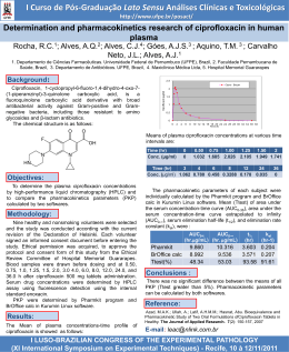
Nutr Hosp. 2011;26(1):144-151
ISSN 0212-1611 • CODEN NUHOEQ
S.V.R. 318
Original
Correlation between serum content of the main COPs (cholesterol oxidation
products) from autoxidation and cardiovascular risk factors
M.ª Menéndez-Carreño1, N. Varo2, C. Mugueta2, P. Restituto2, D. Ansorena1 and I. Astiasarán1
Department of Nutrition, Food Sciences, Physiology and Toxicology. Faculty of Pharmacy. University of Navarra. Pamplona.
Spain. 2Laboratory of Biochemistry. University Clinic of Navarra. Pamplona. Spain.
1
Abstract
Background/aims: Risk factors for cardiovascular disease (CVD) have been proven to be associated with an
increased oxidative stress. Several studies have considered cholesterol oxidation products (COPs) as specific in
vivo markers of oxidative stress. The aim of this study was
to investigate the association between the levels of COPs
derived from autoxidation processes and established cardiovascular risk factors, comparing the levels of serum
COPs in subjects with or without showing values out of
the reference ranges.
Methods: It was a cross-sectional study in which 88
subjects were recruited and individual and total COPs
from autoxidation origin was analyzed in serum by GCMS. The simultaneous correlation of COPs with different
CVD risk factors have been analyzed.
Results and discussion: A great variability of total
COPs concentrations were found. Subjects presented
total COPs values from 0.091 to 2.052 μg/mL. Total COPs
were significantly higher (p < 0.05) in patients with hypertriglycerolemia, hypertension, diabetes and overweight/
obesity status compared to those subjects who did not
present those CVD risk factors. Moreover, 7α and 7βhydroxycholesterol and 7-ketocholesterol were significantly higher (p < 0.05) in patients with hypertension and
diabetes. No significant differences in total COPs were
found between patients with and without hypercholesterolemia.
Conclusions: The obtained results showed that the
analyzed COPs correlate well with at least 4 out of 6 risk
factors of development of CVD.
(Nutr Hosp. 2011;26:144-151)
DOI:10.3305/nh.2011.26.1.4690
Key words: Oxidation. Hypertriglyceridemia. Hypertension. Diabetes. Obesity. Atherosclerosis. GC-MS.
Correspondence: Iciar Astiasarán.
Department of Nutrition, Food Science, Physiology and Toxicology.
Faculty of Pharmacy. University of Navarra.
Irunlarrea, s/n.
31008 Pamplona. Spain.
E-mail: [email protected]
Recibido: 17-II-2010.
Aceptado: 15-IX-2010.
144
CORRELACIÓN ENTRE LOS PRODUCTOS
DE OXIDACIÓN DE COLESTEROL (COPs)
FORMADOS POR AUTOOXIDACIÓN
Y FACTORES DE RIESGO CARDIOVASCULAR
Resumen
Introducción: Se ha demostrado que los factores de
riesgo cardiovascular están estrechamente asociados con
un elevado nivel de estrés oxidativo. Varios estudios consideran a los productos de oxidación del colesterol
(COPs) como marcadores específicos in vivo de estrés
oxidativo. El objetivo de este trabajo fue estudiar la asociación entre los niveles de COPs derivados de procesos
de autooxidación de colesterol y factores de riesgo cardiovascular, comparando el contenido sérico de COPs en
sujetos afectados o no por dichos factores.
Métodos: Se trata de un estudio transversal en el que se
reclutaron 88 personas a las que se analizó el perfil de óxidos de colesterol en suero procedentes de autooxidación,
por cromatografía de gases-espectrometría de masas. Se
valoró la correlación de los niveles de COPs con diferentes factores de riesgo cardiovascular.
Resultados y discusión: Se encontró una gran variabilidad en el contenido en COPs totales, observándose valores entre 0,091 y 2,052 μg/mL. COPs totales fueron significativamente superiores (p < 0,05) en pacientes con
hipertrigliceridemia, hipertensión, diabetes y sobrepeso/
obesidad con respecto a aquellos sujetos que no presentaron estos factores de riesgo cardiovascular. Además, 7α
y 7β-hidroxicolesterol y 7-ketocolesterol mostraron valores mayores (p < 0,05) en pacientes con hipertensión y
diabetes. No se observaron diferencias en COPs totales
entre pacientes con y sin hipercolesterolemia.
Conclusiones: Los resultados de este estudio mostraron
que los COPs analizados presentan altos niveles de correlación con, al menos, 4 de 6 factores de riesgo cardiovascular considerados.
(Nutr Hosp. 2011;26:144-151)
DOI:10.3305/nh.2011.26.1.4690
Palabras clave: Oxidación. Hipertrigliceridemia. Hipertensión. Diabetes. Obesidad. Aterosclerosis. GC-MS.
Abbreviations
COPs: Cholesterol oxidation products.
CVD: Cardio vascular disease.
Introduction
Cholesterol is an unsaponificable lipid prone to oxidation leading to the formation of cholesterol oxidation
products (COPs). Certain oxysterols of biomedicalinterest (e.g., 26-OH-cholesterol, 24-OH-cholesterol, and
22-OH-cholesterol) are generally considered to not represent significant products of the autoxidationof cholesterol, while others (including 7-ketocholesterol, 7β-OHcholesterol, 7α-OH-cholesterol, 25-OH-cholesterol,
5β,6β-epoxycholesterol, 5α,6α-epoxycholesterol, and
5α,6β-diOH-cholesterol) are recognized products of
cholesterol autoxidation.1 These last ones could be both
from endogenous origin or also they can be absorbed
from the diet.2,3
The presence of COPs in plasma or serum has been
evident, and it has been demonstrated that they are
potentially involved in the initiation and progression of
major chronic diseases.4 They are present in high concentrations in atherosclerotic plaques, contributing to
the development of atherosclerosis, the most common
cause of death in Western world.5
Chronic and acute over-production of reactive oxygen species (ROS) under pathophysiologic conditions
are associated with the development of CDV.6 There is
growing evidence that oxidized LDL (oxLDL) plays a
major role in the injury of endothelium, being its content in COPs the reactive mediator of structural and
functional changes of the vascular entothelium
affected by atherosclerotic process.7,8 oxLDL is not recognized by LDL receptors, but it is instead taken up by
scavenger receptors on macrophagues of arterial walls.
The macrophagues then develop into foam cells that
form a fatty streak that ultimately turn into an atherosclerotic plaque.9
Zhou et al.10 found that plasma from catheterized
patients showed much higher total free oxysterols than
control ones, being the most abundant those from
autoxidation origin. Larsson et al.11 showed that
increased levels of 7β-hydroxycholesterol and 7-ketocholesterol may play an important role in the induction
of oxidative stress in atheroma plaques by stimulating
ROS production and decreasing cellular antioxidants.
Several diseases are associated with local substained
imbalance of the ratio between oxidative and reductive
biochemical reactions towards oxidation, causing
oxidative stress. Increased COPs levels (specially 7ketocholesterol and 7β-hydroxycholesterol) have been
reported in disease states where oxidative stress was
increased such as diabetes mellitus12 or familial combined hyperlipidemia.13. For instance, functional
impairment of the vascular endothelium is one of the
first steps in the development of atherosclerosis, and
Oxysterols and cardiovascular disease
vascular adhesion molecules in plasma are indicators
of endothelial damage in diabetes mellitus showing
significant correlation with 7-ketocholesterol.14 Significantly higher concentrations of blood COPs were
found in the blood of diabetic and hypercholesterolemic patients than in blood of control subjects.15
Other authors cannot exclude the role of COPs (7oxysterols) in pathogenesis of arterial hypertension
and non-insulin dependent diabetes mellitus in morbidity obese patients.16 Obesity is linked with enhanced
inflamatory stress and increased atherosclerosis, which
are associated with oxidative stress and greater formation of COPs. A recent study has established that serum
oxysterol concentrations in adolescents increase with
obesity, insulin, and ApoB indicating its relevance as
potential indicators for assessing certain metabolic
derangements.17
The ultimate goal of research on COPs should be to
link findings regarding the biological roles of COPs to
the prevalence of COPs in tissues or fluids, contributing with interesting data on the “focused” lipidomics
research area.18 In this context, the aim of this study was
to investigate the association between the levels of
COPs derived from autoxidation processes and established cardiovascular risk factors,19 comparing the levels of serum COPs in subjects with or without showing
values out of the reference ranges.
Materials and Methods
Materials and reagents
7α-Hydroxycholesterol, 7β-hydroxycholesterol, 5,6βepoxycholesterol, 5,6α-epoxycholesterol, cholestanetriol,
25-hydroxycholesterol, 7-ketocholesterol and 19hydroxycholesterol were purchased from Steraloids
(Wilton, NH, U.S.A.). Tri-Sil reagent was obtained
from Pierce (Rockford, IL, U.S.A.). Acetone, chloroform, diethyl ether, methanol, hexane, sodium sulphate
anhydrous and potassium hydroxide were obtained
from Panreac (Barcelona, Spain). Hexane for gas chromatography and butylhydroxytoluene (BHT) were
from Merck & Co., Inc (Whitehouse Station, NJ,
U.S.A.). Sep-pack Vac 6cc silica 1g cartridges were
obtained from Waters (Milford, Massachussets,
U.S.A.).
Study population
This cross sectional study was performed in 88 subjects (55.7 % males; 63.5 ± 14.7 yr) attending the Cardiovascular Risk Area of the University of Navarra for
a general check-up.
All participants underwent a complete medical
examination and anthropometric measurements were
taken. Subjects were free from clinically apparent atherosclerotic disease on the basis of absence of history
Nutr Hosp. 2011;26(1):144-151
145
of coronary disease, stroke, or peripheral artery disease
and normal electrocardiogram. Exclusion criteria
were: impaired renal or liver function, cancer, arteritis,
inflammatory diseases and connective tissue diseases.
The mean body mass index (BMI) was 28.8 kg/m2, and
it was calculated using the following formula: weight
(kg)/height2 (m). Blood pressure was measured on the
right arm, with the subjects in a seated position and
after a 5-min rest, with a mercury sphygmomanometer.
The average of two measurements, at the beginning
and end of the visit, was considered.
Risk factors for CVD were diagnosed according to
the National Cholesterol Education Program’s Adult
Treatment Panel III guidelines with modification of
waist criterion into body mass index (BMI).
The local committee on human research approved
the study, performed in accordance with the Declaration of Helsinki, and all participants gave written
informed consent.
Seven different cholesterol oxidation products were
analyzed as it has been demonstrated that 7α-hydroxycholesterol, 7β-hydroxycholesterol, 5,6α-epoxide,
5,6β-epoxide, triol, 25-hydroxycholesterol and 7-ketocholesterol are the most predominant COPs derived
from autooxidation in vivo.21
Statistical analysis
The statistical analysis was performed with version 15.0; SPSS (Chicago, IL). The normal distribution of variables was tested with the Shapiro Wilks
test. Differences between groups were evaluated
with the Student t test and with the Mann-Whitney U
test for non-normally distributed variables. Spearman correlation coefficients for continuous variables
were used to assess univariate correlations. Results
are presented as mean ± EEM, p < 0.05 was considered significant.
Biochemical analysis
Results
Following an overnight fast, serum and plasma were
collected by venous puncture into Vacutainer tubes.
Fasting serum glucose, cholesterol, triglycerides (TG),
and high-density lipoprotein (HDL) and low-density
lipoprotein (LDL) cholesterol were measured by standard laboratory techniques.
Serum COPs analysis
A validated method for the analysis of COPs in
serum was used, by means of GC-MS Menéndez-Carreño et al.20 Gas chromatography-Mass spectrometry
analysis was performed on a GC 6890N Hewlett
Packard coupled to a 5975 Mass Selective Detector
(Agilent Technologies, Inc., CA, USA). The TMSethers derivates of sterol oxides were separated on a
capillary column Varian VF-5ms CP8947 (50 m x 250
m x 0.25 m film thickness) (Varian, France). Identification of the peaks was made by the characteristic ion
fragmentation of the standard substances and the quantification was made using selected ion monitoring
(SIM) analysis. Integration was performed with Agilent G1701DA GC/MSD ChemStation (Agilent Technologies, Inc., CA, U.S.A.).
Regarding demographic and clinical characteristics of
the study population, the most prevalent risk factor was
dislipemia (79.4%) (data not shown). Mean cholesterol
was 200 ± 49 mg/dL (HDL = 56 ± 15 mg/dL, LDL = 124
± 47 mg/dL and VLDL = 21 ± 9 mg/dL) and 47% of the
study participants were normocholesterolemic. There
were 37.5% of hypertensive patients, most of them (30.7
% of the volunteers) under treatment with antagonists of
the angiotensin II receptor or ACE inhibitors. Mean SBP
was 136 ± 20 mmHg and mean DBP was 77 ± 11
mmHg. The prevalence of diabetes was 18.6%. 47.7%
of the participants were smokers.
The minimum and maximum concentrations of total
and individual serum COPs observed are shown in
table I. Total analyzed COPs results ranged from 0.091
to 2.053 μg/mL. Various authors have reported total
plasma/serum COPs values in normal volunteers ranging from 0.02 μg/mL to 9.6 μg/mL.2,22,23 The main cholesterol oxidation products found in serum were 7αhydroxycholesterol and 7β-hydroxycholesterol (0.304
and 0.247 μg/mL, respectively), followed by 7-ketocholesterol and 5,6β-epoxycholesterol (0.062 and 0.060
μg/mL, respectively). No 25-hydroxycholesterol was
detected in serum samples.
Table I
Serum concentration of individual COPs [μg/mL] (n = 88)
COPs (μg/mL)
7α-hydroxy
cholesterol
7β-hydroxy
cholesterol
5,6β-epoxycholesterol
5,6α-epoxycholesterol
Cholestanetriol
25-hydroxycholesterol
7-ketocholesterol
Minimum concentration
0.011
0.016
0.002
0.003
0.003
n.d.
0.006
Maximum concentration
Mean
1.092
0.304
0.762
0.247
0.241
0.06
0.264
0.046
0.162
0.034
n.d.
n.d.
0.242
0.062
n.d.: non detectable.
146
Nutr Hosp. 2011;26(1):144-151
M.ª Menéndez-Carreño et al.
Table II
Serum COPs concentrations [μg/mL] according to total lipids levels
COPs (μg/mL)
CVD risk factor
Total cholesterol
< 240 mg/dL (n = 56)
> 240 mg/dL (n = 29)
p
Low HDL
HDL > 40 mg/dL (women);
HDL > 50 mg/dL (men) (n = 63)
HDL < 40 mg/dL (women);
HDL < 50 mg/dL (men) (n = 22)
p
Total triglycerides
< 150 mg/dL (n = 71)
> 150 mg/dL (n = 11)
p
Total COPs
7α-hydroxy
cholesterol
7β-hydroxy
cholesterol
5,6β-epoxycholesterol
5,6α-epoxycholesterol
Cholestanetriol
7-ketocholesterol
0.70 ± 0.50
0.86 ± 0.57
n.s.
0.29 ± 0.27
0.32 ± 0.32
n.s.
0.22 ± 0.19
0.30 ± 0.21
n.s.
0.06 ± 0.05
0.07 ± 0.05
n.s.
0.04 ± 0.03
0.05 ± 0.05
n.s.
0.03 ± 0.03
0.04 ± 0.03
n.s.
0.06 ± 0.05
0.08 ± 0.06
p = 0.08
0.70 ± 0.49
0.27 ± 0.25
0.23 ± 0.20
0.06 ± 0.05
0.05± 0.04
0.03 ± 0.03
0.06 ± 0.06
0.91 ± 0.61
0.41 ± 0.36
0.29 ± 0.21
0.06 ± 0.04
0.04 ± 0.03
0.04 ± 0.03
0.06 ± 0.04
n.s.
n.s.
n.s.
n.s.
n.s.
n.s.
n.s.
0.71 ± 0.50
1.02 ± 0.71
*
0.28 ± 0.28
0.44 ± 0.37
p = 0.08
0.23 ± 0.19
0.37 ± 0.26
*
0.06 ± 0.05
0.06 ± 0.05
n.s.
0.04 ± 0.04
0.04 ± 0.03
n.s.
0.03 ± 0.03
0.03 ± 0.03
n.s.
0.06 ± 0.05
0.07 ± 0.05
n.s
Results are expressed as mean ± standard deviations. * p < 0.05; n.s.: non significant.
Subjects were classified into two groups depending
on the presence or absence of different CVD risk factors, following the criteria described by the ATP III
guidelines.19 Hypercholesterolemia was defined as
cholesterol levels higher than 240 mg/dL. Low HDL
was characterized as HDL levels lower than 40 mg/dL
for women and lower than 50 mg/dL for men. Hypertriglyceridemia was considered when triglycerides levels were higher than 150 mg/dL. In relation to hypertension, the Sixth Report of the Joint National
Committee on Prevention, Detection, Evaluation, and
Treatment of High Blood Pressure defined categorical
hypertension as a blood pressure ≥ 140 mm Hg systolic
or ≥ 90 mm Hg diastolic.24 Diabetes was described as
fasting blood glucose levels higher than 126 mg/dL.25
Finally, overweight and obesity were classified according to body mass index (BMI) criteria described by the
National Institutes of Health (BMI > 25 kg/m2 for overweight and > 30 kg/m2 for obesity.26
Regarding hypercholesterolemia, there were no significant differences between the two groups of subjects, with and without hypercholesterolemia, for total
COPs levels, 7-ketocholesterol being the only oxide
that showed some trend to signification (p = 0.08)
(table II). In relation to HDL levels, total COPs concentrations were higher in patients with low HDL levels
(0.91 μg/mL) than in subjects with high HDL levels
(0.70 μg/mL),but no statistical significance was
noticed (table II).
Subjects with hypertriglyceridemia showed significantly higher total COPs levels (1.02 μg/mL) than
those without hypertriglyceridemia (0.71 μg/mL)
(table II). These differences were due to 7α-hydroxycholesterol, which showed significant differences
between both groups, and also to the quantitatively
Oxysterols and cardiovascular disease
great differences (although not statistically significant
p = 0.08) found for 7β-hydroxycholesterol.
Results regarding COPs concentrations depending on
the blood pressure of subjects are shown in table III.
There were significant differences between the two
groups for total COPs levels (0.89 μg/mL for hypertensive patients and 0.61 μg/mL for normal subjects),
detecting significant increased levels of cholesterol
oxides at C7 position. The rest of the COPs, 5,6β-epoxycholesterol, 5,6α-epoxycholesterol and cholestanetriol
did not show significant differences between both
groups. Similar results were found when COPs were
analyzed in subjects with and without diabetes (table
III). 7α-hydroxycholesterol, 7β-hydroxycholesterol
and 7-ketocholesterol showed significant increases in
subjects with diabetes in relation to subjects which did
not suffer from this disease. Total COPs were also significantly higher in those patients (1.00 and 0.70 μg/mL,
respectively).
In relation to subjects which were classified
according to their BMI, significant differences were
found between subjects with BMI higher or lower
than 30 kg/m2, showing 1.05 μg/mL in the first case
and 0.71 μg/mL in the latest. In obese population, 7αhydroxycholesterol and 7β-hydroxycholesterol also
showed a trend to signification, with p values of 0.07
and 0.08, respectively (table IV). An evaluation of the
COPs content in overweight + obesity subjects was
also done. In this case, significantly higher values
were found for total COPs in the group with a BMI >
25 kg/m2, with 0.84 μg/mL, compared to 0.60 μg/mL
that was observed for the group with BMI < 24.9
kg/m2. 7α-hydroxycholesterol was the only COP that
presented significant differences between these two
groups.
Nutr Hosp. 2011;26(1):144-151
147
Table III
Serum COPs concentrations [μg/mL] regarding hypertension and diabetes
COPs (μg/mL)
Total COPs
7α-hydroxy
cholesterol
7β-hydroxy
cholesterol
5,6β-epoxycholesterol
5,6α-epoxycholesterol
Cholestanetriol
7-ketocholesterol
Hypertension
No (n = 42)
Yes (n = 45)
p
0.61 ± 0.41
0.89 ± 0.59
*
0.23 ± 0.21
0.37 ± 0.33
*
0.20 ± 0.15
0.29 ± 0.24
*
0.06 ± 0.05
0.06 ± 0.05
n.s.
0.04 ± 0.04
0.05 ± 0.04
n.s.
0.03 ± 0.03
0.04 ± 0.03
n.s.
0.05 ± 0.04
0.08 ± 0.06
*
Diabetes
No (n = 70)
Yes (n = 16)
p
0.70 ± 0.50
1.00 ± 0.8
*
0.27 ± 0.27
0.44 ± 0.32
*
0.23 ± 0.20
0.33 ± 0.21
*
0.06 ± 0.05
0.06 ± 0.03
n.s.
0.04 ± 0.04
0.05 ± 0.03
n.s.
0.03 ± 0.03
0.03 ± 0.02
n.s.
0.06 ± 0.05
0.09 ± 0.07
*
Results are expressed as mean ± standard deviations. * p < 0.05; n.s.: non significant.
Table IV
Serum COPs concentrations [μg/mL] regarding obesity (IMC > 30.0 kg/m2) and overweight + obesity (IMC > 25.0 kg/m2)
COPs (μg/mL)
Total COPs
7α-hydroxy
cholesterol
7β-hydroxy
cholesterol
5,6β-epoxycholesterol
5,6α-epoxycholesterol
Cholestanetriol
7-ketocholesterol
Obesity
BMI < 29.9 kg/m2 (n = 71)
BMI > 30 kg/m2 (n =13)
p
0.71 ± 0.80
1.05 ± 0.60
*
0.28 ± 0.28
0.44 ± 0.32
P = 0.07
0.23 ± 0.18
0.37 ± 0.27
P = 0.08
0.06 ± 0.06
0.06 ± 0.04
n.s.
0.04 ± 0.04
0.05 ± 0.03
n.s.
0.03 ± 0.03
0.03 ± 0.03
n.s.
0.06 ± 0.05
0.07 ± 0.06
n.s.
Overweight + Obesity
BMI < 24.9 kg/m2 (n = 26)
BMI > 25 kg/m2 (n = 58)
p
0.60 ± 0.40
0.84 ± 0.57
*
0.22 ± 0.22
0.35 ± 0.31
*
0.20 ± 0.15
0.28 ± 0.22
P = 0.06
0.05 ± 0.04
0.06 ± 0.05
n.s.
0.04 ± 0.02
0.05 ± 0.04
n.s.
0.03 ± 0.03
0.04 ± 0.03
n.s.
0.06 ± 0.05
0.07 ± 0.05
n.s.
Results are expressed as mean ± standard deviations. *p < 0.05; n.s.: non significant.
Figure 1 shows COPs concentrations in relation to the
different CVD risk factors evaluated in this work.
Analysis of COPs concentrations revealed a significant
increase in total COPs levels in patients presenting
hypertriglycerolemia, hypertension, diabetes and over-
1.2
Total COPs
* *
*
1
*
0.8
0.6
0.4
*
Cholesterolemia
Low HDL
Total triglycerides
Hypertension
Diabetes
Obesity
Overweight + Obesity
0.2
0
1
2
CVD risk factors
Statistical significance at *p < 0.05.
1: Volunteers without risk factors (from the left to the right, the bars represent cholesterolemia, low HDL, total triglycerides, hypertension, diabetes, obesity and overweight+obesity).
2: Volunteers with risk factors (from the left to the right, the bars represent cholesterolemia, low HDL, total triglycerides, hypertension, diabetes, obesity and overweight
+ obesity).
Fig. 1.—Concentration of Total Cholesterol Oxidation Products
(μg/mL serum) in relation to the absence (1) or presence (2) of
different CVD risk factors in the studied volunteers (n = 88).
148
Nutr Hosp. 2011;26(1):144-151
weight/obesity status. Regarding hypercholesterolemia
and HDL levels, COPs did not show significant differences with the presence of the risk factor although their
levels were increased in both cases.
Significant positive Spearman correlations between
total COPs and total triglycerides (p = 0.012), between
total COPs and systolic and diastolic arterial pressure
(p = 0.038 and p = 0.06, respectively) and between total
COPs and BMI (p = 0.015) were obtained (table V). On
the contrary, as it could have been expected, a negative
and significant Spearman correlation between total
COPs and HDL was found (p = 0.029). No significant
correlations between total COPs and glucose levels
were found. Regarding correlations between serum
cholesterol levels and different CVD, only negative
Spearman significant correlations between cholesterol
and HDL (p < 0.001) were found (table V).
Discussion
The high variability found for the amount of COPs in
the analyzed subjects serum shows that their level is
probably affected by several factors. The major oxysterol detected in this study, 7α-hydroxycholesterol, is
partially formed in vivo by the liver specific cholesterol
M.ª Menéndez-Carreño et al.
Table V
Correlation between serum COPs and cholesterol levels and CDV risk factors
HDL
cholesterol
Triglycerides
Sistolic
blood pressure
Diastolic
blood pressure
BMI
Glucose
Correlation with serum COPS
R Spearman
p
-0.236
0.029
0.273
0.012
0.31
0.038
0.291
0.006
0.264
0.015
0.092
n.s.
Correlation with serum cholesterol
R Spearman
p
-0.226
0.037
0.197
n.s.
-0.03
n.s.
0.176
n.s.
-0.096
n.s.
-0.288
0.011
CVD risk factor
n.s.: non significant (p > 0.05).
7α-hydroxylase (CYP7A), a P-450 enzyme of liver
microsomes limiting step in the synthesis of primary
bile acids,27 but it is also a common non-enzymatic cholesterol oxidation product that may be formed by secondary lipid peroxidations.28 7β-hydroxycholesterol is
generally regarded as COPs formed in vivo by a nonenzymatic mechanism because no specific enzymes
responsible for their formation have yet discovered in
humans.29 25-hydroxycholesterol, the only nondetected COPs among those analyzed, it is a minor
cholesterol oxidation product in human plasma and in
atherosclerotic lesions,5 Diczfalusy 2009 et al.30 have
described values around 5 ng/ml for this oxysterol.
Increased plasma cholesterol levels, particularly
LDL, are considered one of the most important risk
factor for CVD by several institutions as the Framingham Heart Study,31 the Multiple Risk Factor Intervention Trial (MRFIT)32 or the Lipid Research Clinics trials.33 Nevertheless, the mechanisms by which
cholesterol contributes to the initiation and progression
of atherosclerotic lesions are not still clear owing to its
lack of reactivity per se.8 Although the concentration of
LDL in circulation is important in determining its
uptake into the endothelium during the development of
atherosclerosis, LDL particles are taken especially
after being oxidized and may be then deposited in the
arterial intima, thus leading to the formation of
atheroma. In a previous paper it was also found that no
correlation exists between serum cholesterol and levels
of serum COPs when analyzing the effects of feeding
on rats with different diets.3 In this sense, the fact that
levels of serum COPs did not show correlations with
hypercholesterolemia, which is in agreement with
other researchers,34,35 has induced to some authors to
hypothesize that COPs could be used as more appropriate markers for the development of atherosclerosis than
cholesterol or LDL in normocholesterolemic patients
with atherosclerosis of the lower limbs.36
In addition, HDL has been proposed to protect
against the development of CDV by facilitating the
transport of cholesterol in periphericall cells back to
the liver for removal from the body (reverse cholesterol
transport). It has been demonstrated that there is an
inverse association between plasma HDL level and
incidence of CVD. Constantly low HDL concentration
Oxysterols and cardiovascular disease
in young healthy men free from other coronary risk factors, is associated with increased in vivo LDL oxidation
and with arterial endothelial vasodilatory dysfunction.37 HDL was found to protect macrophages from
apoptosis induced by oxidized LDL or by loading with
free cholesterol.38 7-ketocholesterol and related oxysterols modified at the C7 position are selectively
exported to HDL by macrophages ATP-binding cassette transporter ABCG1 having a protective role in
advanced atherosclerotic plaques. The negative correlation found between total COPs and HDL cholesterol
pointed out the potential importance of HDL levels in
preventing oxidative mechanisms which occur during
the development of atherosclerosis. However, these
results were not confirmed by the differences observed
for COPs between both groups, high and low HDL
cholesterol, which did not reach statistical significance.
Many studies have shown an association between
high level of plasma triglycerides (TG) and CVD.39 In
contrast, there is a controversial debate whether hypertriglycerolemia represents a risk factor independent of
other factors that are often related to it.40 One remarkable finding of the present work is that total COPs
analysed in this study were significantly higher in
hypertriglycerolemic patients, showing also a positive
and significant correlation between both variables.
This study also showed that hypertensive patients
showed significantly higher COPs concentrations in
serum than non-hypertensive ones. High blood pressure is a reversible risk factor and even small reductions in blood pressure can imply large beneficial
effects on the risk of CVD.41 In hypertension, there is an
enhancement of lipid peroxidation and antioxidant
consumption in plasma. Furthermore, COPs inhibit
nitric oxide radical production, considered as the major
endothelium-derived relaxing factor.42 Moriel et al.43
found higher concentrations of 7-ketocholesterol, 5αcholestane-3β,5,6β-triol and 5,6α-epoxy-5α-cholestan3α-ol in LDL particles of hypertensive patients than in
those of normotensive subjects, despite the normal
concentrations of cholesterol and triglycerides found in
hypertensive patients. Studies in rabbits showed that
induction of hypertension by coarctation of the aorta
gave rise to an enhancement of COPs in plasma and
Nutr Hosp. 2011;26(1):144-151
149
aortic tissue.44 There were significant differences
between the two groups for total COPs levels, detecting significant increased levels of cholesterol oxides at
C7 position.
Diabetes is a major independent CVD risk factor.45
The finding that COPs levels were significantly
increased in diabetic patients is relevant because COPs,
in addition to be biomarkers of oxidative stress, also
have cytotoxic and proinflamatory effects, which can
be related to the implications of diabetes.8 Other
authors46,47 also found high concentrations of plasma
oxysterols in patients with diabetes mellitus. Yoshioka
et al.48 studied whether diabetes enhanced lipid peroxidation in diabetic Wistar rats detecting higher levels of
7α-hydroxycholesterol, 7β-hydroxycholesterol and 7ketocholesterol in diabetic rats than in control ones.
Overweight/obesity is a major, modifiable risk factor for CDV although the link between increased fat
mass and atherosclerosis is still unknown.49 This study
revealed that subjects with BMI > 25 kg/m2 had significantly increased serum COPs levels, and even higher
quantitative differences were detected for COPs values
between those subjects suffering obesity (BMI > 30
kg/m2) compared to non obese subjects. Alkazemi et
al.17 also found increased serum COPs concentration
with obesity, detecting increased levels of 7-oxysterols. The increases of serum COPs from autoxidation
origin with obesity could be related with data suggesting that oxidative stress is a risk factor for obesity.50,51.
In summary, serum levels of COPs with patophysiologic interest are positively related to five CVD risk
factors (hypertriglyceridemia, hypertension, diabetes,
obesity and overweight). It could be interesting to
determine in which extent these oxysterols are originated in the organism or proceed from the diet. Also the
study of the viability of the use of these compounds as
potential biomarkers of development of CVD would be
of great interest.
Acknowledgements
We thank Plan Investigador de la Universidad de
Navarra (PIUNA), Asociación de Amigos de la Universidad de Navarra (ADA), for their contribution to
the financial support of this research work.
References
1. Schroepfer GJ. Oxysterols: Modulators of cholesterol metabolism and other processes. Physiol Rev 2000; 80 (1): 361-554.
2. Guardiola F, Tres A, Codony R, Addis PB, Bergmann SD,
Zavoral JH. Lack of effect of oral supplementation with
antioxidants on cholesterol oxidation product concentration of
human plasma, as revealed by an improved gas chromatography method. Anal Bioanal Chem 2007; 389: 277-289.
3. Menéndez-Carreño M, Ansorena D, Milagro FI, Campión J,
Martínez JA, Astiasarán I. Inhibition of serum cholesterol oxidation by dietary vitamin C and selenium intake in high fat fed
rats. Lipids 2008; 43: 383-390.
150
Nutr Hosp. 2011;26(1):144-151
4. Sottero B, Gamba P, Gargiulo S, Leonarduzzi S, Poli G. Cholesterol oxidation products and disease: an emerging topic of
interest in medicinal chemistry. Curr Med Chem 2009; 16 (6):
685-705.
5. Brown AJ, Jessup W. Oxysterols and atherosclerosis. Atherosclerosis 1999; 142: 1-28.
6. Singh U, Jialal I. Oxidative stress and atherosclerosis. Pathophysiology 2006; 13 (3): 129-142.
7. Guardiola F, Codony R, Addis PB, Rafecas M, Boatella J. Biological effects of oxysterols: Current status. Food Chem Toxicol 1996; 34 (2): 193-211.
8. Leonarduzzi G, Sottero B, Poli G. Oxidized products of cholesterol: dietary and metabolic origin, and proatherosclerotic
effects (review). J Nutr Biochem 2002; 13: 700-710.
9. Stocker R, Keaney JF Jr. Role of Oxidative Modifications in
Atherosclerosis. Physiol Rev 2004; 84: 1381-1478.
10. Zhou Q, Wasowicz E, Handler B, Fleischer L, Kummerow FA.
An excess concentration of oxysterols in the plasma is cytotoxic to cultures endothelial cells. Atherosclerosis 2000; 149:
191-197.
11. Larsson DA, Baird S, Nyhalah JD, Yuan XM, Li W. Oxysterols
mixtures, in atheroma-relevant prportions, display synergistic
and proapoptotic effects. Free Rad Biol Med 2006; 41: 902910.
12. Abo K, Mio T, Sumino K. Comparative analysis of plasma and
erythrocyte 7-ketocholesterol as a marker for oxidative stresss
in patients with diabetes mellitus. Clin Biochem 2000; 33 (7):
541-547.
13. Arca M, Natoli S, Micheletta F, Riggi S, Di Angelantonio E,
Montli A, Antonini MT, Antonini R, Diczfalusy U, Iuliano L.
Increased plasma levels of oxysterols, in vivo markers of oxidative stress, in patients with familial combined hyperlipidemia:
Reduction during atorvastatin and fenofibrate therapy. Free
Rad Biol Med 2007; 42: 698-705.
14. Murakami H, Tamasawa N, Matsui J, Yamato K, JingZhi G,
Suda S. Plasma levels of soluble vascular adhesion molecule-1
and cholesterol oxidation products in type 2 diabetic patients
with nephropathy. J Atheroscl Thromb 2002; 8 (1): 21-24.
15. Szuchman A, Aviram M, Musa R, Khatib S, Vaya J. Characterization of oxidative stress in blood from diabetic vs. hypercholesterolaemic patients, using a novel synthesized marker. Biomarkers 2008; 13 (1): 119-131.
16. Zwirska-Korczala K, Jagodzinska J, Wielkoszynski T, Jochem
J, Bodzek D, Wylezol M, Pardela M. Assessment of plasma
lipid profile oxysterols and vitamin E concentration in morbidity obese patients with coexisting arterial hypertension and
non-insulin dependent diabetes mellitus. Pol Arch Med Wewn
2002; 107 (2): 141-147.
17. Alkazemi D, Egeland G, Vaya J, Meltzer S, Kubow S. Oxysterol as a marker of atherogenic dyslipemia in adolescence.
J Clin Endocrinol Metabol 2008; 93 (11): 4282-4289.
18. Griffiths WJ, Wang Y. Sterol lipidomics in health and disease:
Methodologies and applications. Eur J Lipid Sci Technol 2009;
111: 14-38.
19. National Cholesterol Education Program’s Adult Treatment
Panel III: Third Report of the National Cholesterol Education
Program (NCEP) Expert Panel on Detection, Evaluation, and
Treatment of High Blood Cholesterol in Adults (Adult Treatment Panel III) Final Report. Circulation 2002; 106: 31433441.
20. Menéndez-Carreño M, García-Herreros C, Astiasarán I,
Ansorena D. Validation of a gas chromatography–mass spectrometry method for the analysis of sterol oxidation products in
serum. J Chromat B 2008; 864: 61-68.
21. Rozner S, Garti N. The activity and absorption relationship of
cholesterol and phytosterols. Colloids Surfaces A: Physicochem Eng Aspects 2006; 82, 283: 435-456.
22. Sevanian A, Hodis HN, Hwang J, Peterson H. In: Bellomo G,
Finardi G, Maggi E, Rice Evans C (eds). Free radicals, lipoprotein oxidation and atherosclerosis. Richelieu, London, 1995,
pp. 139-161.
23. Micheletta F, Natoli S, Misuraca M, Sbarigia E, Diczfalusy U,
Iuliano L. Vitamin e supplementation in patients with carotid
M.ª Menéndez-Carreño et al.
24.
25.
26.
27.
28.
29.
30.
31.
32.
33.
34.
35.
36.
atherosclerosis - Reversal of altered oxidative stress status in
plasma but not in plaque. Arterioscler Thromb Vasc Biol 2004;
24 (1): 136-140.
JNC VI: The Sixth Report of the Joint National Committee on
Prevention, Detection, Evaluation, and Treatment of High
Blood Pressure. Arch Intern Med 1997; 157: 2413-2446.
Gavin III JR, Alberti KGMM, Davidson MB, DeFronzo RA,
Drash A, Gabbe SG, Genuth S, Harris MI, Kahn R, Keen H,
Knowler WC, Lebovizt H, Maclaren JK, Palmer JP, Raksin P,
Rizza RA, Stern MP. Report of the Expert Committee on the
Diagnosis and Classification of Diabetes Mellitus. Diabetes
Care 1998; 21 (Suppl.):S5-S19.
National Institutes of Health: Clinical guidelines on the identification, evaluation, and treatment of overweight and obesity in
adults-the evidence report. Obes Res 1998; (6 Suppl.): 51S209S.
Crosignani A, Del Puppo M, De Fabiani E, Caruso, Gallisai D,
Mela MG, Melzi M, Galli Kienle M, Colombo C. Plasma oxysterols in normal and cholestatic children as indicators of the two
pathways of bile acid synthesis. Clin Chim Acta 2008; 395: 8488.
Smith L. Cholesterol autoxidation 1981-1987. Chem Phys
Lipids 1997; 44: 87-125.
Diczfalusy U. Oxysterols in biological samples. In Cholesterol
and phytosterol oxidation products: analysis, occurrence, and
biological effects. Ed. Guardiola F, Dutta P, Codony R, Savage
GP. AOCS Press. 2002, pp. 204-240.
Diczfalusy U, Olofsson KE, Carlsson AM, Gong M, Golenbock DT, Rooyackers O, Flaring U, Bjorkbacka H. Marked
upregulation of cholesterol 25-hydroxylase expression by lipopolysaccharide. J Lipids Res 2009; 50: 2258-2264.
Wilson PWF, D’Agostino RB, Levy D, Belanger AM, Silberhatz H, Kannel WB. Prediction of coronary heart disease using
risk factor categories. Circulation 1998; 88: 1837-47.
Stamler J, Wentworth D, Neaton JD, for the MRFIT Research
Group. Is relationship between serum cholesterol and risk of
premature death from coronary heart disease using continuous
and graded. Findings in 356 222 primary screenees of the Multiple Risk Factor Intervention Trial (MRFIT). J Am Med Assoc
1986; 256: 2823-2838.
Lipid Research Clinics Program. The Lipid Research Clinics
Coronary Primary Prevention Trial results. I: Reduction in the
incidence of coronary heart disease. JAMA 25: 351-364.
Iuliano L, Micheletta F, Natoli S, Ginanni-Corradini S, Iappelli
M, Elisei W, Giovannelli L, Violi F, Diczfalusy U. Measurement of oxysterols and α-tocopherol in plasma and tissues samples as indices of oxidant stress status. Anal Biochem 2003;
312: 217-223.
Rimner A, Al Makdessi S, Sweidan H, Wischhusen J, Rabenstein B, Shatat K, Spyridopoulos I. Relevance and mechanism
of oxysterol stereospecifity in coronay artery disease. Free Rad
Bio Med 200; 535: 535-544.
Prunet C, Petit JM, Ecarnot-Laubriet A, Athias A, Miguet-Afonsi
C, Rohmer JF, Steinmetz E, Néel D, Lizard G. High circulating
levels of 7β- and 7α-hydroxycholesterol and presence of apoptotic and oxidative markers in arterial lesions of normocholesterolemic atherosclerotic patients undergoing endarterectomy.
Pathol Biol 2006; 54: 22-32.
Oxysterols and cardiovascular disease
37. Toikka JO, Ahotupa M, Viikari JSA, Niinikoski H, Taskinen
MR, Irjala K, Hartiala JJ, Raitakari OT. Constantly low HDLcholesterol concentration relates to endothelial dysfunction and
increased in vivo LDL-oxidation in healthy young men. Atherosclerosis 1999; 147: 133-138.
38. Tall AR. Cholesterol efflux pathways and other potential mechanism involved in the athero-protective effect of high density
lipoproteins. J Intern Med 2008; 263: 256-273.
39. Austin MA, Hokanson JE, Edwards KL. Hypertriglyceridemia
as a cardiovascular risk factor. Am J Cardiol 1998; 81: 7B-12B.
40. Hulley SB, Rosenman RH, Bawol RD, Brand RJ. Epidemiology a a guide of clinical decisions: the association between
triglyceride and coronary heart disease. N Engl J Med 1998;
302: 1383-1389.
41. MacMahon S, Peto R, Cutler J, Collins R, Sorlie P, Neaton J,
Abbot R, Godwin J, Dyer A, Stamler J. Blood pressure, stroke,
and coronary heart disease: part 1: prolonged differences dilution bias. Lancet 1990; 335: 765-774.
42. Deckert V, Brunet A, Lantoine F, Lizard G, Millanvoye-van
Brussel E, Monier S, Larost L, David-Dufilho M, Gambert P,
Devynk MA. Inhibition by cholesterol oxides of NO release
from human vascular endothelial cells. Arterioscl Thromb Vasc
Biol 1998; 18: 1054-1060.
43. Moriel P, Sevanian A, Ajzen S, Zanella MT, Plavnik FL,
Rubbo H, Abdalla DSP: Nitric oxide, cholesterol oxides and
endothelium-dependent vasodilation in plasma of patients with
essential hypertension. Braz J Med Biol Res 2002; 35: 13011309.
44. Hodis HN, Hashimoto S, Mack WJ, Sevanian A. Probucol
reduces oxysterol formation in hypertensive rabbits. Free Rad
Biol Med 2000; 25: 621-628.
45. Bierman EL. George Lyman Duff Memorial Lecture. Arteriosclerosis Thrombosis 1992; 12: 647-656.
46. Ferderbar S, Pereira EC, Apolinário E, Bertolami MC, Faludi
A, Monte O, Calliari LE, Sales JE, Gagliardi AR, Xavier HT,
Abdalla DSP. Cholesterol oxides as biomarkers of oxidative
stress in type 1 and type 2 diabetes mellitus. Diabetes/Metabolism Research and Reviews 2007; 23: 35-42.
47. Endo K, Oyama T, Saiki A, Ohira M, Koide N, Murano T,
Watanabe H, Nishii M, Miura M, Sekine K, Miyashita Y, Shirai
K. Determination of serum 7-ketocholesterol concentrations
and their relationships with coronary multiple risks in diabetes
mellitus. Diab Res Clin Pract 2008; 80: 63-68.
48. Yoshioka N, Adachi J, Ueno Y, Yoshida K. Oxysterol
increased in diabetic rats. Free Rad Res 2005; 39 (3): 299-304.
49. Calle EE, Thun MJ, Petrelli JM, Rodríguez C, Heath CW.
Body-mass index and mortality in a prospective cohort of U.S.
adults. N Engl J Med 1999; 341: 1097-1105.
50. Furukawa S, Fujita T, Shimabukuro M, Iwaki M, Yamada Y,
Nakajima Y, Nakayama O, Makishima M, Matsuda M, Shimomura I. Increased oxidative stress in obesity and its impact
on metabolic syndrome. J Clin Invest 2004; 114 (12): 17521761.
51. Njajou OT, Kanaya AM, Holvoet P, Connelly S, Strotmeyer
ES, Harris TB, Cummings SR, Hsueh WC. Association
between oxidized LDL, obesity and type 2 diabetes in a population-based cohort, the Health, Aging and Body Composition
Study. Diab Metab Res Rev 2009; 25 (8): 733-739.
Nutr Hosp. 2011;26(1):144-151
151
Download