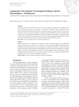
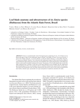
XVIII Simpósio Brasileiro de Química Teórica – SBQT 2015
Pirenópolis – GO, 22-25 Novembro de 2015
Experimental and Theoretical Infrared Study of Homogalacturonans in the Psidium
myrtoides – Nothotrioza myrtoidis system
Renê G. S. Carneiro a (PQ), Rosy M. S. Isaias a (PQ), Bárbara D. L. Ferreira b (PG), Breno R.
L. Galvãoc (PQ), Cláudio L. Donnicib (PQ), Rita C. O. Sebastiãob* (PQ)
a
Departamento de Botânica - Instituto de Ciências Biológicas
b
Departamento de Química - Instituto de Ciências Exatas
Universidade Federal de Minas Gerais-UFMG
Av. Antônio Carlos, 6627, Belo Horizonte – MG, Brazil
c
Centro Federal de Educação Tecnológica de Minas Gerais- CEFET-MG
Av. Amazonas ,5253, Belo Horizonte – MG, Brazil
* [email protected]
Keywords: Acetylation, Homogalacturonans, Infrared spectroscopy, Methylation.
INTRODUCTION
Structural and functional properties of plant
cells rely on the composition of cell walls, which
may be altered by the influence of galling
herbivores1,2. The alteration of chemical
compounds in plant cell walls may be detected by
different techniques as the absorption of infrared
light and nuclear magnetic resonance (NMR)3,4.
The use of infrared absorption spectra allows the
identification of molecular structures by the
fingerprint of chemical bonds and functional
groups, and by chemometric methods analyzing
the region of 1000-500 cm-1 5,6. The fingerprints
of chemical compounds may be successfully
assessed in complex mixtures, which is the case of
plant cell walls7.
Plant epidermis mediates the interactions
between plant organs and the surrounding
environment by lining on both leaf surfaces, and
on the inner and outer surface of galls8. In the
Psidium myrtoides (Myrtaceae) – Nothotrioza
myrtoidis (Psylloidea) system, the leaf epidermis
undergoes distinct degrees of alterations during
gall formation, as evidenced by anatomical,
cytological and immunocytochemical analyses9,10.
The set of structural alterations in the galls of N.
myrtoidis indicate that the inner epidermis is
functionally altered11. Even though the inner and
outer gall surfaces have the same ontogenetic
origin11,12, their structure is distinct and so is the
molecular composition of their cell walls. Based
on this premise, current study proposes the
investigation of the surfaces of non-galled leaves
of P. myrtoides and leaf galls of N. myrtoidis
using IR-ATR spectroscopy. Furthermore, we
analyze
the
optimized
structure
of
homogalacturonans (HGAs), which are the main
pectins in the cell walls11-13, with different degrees
of methylation/acetylation. The main objective is
to check if gall establishment distinctly affects the
molecular structure of the epidermal cell walls on
both gall surfaces. We expect distinct fingerprint
patterns for the gall and leaf epidermises related to
the alterations in the structure of HGAs, with
greater cell alterations occurring in the inner
epidermis when compared to the outer epidermis
of the galls. In addition, we evaluate the efficiency
of experimental and theoretical chemical analyses
in assessing the structure of HGAs, which should
allow new insights on the biology of plant cell
walls.
METHODS
We have experimentally analyzed fresh nongalled leaves and galls using a zinc-selenium
crystal ATR apparatus (Smart orbit ATR, Thermo
Fisher Scientific Inc., Madison, USA), coupled
with the spectrophotometer FTIR Nicolet 380
(Thermo Fisher Scientific Inc., Madison, USA),
and submitted the samples to infrared radiation
between 4000 - 525 cm-1. Series of 32 scannings
with the resolution of 2cm were performed.
Additionally, we theoretically simulated the
infrared spectrum of HGAs of several degrees of
methylation, with and without acetyl groups.
Given the large number of atoms involved, we
have started the analysis at the Hartree-Fock level.
However, we currently worked on higher level
calculations, the DFT level, using the B3LYP
functional, and including Grimme's empirical
dispersion correction in order to improve the
description of intra-molecular interactions13,14.
Geometry optimizations are followed by
calculations of the Hessian matrix, in order to
extract the vibrational frequencies. All electronic
XVIII Simpósio Brasileiro de Química Teórica – SBQT 2015
Pirenópolis – GO, 22-25 Novembro de 2015
structure calculations were performed using the
GAMESS package16.
RESULTS AND DISCUSSION
The experimental ATR-IR spectra of the
adaxial epidermis of non-galled leaves and the
inner epidermis of galls are remarkably different,
especially at the 3500-3000 cm-1 and 1750-1600
cm-1 regions. In the spectra of non-galled leaves,
three intense bands were found at 3360, 1727 and
1642 cm-1, being the second band more intense (~
87%, 75%, and 84% transmittance = T). In the
spectra of galls, the band at 3360 cm-1 is more
intense than the corresponding band in the nongalled leaves (~ 57% T). The band at 1727 cm-1 is
less intense in galls (~ 89% T), and the band at
1642 cm-1 is more intense in galls (~ 68% T) when
compared to the bands of non-galled leaves.
Immuncytochemical investigations using specific
antibodies for homogalacturonans (HGAs) with
different levels of methylation revealed that the
adaxial epidermis of non-galled leaves have low
methylated HGAs, and the inner epidermis of
galls have high methylated HGAs in their cell
walls1. The reduced intensity of bands in the
spectra of galls could be attributed to Hydrogen
bonds (H bonds) between equatorial carbonyl and
hydroxyl groups occurring exclusively in the low
methylated HGAs. Nevertheless, this hypothesis
needed to be proven, and led to the theoretical
study.
For the theoretical study, ten molecular
structures of HGAs were simulated.
Each
structure is composed by six monomers of
galacturonic acid linked by glucoside bonds.
Figure 1 represents the structure of the monomer,
with H as R and R1 groups.
Figure 1. Galacturonic acid monomer.
The structures were divided into two sets: (a)
with acetyl group in the R1 position of the fourth
and sixth monomers and, (b) without acetyl
groups. In both sets, the structures were methylesterified in the R position of the monomers: (i)
1,6; (ii) 1,2,6; (iii) 2,3,4,5; (iv) 3,4,5 and (v) 1,3,5.
The OH band analysis of simulated infrared
spectra is presented in Table 1. The OH bands in
acetylated structures always present stronger
intensity than in the corresponding non-acetylated.
The experimental infrared spectra of galls present
similar behavior, i.e. show stronger intensity for
OH band in galls when compared to non-galled
leaves. This theoretical result suggests that the
structure of HGAs in galls may be composed by
acetylated monomers with high degree of
methylation. This result adds new information to
the experimental detections of high and low
methylated HGAs in the plant cell walls using the
monoclonal antibodies JIM5 and JIM7, which
cannot assess the degree of acetylation of HGAs.
Together, the theoretical and experimental studies
show that the formation of galls affects both the
methylation and acetylation of HGAs, with
implications for the functional aspects of plant
cell walls.
Table 1. Simulated infrared OH band with H
bond analysis after geometry optimization.
Monomers
methylation
and/or
acetylation
1,6
methylated
1,6
methylated
and 4,6
acetylated
1,2,6
methylated
1,2,6
methylated
and 4,6
acetylated
1,3,5
methylated
1,3,5
methylated
and 4,6
acetylated
2,3,4,5
methylated
2,3,4,5
methylated
and 4,6
acetylated
3,4,5
methylated
3,4,5
methylated
and 4,6
acetylated
OH band
Intra
Molecular
cm-1
Intensity
H bond
4263.45
53.68417
HO---OH
CO---OH
4238.58
66.93222
HO---OH
CO---OH
4266.84
53.63021
HO---OH
CO---OH
4243.64
65.38409
HO---OH
CO---OH
4342.67
16.59795
HO---OH
CO---OH
4306.84
28.14144
HO---OH
CO---OH
4252.92
14.62813
4247.83
15.86863
4251.10
13.90583
4256.87
15.78891
HO---OH
HO---OH
HO---OH
HO---OH
Given that this study was performed on the
isolated molecule (gas phase), the role of
intermolecular H bond is not taken into account.
XVIII Simpósio Brasileiro de Química Teórica – SBQT 2015
Pirenópolis – GO, 22-25 Novembro de 2015
Nevertheless, intramolecular H bond is an
important factor that seems to be crucial on
elucidating the experimental results. It should also
be emphasized that the wave numbers presented
in Table 1 are at the Hartree-Fock level, and thus
cannot quantitatively describe the system. Our
analysis are of qualitative nature, aiming to
rationalize the experimental findings. In the
structures with higher degree of methylation
and/or acetylation, the CO-OH interaction
between the monomers does not occur. The spatial
aspect of the highly methylated and acetylated
structure is linear and does not present helical
configuration. Figure 2 shows the optimized
structure for the (a) 1,6 methylated and (b) 3,4,5
methylated HGAs.
(a)
(b)
Figure 2. HGAs (a) 1,6 methylated and (b) 3,4,5
methylated.
The analyses of the CO band in the theoretical
IR spectra show a general behavior for the
structures (a), in which the acetylation determines
a reduction in the calculated wavenumber, thus
corroborating the experimental results.
CONCLUSIONS
Experimental and theoretical studies on the
structure of HGAs represent new frontiers to help
understanding the biology of plant cell walls, as
far as the intramolecular and intermolecular forces
of polymers are concerned. The simulated infrared
absorption of HGAs showed that acetylation
affects intramolecular bonds the most, thus being
complementary to the immunocytochemical
analyses for the degree of HGAs methylation.
Further analyses including with NMR considering
the complexity of the cell wall matrix should help
elucidating the role of fine structural details of
polymers on the functional aspects of the cell
walls.
ACKNOWLEDGMENTS
The authors thank FAPEMIG and CNPQ for
the financial support.
1
P. Albersheim, A. Darvill, K. Roberts, R.
Sederoff, A. Staehelin, Plant cell walls: from
chemistry to biology, Garland Science, New
York, (2011).
2
D. Burckhardt, Biology, ecology and evolution
of gall-inducing psyllids (Hemiptera: Psylloidea).
In: A. Raman, C. W. Schaefer, T. M. Withers,
Biology, ecology, and evolution of gall-inducing
arthropods, Science Publishers, Plymouth, (2005).
3
R. C. O. Sebastião, J. P. Braga, J. Magn.
Reson.,177, 146, (2005).
4
R. C. O. Sebastião, C. N. Pacheco, J. P.
Braga, D. Piló-Veloso, J. Magn. Reson., 182, 22,
(2006).
5
R. G. S. Carneiro, R. M. S. Isaias, Protoplasma,
252, 637, (2014).
6
R. G. S. Carneiro, D. C. Oliveira, R. M. S. Isaias,
Pl. Cell. Rep., 33, 2093, (2014).
7
R. G. S. Carneiro, P. Pacheco, R. M. S. Isaias,
Plos One, 10, e0129331, (2015).
8
A. A. Christy, S. Kasemsumran, Y. Du, Y.
Ozaki, Analytical Sci 20, 935, (2004).
9
R. F. Evert Esau’s Plant Anatomy: Meristems,
cells, and tissues of the plant body: their structure,
function and development, John Wiley & Sons,
Inc., Hoboken, (2006).
10
A. Fahn, Plant anatomy. Pergamon Press,
Oxford, (1990).
11
A.T. Formiga, D. C. Oliveira, B. G. Ferreira, T.
A. Magalhães, A. C. Castro, G. W. Fernandes, R.
M. S. Isaias, Protoplasma, 250, 899, (2013).
12
D. C. Oliveira, T.A. Magalhães, B. G. Ferreira,
C. T. Teixeira, A. T. Formiga, G. W. Fernandes,
R. M. S. Isaias, Plos One, 9, e94588, (2014a).
13
C. Pasquini, J. Braz. Chem. Soc., 14, 198,
(2003).
14
P. J. Stephens, F. J. Devlin, C. F. Chabalowski,
M. J. Frisch, J. Phys. Chem., 98, 11623, (1994).
15
S. Grimme, J. Antony, S. Ehrlich, H. Krieg, J.
Chem. Phys., 132, 154104, (2010).
16
M. W. Schmidt, K. K. Baldridge, J. A. Boatz, S.
T. Elbert, M. S. Gordon, J. H. Jensen, S. Koseki,
N. Matsunaga, K. A. Nguyen, S. Su, T. L.
Windus, M. Dupuis, J. A. Montgomery, J.
Comput. Chem., 14, 1347, (1993). - COMUT
Download