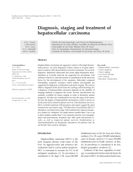
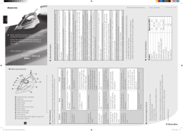
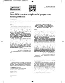
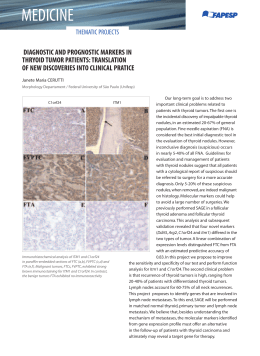
RELATO DE CASO
Hemocromatose associada a carcinoma hepatocelular
e neoplasias extra-hepáticas
Hemochromatosis combined with hepatocellular carcinoma and extrahepatic neoplasms
Eduardo Cambruzzi1, Karla Lais Pêgas2, Cláudio Galeano Zettler3, Márcio Balbinotti Ferrari4
RESUMO
A hemocromatose caracteriza-se pelo acúmulo excessivo de ferro no organismo, que é depositado predominantemente no fígado, e resulta
ou de um defeito genético determinando uma absorção excessiva de ferro ou da administração parenteral deste íon. O ferro em excesso
determina alterações celulares através da peroxidação lipídica, estímulo da deposição de colágeno e interação com o oxigênio reativo e DNA.
Os autores relatam um caso de hemocromatose em paciente portador de cirrose hepática associada ao desenvolvimento de hepatocarcinoma, hemangioma hepático, adenocarcinoma prostático e carcinoma renal, e apresentam uma discussão geral deste processo, frequentemente
associado ao desenvolvimento de neoplasias.
UNITERMOS: Doenças do Metabolismo do Ferro, Hemocromatose, Carcinoma Hepatocelular, Carcinoma de Células Renais, Adenocarcinoma, Cirrose Hepática.
ABSTRACT
Hemochromatosis is characterized by excessive accumulation of iron in the body, which is deposited primarily in the liver. It results either from a genetic defect determining an excessive absorption of iron or from parenteral administration of this ion. The excess iron determines cellular changes through lipid peroxidation, stimulation
of collagen deposition, and interaction with reactive oxygen and DNA. The authors report a case of hemochromatosis in a patient with liver cirrhosis associated with
development of hepatocellular carcinoma, hepatic hemangioma, prostate adenocarcinoma and renal cell carcinoma, and provide a general discussion of this process often
associated with the development of neoplasias.
.
KEYWORDS: Iron Metabolism Disorders, Hemochromatosis, Hepatocellular Carcinoma, Renal Cell Carcinoma, Adenocarcinoma, Liver Cirrhosis.
INTRODUCTION
Hemochromatosis defines a state of body iron overload
that results in tissue injury, and the term hemochromatosis,
from the Greek haima (blood) and Chromatos (color), created
by Von Recklinghausen in 1889. Hereditary hemochromatosis is an inherited recessive disorder associated with increased absorption of iron due to the presence of C282Y and/or
H63D hemochromatosis gene (HFE), which is allocated on
chromosome 6. The secondary hemochromatosis is an acquired disorder that occurs in the context of diseases with known
causes for metabolic overload of iron, such as hemolytic anemia, blood transfusions and liver cirrhosis (1, 2, 3).
1
2
3
4
The iron corresponds to the most abundant transition
metal ion in the body, and its overhead determines tissue
injury through lipid peroxidation by free radicals, stimulation of collagen deposition and reactive oxygen interactions with DNA. The development of liver cirrhosis related to iron overload is associated with development of
hepatocellular carcinoma (1, 2, 3, 4).
The authors report a case of hereditary hemochromatosis associated with liver cirrhosis, hepatocellular carcinoma,
hepatic hemangioma, renal carcinoma and prostatic adenocarcinoma, and present a general discussion of this process.
Doutor. Médico Patologista, Professor Adjunto.
Mestre. Médica Patologista.
Doutor. Médico Patologista.
Acadêmico de Medicina (ULBRA).
Revista da AMRIGS, Porto Alegre, 56 (1): 67-70, jan.-mar. 2012
miolo#1_2012.indd 67
67
10/4/2012 12:04:15
HEMOCROMATOSE ASSOCIADA A CARCINOMA HEPATOCELULAR E NEOPLASIAS EXTRAHEPÁTICAS Cambruzzi et al.
Male patient, 63 years, white, reported low back pain.
The medical history revealed arterial hypertension, cerebral
ischemic attack, family history of brain tumor and renal
cell carcinoma, and prior diagnosis of prostate adenocarcinoma of Gleason score 9 (Figure 1). He denied alcohol
consumption, and had negative serology for HBV and
HCV. Bone scintigraphy showed multiple areas of increased uptake involving almost the entire skeleton. Bone
biopsy revealed metastatic adenocarcinoma. The CT scan
identified two liver nodules, one compromising segments
VI and VII, and one in segment IV, which measured 4.3
cm and 2.3 cm, and an expansive nodule in the upper pole
of right kidney, measuring 5.7 cm. The microscopic evaluation of the resected liver parenchyma showed two distinct
liver lesions, the biggest one corresponding to a trabecular
pattern of hepatocellular carcinoma, grade II / III of Edmondson-Steiner (Figure 2), and the smallest corresponding to a hemangioma (Figure 3). The kidney lesion corresponded to a renal clear cell carcinoma (Figure 4), grade
2 of Fuhrman. Table 1 shows the immunohistochemical
FIGURE 1
FIGURE 3
FIGURE 2
FIGURE 4
CASE REPORT
TABLE 1 – Immunohistochemical profile of tumors
NEOPLASMS / ANTIBODY
PSA
CD10
HSA
CK8/18
VIMENTIN
S100
CA19.9
HEPATOCELLULAR CARCINOMA
--++--RENAL CARCINOMA
-+-++- PROSTATIC ADENOCARCINOMA
+--- ---
68
miolo#1_2012.indd 68
Revista da AMRIGS, Porto Alegre, 56 (1): 67-70, jan.-mar. 2012
10/4/2012 12:04:16
HEMOCROMATOSE ASSOCIADA A CARCINOMA HEPATOCELULAR E NEOPLASIAS EXTRAHEPÁTICAS Cambruzzi et al.
FIGURE 5
FIGURE 6
profile of tumors (using streptavidin-biotin-DAKO). The
liver also showed micronodular cirrhosis (Figure 5) with
accumulation of hemosiderin (4 + / 4 + on a scale of
Scheuer – Figure 6). Perls staining was positive (1 + / 4
+) in renal cell carcinoma. Serum iron was 174ug/dl (normal = 50-150ug/dl), transferrin saturation corresponded
to 96% (normal = 20% -50%), and glucose was equal to
82.0 mg / dl. The diagnosis of hemochromatosis was then
suggested.
DISCUSSION
Iron accumulates as ferritin and hemosiderin in almost
all body cells. In hereditary hemochromatosis, the liver is
the target organ because iron absorbed from the intestinal tract crosses the hepatic parenchyma before reaching
the systemic circulation. Initially the deposit occurs in the
Revista da AMRIGS, Porto Alegre, 56 (1): 67-70, jan.-mar. 2012
miolo#1_2012.indd 69
hepatocytes of zone 1 of Rappaport, progressing to the
surrounding parenchyma. With progressive accumulation,
occurs formation of fibrous septa from the portal spaces,
which may progress to cirrhosis (1, 2, 3).
Iron acts as a catalyst in the generation of reactive oxygen species in pathological conditions, which are associated with carcinogenesis by inducing tissue damage through
lipid peroxidation and DNA damage. Lipid peroxidation
is a critical step for fibrosis (the activation of stellate cells)
and hepatocarcinogenesis, and iron may initiate these mechanisms. Iron overload increases the number and activity
of CD8 T lymphocytes and inhibits the proliferation and
activity of CD4 T lymphocytes, determining the generation
of cytotoxic T cells, changes in immunoglobulin secretion
and suppression of the complement system (1, 2, 4).
The diagnosis of hereditary hemochromatosis is based
on the accumulation of iron excess in parenchymal cells
in the absence of known causes of iron overload and in
the presence of C282Y mutation in HFE. Liver biopsy
quantifies the iron deposit and determines the presence of
fibrosis or cirrhosis. The determination of liver iron content is also a safe diagnostic method, exceeding 1.9 mmol
/ kg / year. The factors that predate the clinical expression
of hemochromatosis are: male sex, alcoholism, infection
with HBV / HCV, food rich in iron and vitamin C and
deficiency of alpha-1-antitrypsin. Up to five individuals in
1000 are homozygous for the disease in the USA, France,
Sweden and the UK, where about 10% of the Caucasian
population is heterozygous. From 64% to 100% of patients with hereditary hemochromatosis in Caucasian descent are homozygous for C282Y, being the heterozygous
state more frequent in men (1, 2, 3, 5, 6).
Hepatocellular carcinoma is a possible complication
of cirrhosis in hereditary hemochromatosis, accounting
for 45% of the deaths. The relative risk of developing hepatocellular carcinoma in patients with hemochromatosis
and cirrhosis is approximately 200 times. The follow-up of
patients with alcoholic cirrhosis or hepatitis B has shown
that patients with elevated serum ferritin or liver iron concentration have a higher risk of liver cancer than patients
with normal or low stocks of iron. Foci of hepatocellular
hyperplasia are more frequent in macro-regenerative iron-positive nodules (73%) than macro-regenerative iron-negative nodules (21%). Foci free of iron can be found in
hereditary hemochromatosis, being those areas associated
with the early stages of hepatocellular carcinoma. Even
with iron overload, the precancerous lesions dont seem
to show ion accumulation, suggesting that their depletion
is one of the early changes of cancer cells. The iron also
has an effect on promotion of hepatocellular carcinoma in
the presence of HBV infection, because it promotes viral
replication and tumor cell growth. HBV is more likely to
infect and replicate in hepatocytes with increased ferritin
(1, 2, 4, 7). The possible relationship between hepatocellular carcinoma and heterozygosity for the C282Y mutation
is not defined, although the homozygous for C282Y in the
69
10/4/2012 12:04:16
HEMOCROMATOSE ASSOCIADA A CARCINOMA HEPATOCELULAR E NEOPLASIAS EXTRAHEPÁTICAS Cambruzzi et al.
absence of blood loss, always develop iron overload, while
in heterozygotes that risk is lower (3, 4, 5, 6, 7).
If the relationship between hemochromatosis and risk
of hepatocellular carcinoma appears to be well documented, the association of hepatocellular neoplasms and hereditary hemochromatosis seem indefinite (1, 3, 4, 8, 10,
11). It is suggested the iron as a promoter of carcinogenesis by altering the immune system and promoting lipid
peroxidation (3, 8, 9, 10, 11). Geier et al found 13 cases
of non-hepatocellular carcinoma in 59 patients with hemochromatosis, being the colon, stomach, prostate, breast
and hematopoietic tissue the primary sites (10). Elmberg
et al., among 1847 patients with hereditary hemochromatosis, found 62 cases of hepatocellular carcinoma and 128
non-hepatobiliar neoplasm (8). Shaheen et al. suggested
that mutations in the HFE gene are associated with increased risk for colon carcinoma (11). Osborne et al. reported that HFE C282Y homozygotes have twice the risk of
colorectal and breast cancer compared with those without
the C282Y variant, although male C282Y homozygotes
were not at increased risk for prostate cancer (12). Gannon
et al described that C82Y mutation may increase the risk
of developing ovarian carcinoma, and may be associated
with poor outcomes. Dorak et al described a strong association between C282Y mutation and childhood acute
lymphoblastic leukemia. Kallianpur et al reported a high
prevalence of C282Y alleles in female breast cancer, and its
association with more aggressive forms of the tumor (15).
The HFE gene is a major histocompatibility class I-like molecule, that, when mutated, may cause hereditary
hemochromatosis. HFE associated with the major protein
responsible for cellular iron uptake, namely the transferrin receptor (TfR). The association of HFE with TfR at
the cell surface lowers TfR affinity for the circulating iron-transporter transferrin, thereby limiting iron uptake and
thus directly implicating HFE in the modulation of cellular iron levels. A failure to appropriately express HFE at
the cell surface, as is found in the C282Y mutation, may
result in an enhanced ability to capture iron and may induce tumor cell proliferation. In addition, it looks that high
level of free iron may accentuate the effects of other carcinogenic agents, such as ethanol and ionizing radiation.
Iron-catalyzed oxidative stress causes lipid peroxidation
and protein modification, DNA damage with consequent
promotion of mutagenesis, and leads to the depletion of
antioxidant defenses (12, 13).
FINAL COMMENTS
Although it is not possible to attribute a direct cause-effect relationship between hemochromatosis and the ne-
70
miolo#1_2012.indd 70
oplasms, especially after reviewing the literature, the presence of a basic metabolic disease and four tumors induces
the hypothesis of a common cause for the development of
these tumors. At diagnosis of hemochromatosis, done by
the research of iron in the liver tissue and by abnormal levels of iron and transferrin saturation, should perform the
study for genetic mutations in the HFE gene for confirmation of homozygosity. Given the natural history of disease,
it is necessary to track patients with hemochromatosis for
the early detection of complications such as cirrhosis and
cancer.
REFERENCES
1.Gayotto LCC, Alves VAF. Doenças do fígado e vias biliares. São
Paulo: Ed. Atheneu; 2001.
2.Hanson, EH et al. HFE gene and hereditary hemochromatosis: a
huge review. Am J Epidemiol. 2001;154:193-206.
3.Searle J. Iron storage diseases. In: MacSween, RNM; Burt, AD; Portmann, BC; Ishak, KG; Scheuer, PJ (2002). 4th ed. London: Churchill
Livingstone. Cap.5, p 257- 272.
4.Kew MC. Hepatic iron overload and hepatocellular carcinoma. Cancer Lett. 2009;286:38-43.
5.Cauza E et al. Mutations of the HFE gene in patientes with hepatocellular carcinoma. Am J Gastroenterol. 2003;98:442-7.
6.Fargion S et al. Mutations in the HFE gene and their interaction
with exogenous risk factors in hepatocelular carcinoma. Blood Cells
Mol Dis. 2001;27:505-11.
7. Deugnier Y, Turlin B. Iron and hepatocellular carcinoma. J Gastroenterol Hepatol. 2001;16:491.
8.Elmberg M et al. Cancer risk in patients with hereditary hemochromatosis and in their first-degree relatives. Gastroenterology.
2003;125:1733-4.
9.Fracanzani AL et al. Hemochromatosis in Italy in the last 30 years:
role of genetic and acquired factors. Hepatology. 2010;51:1473-4.
10.Geier D et al. Risk of primary non-hepatocellular malignancies in
hereditary hemochromatosis. Anticancer Res. 2002;22:3797-9.
11.Shaheen NJ et al. Association between hemochromatosis (HFE)
gene mutation carrier status and the risk of colon cancer. J Natl
Cancer Inst. 2003;95(11):829-30.
12.Osborne NJ et al. HFE C282Y homozygotes are at increased risk
of breast and colorectal cancer. Hepatology. 2010;51(4):1311-1318.
13.Gannon et al. Impact of hemochromatosis gene (HFE) mutations on epithelial ovarian cancer risk and prognosis. Int J Cancer.
2010;128:2326-2334.
14.Dorak MT et al. Hemochromatosis gene in leukemia and lymphoma. Leuk Lymphoma. 2002;43(3):467-477.
15.Kallianpur AR et al. Increased prevalence of the HFE C282Y hemochromatosis allele in women with breast cancer. Cancer Epid Bio
Prevent. 2004;13(2):205-212.
* Endereço para correspondência
Eduardo Cambruzzi
Av. Loureiro da Silva, 1500/1308
90.050-240 – Porto Alegre, RS – Brasil
( (51) 3357-2164 / (51) 3226-1779 / (51) 9954-5433
: [email protected]
Recebido: 11/3/2011 – Aprovado: 28/4/2011
Revista da AMRIGS, Porto Alegre, 56 (1): 67-70, jan.-mar. 2012
10/4/2012 12:04:16
Download