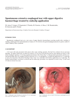
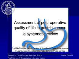
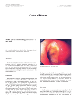
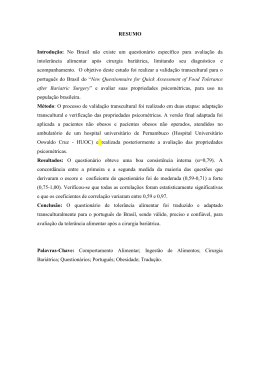
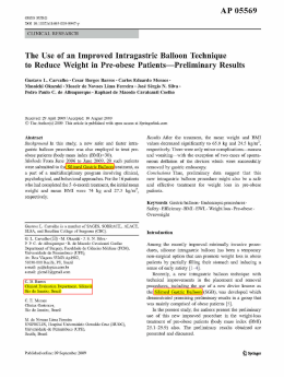
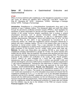
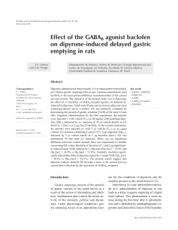
OBES SURG
DOI 10.1007/s11695-011-0444-8
CLINICAL REPORT
Gastrobronchial fistula after sleeve gastrectomy and gastric
bypass: endoscopic management and prevention
Josemberg Marins Campos & Eduardo Franca Pereira & Luis Fernando Evangelista &
Luciana Siqueira & Manoel Galvão Neto & Victor Dib & Marcelo Falcão &
Vitor Arantes & Diego Awruch & Walton Albuquerque & João Ettinger &
Almino Ramos & Álvaro Ferraz
# Springer Science+Business Media, LLC 2011
Abstract Gastrobronchial fistula (GBF) is a serious complication following bariatric surgery, whose treatment by
thoracotomy and/or laparotomy involves a high morbidity
rate. We present the outcomes of endoscopic management
for GBF as a helpful technique for its healing process. This
is a multicenter retrospective study of 15 patients who
underwent gastric bypass (n=10) and sleeve gastrectomy
(n = 5) and presented GBF postoperatively (mean of
6.7 months). Ten patients developed lung abscess and were
treated by antibiotic therapy (n=10) and thoracotomy
(n=3). Abdominal reoperation was performed in nine
patients for abscess drainage (n=9) and/or ring removal
(n=4) and/or nutritional access (n=6). The source of the
GBF was at the angle of His (n=14). Furthermore, 14
patients presented a narrowing of the gastric pouch treated
by 20 or 30 mm aggressive balloon dilation (n=11),
stricturotomy or septoplasty (n=10) and/or stent (n=7).
Fibrin glue was used in one patient. We performed, on
average, 4.5 endoscopic sessions per patient. Endotherapy
led to a 93.3% (14 out of 15) success rate in GBF
closure with an average healing time of 4.4 months
(range, 1–10 months), being shorter in the stent group
J. M. Campos (*) : E. F. Pereira : L. F. Evangelista :
L. Siqueira : M. Falcão : D. Awruch : J. Ettinger : Á. Ferraz
Universidade Federal de Pernambuco,
Rua Vigário Barreto, 127/802-Graças,
52020-140, Recife, PE, Brazil
e-mail: [email protected]
M. G. Neto : V. Dib : A. Ramos
Gastro Obeso Center,
São Paulo, SP, Brazil
V. Arantes : W. Albuquerque
Serviço de Endoscopia, Instituto Alfa de Gastroenterologia,
Belo Horizonte, MG, Brazil
(2.5×9.5 months). There was no recurrence during the
average 27.3-month follow-up. A patient persisted with
GBF, despite the fibrin glue application, and decided to
discontinue it. GBF is a highly morbid complication,
which usually arises late in the postoperative period.
Endotherapy through different strategies is a highly
effective therapeutic option and should be implemented
early in order to shorten leakage healing time.
Keywords Bariatric surgery . Gastrobronchial fistula .
Bronchogastric fistula . Lung abscess . Gastric fistula .
Sleeve gastrectomy . Endoscopy . Balloon dilation . Stents
Introduction
Gastric fistula (GF) following bariatric surgery occurs in up
to 8.3% of the cases [1]. It is a severe complication that
often results in abdominal sepsis and respiratory alterations,
especially in the left lung secondary to subphrenic abscess
[2, 3]. Measures of clinical support, antibiotic therapy, and
abdominal drainage may be sufficient to control systemic
infection and to heal the leakage [4]. Nevertheless, some
patients still evolve unsatisfactorily.
Due to proximity with the respiratory tract, an abdominal
infection can result into a gastrobronchial fistula (GBF),
although its occurrence after bariatric surgery has seldom
been reported (Table 1) [5–11]. GBF treatment can be
challenging when performed by means of major abdominal
and thoracic operations [6–8, 11].
As a severe lung infection with a high morbidity–
mortality rate [10], it is necessary to throw some light upon
the predisposing factors and the clinical aspects of GBF, as
well as to establish preventive, diagnostic, and minimally
invasive approach strategies. Thus, we aim to report a
LSG+LDS –
LAGB
LSG
1
1
1
1
6
5
7
3
2.5
20
CT, UGIS
UGIS
Hemoptysis, recurrent
pneumonia, cough,
dyspnea, fever
Recurrent pneumonia,
empyema, hemoptysis,
purulent sputum
Recurrent pneumonia
Pouch
His
His
DIA
Source of the
leakage
CT, UGIS
Laparoscopic band removal+
interposition of omentum
Total gastrectomy+thoracotomy+
lobectomy
Endoscopic dilatation+
stenostomy
SEMS+SEPS+fibrin glue
Endoscopic dilatation+
stenostomy+fibrin glue
Total gastrectomy
Treatment
of GBF
Paraesophageal Open band removal+
hernia
thoracotomy+lobectomy
Clinical, UGIS,
Pouch
endoscopy
Methylene blue, CT His
Vomica, cough, dyspnea, CT, UGIS
fever
–
–
Lung abscess, vomica,
cough, dyspnea, fever
Lung abscess
Diagnosis
of GBF
2
Healing
time of GBF
(months)
1
1
–
1
–
Jejunostomy
Death (3 m)
Parenteral
Gastrostomy,
6
nasoenteral tube 8
Gastrostomy
1
Nutrition
access
CT computed tomography scan, UGIS upper gastrointestinal X-ray series, ODS open duodenal switch, RYGB Roux-en-Y gastric bypass, GBF gastrobronchial fistula, LSG laparoscopic sleeve
gastrectomy, LDS laparoscopic duodenal switch, LAGB laparoscopic adjustable gastric band, DIA duodenal–ileal anastomosis, SEMS self-expanding metal stent, SEPS self-expanding plastic stent
Garrett KA, Obes 1
Surg 2009 [11]
LAGB
RYGB
LSG
RYGB
2
Campos J, JACS
2007 [9]
Eisendrath P,
Endoscopy
2007 [10]
Chin P, SOARD
2008 [6]
Fuks D, Obes
Surg 2009 [8]
ODS
1
Serra C, Obes
Surg 2006 [7]
Campos J, JBP
2007 [5]
14
Number Bariatric
surgery
Author/year
Respiratory
Onset of
respiratory symptoms
symptoms
(months)
Table 1 Studies involving patients with gastrobronchial fistula (GBF) after bariatric surgery: analysis of clinical and therapeutic aspects
OBES SURG
OBES SURG
multicenter experience on the clinical features, endoscopic
treatment results of GBF, and its prevention.
Methods
Fifteen patients (9 women, 6 men; mean age 36 years), with
an average body mass index of 43 kg/m2, who underwent
either open (n=5) or laparoscopic (n=5) Roux-en-Y gastric
bypass (RYGB) and laparoscopic sleeve gastrectomy (LSG)
(n=5) at 14 Brazilian bariatric surgery institutions took part
in this study, from 2003 to 2010. A silicon ring was used in
RYGB (n=7) and LSG (n=2) (Table 2; Fig. 1).
This is a retrospective study to evaluate the management
of GBF in three institutions of gastrointestinal endoscopy
and surgery in Brazil, with extensive experience in the
management of bariatric surgery postoperative complications. From 2004 to 2010, we enrolled 15 patients who
were referred to us and presented with persistent gastric
leaks and radiologic confirmation of GBF, despite intensive
measures in the original institution, such as antibiotic
therapy, nutritional support, and treatment of the abdominal
and thoracic infection by reoperation or computed
tomography-guided aspiration. The following radiologic
images were used in this study: chest and abdominal
computed tomography (CT) scan, upper gastrointestinal
series, and/or fistulography (Figs. 2, 3 and 4).
After signing terms of informed consent, patients
underwent endoscopic procedures to promote fistula closure. The first aim of our approach was to delineate through
a detailed endoscopic and fluoroscopic study whether upper
gastrointestinal anatomic alterations in the vicinity of the
fistula orifice were present. If positive, we would propose
the appropriate intervention (Fig. 1).
Endoscopic Procedures
The procedures were carried out in endoscopy units with
fluoroscopy under deep sedation or general anesthesia. A
standard one-channel gastroscope (Pentax Medical, Montvale, NJ) was used in all the procedures and a transnasal
gastroscope EG-530N® (Fujinon, Yokohama, Japan) was
used once in the diagnostic fistuloscopy of case 10.
The choice of the specific endoscopic intervention was
based on the following variables: type of bariatric
surgery, presence of the ring, location of the gastric
stricture, presence of perigastric abscess, and anatomy of
the gastric pouch and fistula. Each patient underwent
more than one type of endoscopic procedure (Fig. 1).
The principle of the treatment was the correction of both
the distal gastric stricture and the anatomic defect near the
internal orifice of the fistula (Fig. 5). The following
endoscopic interventions were performed according to the
type of bariatric surgery:
Table 2 Demographic data and clinical conditions of patients with gastrobronchial fistula (GBF)
Patient
number
Age Sex BMI preop.
kg/m2
Bariatric
surgery
Ring Onset of respiratory symptoms
(PO months)
Lung
abscess
Source of the Narrowing of the
GBF
stomach (level)
1
2
3
4
5
6
7
8
9
10
32
44
44
22
38
41
28
18
37
43
W
M
W
W
W
M
M
M
W
W
49
44
37
43
39
45
42
42
41
40
LSG
LRYGB
LSG
ORYGB
LRYGB
LRYGB
LRYGB
ORYGB
ORYGB
ORYGB
Y
N
Y
Y
Y
Y
Y
Y
Y
Y
1.5
3
8
30
2
1
5
1
6
18
Y
Y
Y
Y
N
Y
N
Y
N
Y
Angle
Angle
Angle
Angle
Angle
Angle
Angle
Angle
Angle
Angle
His
His
His
His
His
His
His
His
His
His
Ring
Anastomosis
Angular incisure
Pouch
Ring
Ring
Pouch
Pouch
Anastomosis
Ring
11
12
13
14
15
39
23
53
32
45
M
M
W
W
W
46
45
35
42
55
LSG
LSG
LSGa
ORYGB
LRYGBa
N
N
N
N
N
1
2
15
6
2
Y
Y
N
Y
N
Angle of His
Gastric body
Angle of His
Angle of His
Angle of His
Angular incisure
Angular incisure
Angular incisure
No
Pouch
of
of
of
of
of
of
of
of
of
of
W women, M men, BMI body mass index, preop. preoperatory, PO postoperative, LRYGB laparoscopic Roux-en-Y gastric bypass, ORYGB open
Roux-en-Y gastric bypass, LSG laparoscopic sleeve gastrectomy, Y yes, N no, GBF gastrobronchial fistula
a
Revisional surgery
OBES SURG
Fig. 1 Flowchart of endoscopic
treatment of gastrobronchial
fistula (GBF). SEPS = self-expandable plastic stent
Fifteen patients submitted to gastric by pass (n=10) or sleeve gastrectomy (n=5)
in 14 bariatric surgery institutions
Onset symptoms and signals of GBF (cough,dyspnea,vomica)
Radiologic and endoscopic investigation
GBF diagnosis (n=15)
Endoscopic treatment + Pulmonary management (n=15) in
3 institutions of gastrointestinal endoscopy and surgery
20-30mm balloon dilation
n=11
Stricturotomy or
Septoplasty
n=10
SEPS
n=7
Glue (n=2)
or
Cli p (n=1)
Improvement of the GBF
Yes
No
Radiologic investigation
Closure of the GBF
Persistence
of the GBF
GBF recurrence
Clinical
management
n=1
New endoscopic
treatment
Cure of the GBF (n=14)
Clinical and Endoscopic Follow-up
1. Aggressive endoscopic dilation:
RYGB without ring: The gastrojejunal anastomotic
stricture was dilated up to 20 mm, using a CRE®
balloon (Boston Scientific, Natick, MA), during 3 min
[12].
RYGB with ring: The stricture of the ring in the gastric
pouch was dilated up to 30 mm, using a Rigiflex® (Boston
Scientific, Natick, MA), which was gradually inflated until
either rupturing or stretching the thread running inside the
ring to widen the luminal diameter. The duration of each
dilation session ranged from 5 to 30 min [13].
LSG with or without ring (Figs. 2 and 6): The
narrowed spot of the gastric pouch (ring or angular
incisure) was dilated using a 30-mm pneumatic dilator
(Fig. 5) [14], following the technique for RYGB. The
ideal way to detect the angular incisure stenosis is
through the upper gastrointestinal series. However, the
gastroscope may also identify a longitudinal fold at this
area. So, for the endoscope to pass and reach the antrum,
the tip must be well angled up.
2. Stricturotomy:
RYGB with or without ring: The stenosis of gastrojejunal anastomosis with persistent fibrotic stricture or
OBES SURG
Fig. 2 Images of gastrobronchial fistula (GBF): a) Tomography
showing lung abscess - Case 2; b) Contrast radiography showing a
GBF from the angle of His after sleeve gastrectomy - Case 3; c)
Contrast radiography showing lung (white arrow) and subphrenic
abscess (black arrow) due to GBF at the angle of His (bent arrow)
after sleeve gastrectomy - Case 11; d) Contrast injection through the
subphrenic drain revealing the bronchial tree - Case 4
recurrence was treated by stricturotomy, using Microknife XL® (Boston Scientific, Natick, MA). Afterwards, 20 mm balloon dilation was performed. When
the diameter of the pouch was smaller than the
esophagus, a stricturotomy was also performed, followed by dilation with a 30-mm balloon.
LSG with or without ring: The stenosis in the
angular incisure was treated similarly to the strictur-
otomy technique described for RYGB, followed by
balloon dilation up to 30 mm (Fig. 5).
3. Gastric septoplasty for internal drainage of abscess:
RYGB or LSG: the septum near the internal orifice of
the fistula at the angle of His facilitated the passage of
secretion through the leakage. It was a contributing factor
to abscess formation and hindering the GF healing
process. Therefore, the septum was incised with a Micro-
Fig. 3 Contrast radiographic
images after gastric bypass: a)
Fistulography of the subphrenic
region and bronchial tree - Case
7; b) Contrast swallow showing
esophagus (white arrow), GBF
from the angle of His and
reduced diameter of the gastric
pouch - Case 15
OBES SURG
Fig. 4 Images of patient 14
after gastric bypass: a) Contrast
radiography showing a GBF
from the angle of His, b) Computerized tomographic reconstruction showing a clearer
image of the GBF
knife XL® (Boston Scientific, Natick, MA), followed by
a 30-mm balloon dilation. This technique allows the
internal drainage of the abscess, which leads to the closure
of the fistula, since the gastric outlet is now restored.
4. Placement of self-expandable plastic stent (SEPS):
A Polyflex® (diameter, 25×21 mm; length, 150 mm)
(Boston Scientific, Natick, MA) was indicated mainly
when the diameter of the fistula was >10 mm and/or distal
stenosis of the gastric pouch was persistent.
As soon as the endoscopic treatment was initiated, the
patients received the following additional measures:
–
Enteral nutrition either by gastrostomy, jejunostomy, or
nasoenteral tube, as substitutes for the total parenteral
nutrition.
–
–
Early discharge from the hospital.
Therapeutic endoscopy sessions whenever necessary; it
was carried out in the endoscopy suite in an outpatient
setting. LSG patients underwent additional endoscopic
dilation sessions every 30 days, during 3 months to
prevent GBF recurrence.
Results
Symptoms and signs suggestive of GBF (cough, dyspnea,
chest pain, hemoptysis, fever, or vomit of pus from the
lungs—vomica) appeared, on average, 6.7 months (range,
1–30 months) after the bariatric surgery (Table 2).
Fig. 5 Images of angular incisure stenosis (arrow) and fistula after sleeve gastrectomy: a) Endoscopic balloon dilation - Case 1; b) Schematic
image showing a fistula from the angle of His; c) Radiography showing balloon dilation - Case 3
OBES SURG
Fig. 6 Contrast radiographic
images of patient 13 after
sleeve gastrectomy showing a
GBF (white arrows) from the
angle of His; a) Gastric pouch in
spiral with a proximal stenosis
and another at the angular incisure (curved arrows); b) GBF
and subphrenic abscess
Chest CT diagnosed lung abscess, with diameters ranging
from 4 to 10 cm (Fig. 2), in the lower left lung in ten patients;
three of them were treated by thoracotomy (Fig. 7).
Before referring the patients to our institution for
endoscopic treatment, several abdominal and thoracic
operations were attempted to close the GBF and to manage
its secondary complications (Table 3). Abdominal reoper-
Fig. 7 Patient 10 with scars due to major abdominal and thoracic
surgery and a gastrobronchial-cutaneous fistula. She was the only
patient who persisted with GBF
ation was done in nine patients for the following procedures: drainage and cleansing of the abdominal cavity (n=
9), ring removal (n=4), and to create a nutritional access
(n=6). CT-guided aspiration of subphrenic abscess was
performed in five cases, which avoided reoperation in three
patients. The abdominal drainage tube placed during the
initial bariatric surgery also avoided reoperation in three
other cases. Ten patients developed lung abscess and were
treated by the following procedures: antibiotic therapy (n=
10) and thoracotomy (n=3).
The source of the leakage was seen at the angle of His
(n=14) and gastric body (n=1) by gastroscopy. Those 14
patients developed a narrowing of the gastric pouch at
different spots: at the gastrojejunal anastomosis (n=2), at the
ring (n=4), at the gastric pouch (n=4), and at the angular
incisure (n=4) (Table 2; Figs. 3 and 4).
The following endoscopic procedures were undertaken:
SEPS placement (n=7), balloon dilation (n=11), stricturotomy or septoplasty (n=10), and application of clip (n=1)
and fibrin glue (n=2) (Fig. 5). There were, on average, 4.5
endoscopic sessions per patient. Patients 5 and 6 received a
SEPS after RYGB with ring that were both purposely
removed by endoscopy due to intragastric ring erosion
1 month later on average (Tables 2 and 3).
Minor complications occurred in two patients; case 4
presented a self-limited upper digestive hemorrhage after a
septoplasty, and case 15 presented a distal migration of
SEPS in the post-RYGB gastric pouch, undergoing a new
endoscopy to adjust it (Table 3).
The overall average healing time of the GBF was
4.4 months (range, 1–10 months), after the beginning of
OBES SURG
Table 3 Endoscopic and surgical treatment and results of patients with gastrobronchial fistula (GBF)
Patient Reoperation before
number endoscopic procedures
1
2
3
4
5
6
7
8
9
10
11
12
13
14
15
–
Drain
Drain+ring removal
Drain+ring removal
–
–
–
Thoracotomy+drain+ring
removal
–
Thoracotomy+drain+ring
removal
Drain
Thoracotomy+drain
Drain
Drain+leak suture
–
Nutritional
access
SEPS Balloon Stricturotomy or Endoscopic Minor
dilation septoplasty
sessions
complication
Nasoenteral tube
Gastrostomy
Nasoenteral tube
Gastrostomy
Nasoenteral tube
Nasoenteral tube
Nasoenteral tube
Gastrostomy
N
N
N
N
Y
Y
Y
Y
Y
Y
Y
Y
Y
Y
Y
N
Y
Y
Y
Y
N
Y
Y
N
6
8
6
5
3
3
4
3
–
–
–
Bleeding
–
–
–
6
6
8
6
2
3
1.5
2
Nasoenteral tube Y
Gastrostomy
N
Y
N
Y
N
3
2
–
–
1
–
Jejunostomy
Nasoenteral tube
Jejunostomy
Nasoenteral tube
–
N
N
Y
Y
Y
N
N
Y
Y
Y
3
2
11
5
4
–
–
–
–
Stent migration
10
5
6
2.5
3
N
Y
N
N
Y
Healing time of
GBF (months)
Drain drainage, SEPS self-expandable plastic stent, Y yes, N no, GBF gastrobronchial fistula
the endoscopic treatment. The group with SEPS presented a
shorter recovery time (2.5 months), in comparison with the
group submitted to other endotherapy (9.5 months).
Endotherapy led to a 93.3% (14 out of 15) success rate
in GBF closure without recurrence after an average followup of 27.3 months (range, 2–72 months). The only patient
(case 10) who persisted with a GBF (Fig. 7), despite the
application of fibrin glue (one session), denied to undergo
further treatment.
Discussion
After RYGB and LSG, the closure of GF can be hindered by
the presence of distal stenosis and abdominal infection [8, 9,
15]. The spontaneous communication of the subphrenic
abscess with the bronchial tree suggests the previous
existence of a persistent GF, possibly secondary to a chronic
difficulty in gastric emptying [5, 9]. The GBF of our patients
occurred late in the postoperative period (5.7 months on
average), consistent with other studies [5–9].
GBF may have some possible predisposing factors such
as recurrent abscesses in the upper abdomen without
drainage or early withdrawal of the drain, with resultant
chronic subphrenic inflammation [5, 7–9]. It also may have
occurred after adjustable gastric band erosion [6].
Coughing with phlegm and fever are the main symptoms
of GBF [5, 7, 8]. However, vomica, recurrent pneumonia,
and/or lung abscess were observed mainly in patients with
late diagnosis, due to a delay in evaluation by a specialist in
bariatric surgery [6, 9]. We observed a significant decrease in
oxygen saturation during gastroscopy or contrast radiography due to the passage of air or contrast material into the
airways through the GBF. Obtaining a precise diagnosis of
GBF is a laborious process, requiring imaging exams with
iodinated contrast media [5, 7–9]. The use of contrast with
barium can cause severe bronchial reactions, which needs to
be cleaned by bronchoscopy [6].
In our study, no patient was referred to bronchoscopy.
Although this exam does not reveal the precise location of
the fistula, bronchoscopy can suggest GBF by the observation of methylene blue in the bronchus after oral or drain
administration [8].
Treatment with antibiotics was successful to manage the
majority of our patients with lung infection. In cases of
failure, CT-guided aspiration of the collection and surgical
approach are other options for the management of GBF
[8, 9]. In our series, six patients showed no lung infection
on CT scan. Within this group, we adopted SEPS to bridge
the fistula in three cases (5, 7, and 9), and we suppose that
its early application may have prevented the development
of lung abscess.
Upper digestive endoscopy does not diagnose GBF, but
identifies its internal opening, evaluates the anatomy of the
gastric pouch, and promotes therapy, minimizing the need
of invasive surgery [5, 9]. Reoperation may provide little
benefit because of the difficult access to the region of the
upper abdomen with adhesions and fibrosis [5, 6, 8, 9]. In
OBES SURG
addition to the complexity of major abdominal and thoracic
reoperations with its high morbidity rate [7, 8], it is often
not efficient to completely heal GBF, as it has shown to be
true in three patients of the present series.
Fuks et al. [8] report the complex treatment of a patient
with a GBF after LSG, which failed initially with the clinical
and endoscopic approach. Usually, this may be secondary to
a stenosis at the angular incisure, which increases the
intragastric pressure and promotes the persistent GBF [14,
16]. In this case, the healing of the fistula was achieved only
through a major abdominal and thoracic surgery [8].
Among our five LSG patients, only one had undergone a
thoracotomy. The other four patients had been treated by
antibiotics, combined with endoscopic correction of angular
incisure stenosis. Thoracotomy for cleaning of the lung
infection seems to bring little benefit to patients with GBF,
whereas thoracoscopy is minimally invasive and could
promote lung expansion, helping avoid invasive ventilatory support. However, most patients in the current series
(n=12) and from other reports did not need thoracic
surgery [5–7, 9].
Endoscopic stricturotomy and dilation are minimally
invasive and effective techniques in resolving gastric stenosis
[5, 9, 12]. In LSG, the angular incisure stenosis hinders the
healing of the fistula at the angle of His and some authors
have proposed total gastrectomy for the final settlement of
the GBF [7, 8]. In the present series, healing of GBF was
achieved by a combination of aggressive balloon dilation,
SEPS placement, and stricturotomy or septoplasty. The
systematic dilation every 30 days for a 3-month period,
even in the absence of obstructive symptoms, prevented the
recurrence of GBF.
The endoscopic use of SEPS in fistula related to trauma,
perforation, or esophagogastrectomy has been reported [17,
18]. In the present series, SEPS prevented the passage of
secretion for the GBF and also promoted expansion of the
distal gastric stricture. This led to ring erosion in two cases,
occurring when complete resolution of the stenosis was
achieved, which has been described previously [19, 20].
Through the use of SEPS, the digestive tract is permanently
patent, allowing early food intake and decreasing the number
of endoscopic sessions [21]. The SEPS migration occurred in
patient 15, which can be avoided by anchoring the distal end
of the stent in the gastrojejunal anastomosis.
The use of endoscopic clip and fibrin glue for the closure
of GBF was attempted only at the beginning of this study. It
was discontinued due to unfavorable results [5, 8, 9]. We
hypothesize that, most likely, this failure occurred because
of persistence of the distal stenosis.
In conclusion, the present series demonstrates that
GBF is a severe complication of bariatric surgery that
usually presents late in the postoperative period. GBF
can be healed with an endoscopic approach directed to
open up widely the lumen of the gastric pouch by a
combination of aggressive dilations, SEPS, and stricturotomy or septoplasty. Early endoscopic intervention is
important to shorten the healing time of the fistula and to
prevent lung abscess, particularly with the early use of
SEPS. Recurrence of GBF is avoided through the final
resolution of gastric stenosis by balloon dilation and
stricturotomy.
References
1. Baker RS, Foote J, Kemmeter P, et al. The science of stapling and
leaks. Obes Surg. 2004;14(10):1290–8.
2. Merkle EM, Hallowell PT, Crouse C, et al. Roux-en-Y gastric
bypass for clinically severe obesity: normal appearance and
spectrum of complications at imaging. Radiology. 2005;234
(3):674–83.
3. Varghese JC, Roy-Choudhury SH. Radiological imaging of the GI
tract after bariatric surgery. Gastrointest Endosc. 2009;70
(6):1176–81.
4. Yurcisin BM, DeMaria EJ. Management of leak in the bariatric
gastric bypass patient: reoperate, drain and feed distally. J
Gastrointest Surg. 2009;13(9):1564–6.
5. Campos JM, Siqueira LT, Meira MR, et al. Gastrobronchial fistula
as a rare complication of gastroplasty for obesity: a report of two
cases. J Bras Pneumol. 2007;33(4):475–9.
6. Chin PL. Gastrobronchial fistula as a complication of laparoscopic adjustable gastric banding. Surg Obes Relat Dis. 2008;4
(5):671–3.
7. Serra C, Baltasar A, Perez N, et al. Total gastrectomy for
complications of the duodenal switch, with reversal. Obes Surg.
2006;16(8):1082–6.
8. Fuks D, Dumont F, Berna P, et al. Case report—complex
management of a postoperative bronchogastric fistula after laparoscopic sleeve gastrectomy. Obes Surg. 2009;19(2):261–4.
9. Campos JM, Siqueira LT, Ferraz AA, et al. Gastrobronchial fistula
after obesity surgery. J Am Coll Surg. 2007;204(4):711.
10. Eisendrath P, Cremer M, Himpens J, et al. Endotherapy including
temporary stenting of fistulas of the upper gastrointestinal tract
after laparoscopic bariatric surgery. Endoscopy. 2007;39(7):625–
30.
11. Garrett KA, Rosati C. Gastro-broncho-pleural fistula after laparoscopic gastric band placement. Obes Surg. 2009;19(7):941–3.
12. Galvão Neto M, Moura EGH, Campos JM, et al. Gastrojejunostomy stenosis: endoscopic dilatation with TTS balloons
in 107 patients. Obes Surg 2005;15(Suppl):942.
13. Campos JM, Evangelista LF, Ferraz AA, et al. Treatment of ring
slippage after gastric bypass: long-term results after endoscopic
dilation with an achalasia balloon (with videos). Gastrointest
Endosc. 2010;72(1):44–9.
14. Zundel N, Hernandez JD, Galvao NM, Campos J. Strictures after
laparoscopic sleeve gastrectomy. Surg Laparosc Endosc Percutan
Tech. 2010;20(3):154–8.
15. Falconi M, Pederzoli P. The relevance of gastrointestinal fistulae in
clinical practice: a review. Gut 2001;49(Suppl 4):iv2–10.
16. Yehoshua RT, Eidelman LA, Stein M, et al. Laparoscopic sleeve
gastrectomy—volume and pressure assessment. Obes Surg.
2008;18(9):1083–8.
17. Sakakura C, Hagiwara A, Kato D, et al. Successful treatment of
intractable esophagothoracic fistula using covered self-expandable
stent. Hepatogastroenterology. 2003;50(49):77–9.
OBES SURG
18. Chung MG, Kang DH, Park DK, et al. Successful treatment of
Boerhaave's syndrome with endoscopic insertion of a selfexpandable metallic stent: report of three cases and a review of
the literature. Endoscopy. 2001;33(10):894–7.
19. Hookey LC, Mehdi A, Le MO, et al. Removal of a gastroplasty
ring. Gastrointest Endosc. 2005;61(4):594.
20. Blero D, Eisendrath P, Vandermeeren A, et al. Endoscopic
removal of dysfunctioning rings or bands after restrictive bariatric
procedures. Gastrointest Endosc. 2010;71(3):468–74.
21. Edwards CA, Bui TP, Astudillo JA, et al. Management of
anastomotic leaks after Roux-en-Y bypass using self-expanding
polyester stents. Surg Obes Relat Dis. 2008;4(5):594–9.
Download